

Correlation between ovarian follicular development and Hippo pathway in polycystic ovary syndrome
- PMID: 38216976
- PMCID: PMC10785326
- DOI: 10.1186/s13048-023-01305-z
Correlation between ovarian follicular development and Hippo pathway in polycystic ovary syndrome
Erratum in
-
Correction: Correlation between ovarian follicular development and Hippo pathway in polycystic ovary syndrome.J Ovarian Res. 2024 Nov 20;17(1):230. doi: 10.1186/s13048-024-01557-3. J Ovarian Res. 2024. PMID: 39568054 Free PMC article. No abstract available.
Abstract
Background: For women of childbearing age, the biggest problem caused by polycystic ovary syndrome (PCOS) is infertility, which is mainly caused by anovulation, abnormal follicular development, proliferation of small antral follicles, and cystic follicles. The mechanism underlying its occurrence is not clear. The abnormal proliferation and development of follicles in PCOS patients is a complex process, which is affected by many factors. The objective of this study was to investigate the relationship between the Hippo pathway and follicular development in PCOS, and to further explore this relationship by using the YAP inhibitor verteporfin (VP).
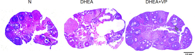
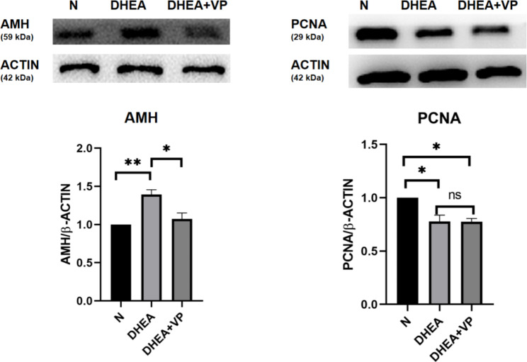
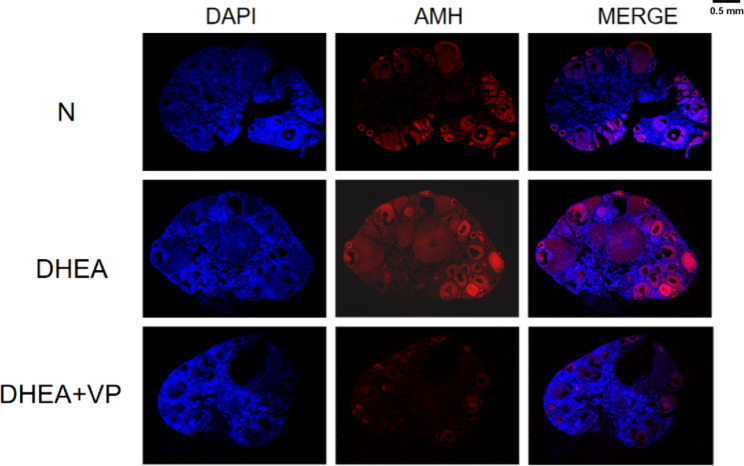
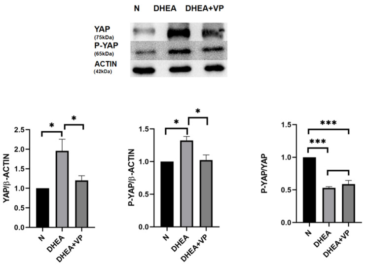
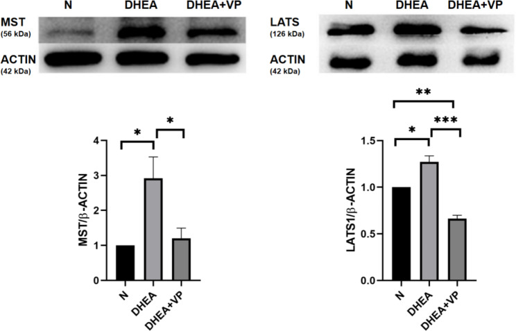
Method: 30 3-week-old BALB/C female rats were randomly divided into control group (n = 10), DHEA group (n = 10) and DHEA + VP group (n = 10). The morphology of ovary and the degree of follicular development were observed by HE staining, and the expression and location of AMH in ovarian follicles were observed by immunofluorescence. The ovarian reserve function index AMH, cell proliferation index PCNA and the ratio of Hippo pathway related proteins MST, LATS, YAP, P-YAP and P-YAP/YAP were detected by Western blot.
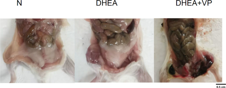
Results: After dividing 30 3-week-old female mice into control, dehydroepiandrosterone (DHEA; model of PCOS), and DHEA + VP groups, we found that the number of small follicles increased in the DHEA group compared to the control group. Additionally, in the DHEA group compared to the control group, anti-müllerian hormone (AMH; ovarian reserve index) increased, proliferating cell nuclear antigen (PCNA; cell proliferation index) decreased, and upstream (MST and LATS) and downstream (YAP and p-YAP) proteins in the Hippo pathway increased, though the p-YAP/YAP ratio decreased. VP ameliorated the increases in AMH, MST, LATS, YAP and p-YAP, but did not ameliorate the decrease in the p-YAP/YAP ratio.
Conclusions: This study indicates that the increased small follicles in the ovaries and changes in ovarian reserve and cell proliferation may be closely related to Hippo pathway activation. This suggests that the Hippo pathway may be an important pathway affecting the proliferation and development of follicles and the occurrence of PCOS.
Keywords: Follicular development; Hippo pathway; Polycystic ovary syndrome.
© 2024. The Author(s).
Conflict of interest statement
The authors declare no competing interests.
Figures
Similar articles
-
Anti-Müllerian hormone regulates ovarian granulosa cell growth in PCOS rats through SMAD4.Int J Gynaecol Obstet. 2025 Jul;170(1):209-221. doi: 10.1002/ijgo.16184. Epub 2025 Jan 24. Int J Gynaecol Obstet. 2025. PMID: 39865361
-
Long-term treatment with dehydroepiandrosterone may lead to follicular atresia through interaction with anti-Mullerian hormone.J Ovarian Res. 2014 Apr 30;7:46. doi: 10.1186/1757-2215-7-46. eCollection 2014. J Ovarian Res. 2014. PMID: 24851135 Free PMC article.
-
[Mechanism of resveratrol in protecting ovary in poor ovarian response mice by regulating Hippo signaling pathway].Zhongguo Zhong Yao Za Zhi. 2024 Feb;49(3):744-753. doi: 10.19540/j.cnki.cjcmm.20230915.401. Zhongguo Zhong Yao Za Zhi. 2024. PMID: 38621878 Chinese.
-
Interactions between androgens, FSH, anti-Müllerian hormone and estradiol during folliculogenesis in the human normal and polycystic ovary.Hum Reprod Update. 2016 Nov;22(6):709-724. doi: 10.1093/humupd/dmw027. Epub 2016 Aug 27. Hum Reprod Update. 2016. PMID: 27566840 Review.
-
[Anti-Mullerian hormone- its role in the pathogenesis of the polycystic ovary syndrome].Akush Ginekol (Sofiia). 2012;51(6):22-6. Akush Ginekol (Sofiia). 2012. PMID: 23390860 Review. Bulgarian.
Cited by
-
Hyperandrogenism-mediated YAP activation drives ovarian inflammation and pyroptosis in PCOS: implications for follicular dysfunction.J Ovarian Res. 2025 Jul 30;18(1):170. doi: 10.1186/s13048-025-01757-5. J Ovarian Res. 2025. PMID: 40739575 Free PMC article.
-
The Role of Mannitol and Vitamin D in Ovarian Ischemia/Reperfusion Injury in Rats with Acute Abdominal.Curr Issues Mol Biol. 2024 Aug 15;46(8):8903-8913. doi: 10.3390/cimb46080526. Curr Issues Mol Biol. 2024. PMID: 39194743 Free PMC article.
-
Correction: Correlation between ovarian follicular development and Hippo pathway in polycystic ovary syndrome.J Ovarian Res. 2024 Nov 20;17(1):230. doi: 10.1186/s13048-024-01557-3. J Ovarian Res. 2024. PMID: 39568054 Free PMC article. No abstract available.
-
Effects of melatonin in polycystic ovary syndrome: is there Hippo pathway crosstalk?J Ovarian Res. 2025 May 14;18(1):101. doi: 10.1186/s13048-025-01642-1. J Ovarian Res. 2025. PMID: 40369589 Free PMC article. Review.
References
-
- Lizneva D, Suturina L, Walker W, Brakta S, Gavrilova-Jordan L, Azziz R. Criteria, prevalence, and phenotypes of polycystic ovary syndrome. Fertil Steril. 2016;106:6–15. 10.1016/j.fertnstert.2016.05.003. PMID 27233760. - PubMed
-
- Hoeger KM, Dokras A, Piltonen T. Update on PCOS: consequences, challenges, and guiding treatment J Clin Endocrinol Metab. 2021;106:e1071-83. 10.1210/clinem/dgaa839, PMID 33211867. - PubMed
-
- Tsai-Turton MT, Luong BT, Tan Y, Luderer U. Cyclophosphamide-induced apoptosis in COV434 human granulosa cells involves oxidative stress and glutathione depletion. Toxicol Sci. 2007;98:216–30. 10.1093/toxsci/kfm087. PMID 17434952. - PubMed
-
- Jakimiuk AJ, Weitsman SR, Navab A, Magoffin DA. Luteinizing hormone receptor, steroidogenesis acute regulatory protein, and steroidogenic enzyme messenger ribonucleic acids are overexpressed in thecal and granulosa cells from polycystic ovaries. J Clin Endocrinol Metab. 2001;86:1318–23. - PubMed
MeSH terms
Substances
LinkOut - more resources
Full Text Sources
Medical
Miscellaneous
