Review
. 2024 Jan 13;43(1):19.
doi: 10.1186/s13046-023-02925-5.

Modulating ferroptosis sensitivity: environmental and cellular targets within the tumor microenvironment

Affiliations
Review

Modulating ferroptosis sensitivity: environmental and cellular targets within the tumor microenvironment

Yuze Hua et al. J Exp Clin Cancer Res. .

Abstract

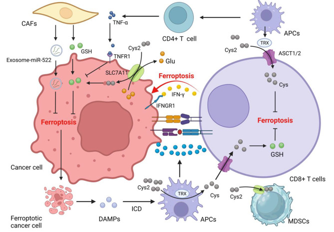
Ferroptosis, a novel form of cell death triggered by iron-dependent phospholipid peroxidation, presents significant therapeutic potential across diverse cancer types. Central to cellular metabolism, the metabolic pathways associated with ferroptosis are discernible in both cancerous and immune cells. This review begins by delving into the intricate reciprocal regulation of ferroptosis between cancer and immune cells. It subsequently details how factors within the tumor microenvironment (TME) such as nutrient scarcity, hypoxia, and cellular density modulate ferroptosis sensitivity. We conclude by offering a comprehensive examination of distinct immunophenotypes and environmental and metabolic targets geared towards enhancing ferroptosis responsiveness within the TME. In sum, tailoring precise ferroptosis interventions and combination strategies to suit the unique TME of specific cancers may herald improved patient outcomes.

Keywords: Cellular metabolism; Ferroptosis; Tumor Microenvironment; Tumor immunity.


Conflict of interest statement

The authors declare no competing interests.

Figures

Fig. 1
Fig. 1
Regulation of ferroptosis [242]: cells require PUFAs for maintaining energy metabolism, with tremendous ROS production to oxidize PUFAs, especially AAs into PLOOH, which leads to cell membrane rupture and cell death. Iron catalyzes this process. System Xc transports Cys2 for the synthesis of GSH. GPX4 reduces the PLOOH into PLOH by GSH to resist ferroptosis. TXN pathway plays as an alternative way when GPX4 is inhibited. BH4 and CoQ also act as anti-ferroptosis way due to the function of eliminating ROS. In mitochondria, where ROS proliferates, DHODH inhibits mitochondrial ferroptosis cooperating with mitochondria GPX4
Fig. 2
Fig. 2
TANs are attracted by necrosis cancer tissue releasing HMGB1 and move to TME, where they are polarized and activated as N2-type TANs by TGF-β and G-CSF secreted from cancer cells. These TANs promote the ferroptosis of cancer cells by MPO, which fuels tumor progression. Ferroptotic cancer cells attract TAMs by other DAMPs, including 4-HNE, 8-OHG, and SAPE-OOH, and these TAMs are polarized into pro-tumor M2-type TAMs. Ferroptotic cancer cells amplify immune response by inducing ICD to mature and activate APCs
Fig. 3
Fig. 3
The complex interaction of cells in the TME: CD8+ T cells kill cancer cells by promoting ferroptosis. Ferroptotic cancer cells release damage-associated molecular patterns (DAMPs), inducing the maturation of DCs and enhancing immune response. Activated APCs like DCs and macrophages reduce Cys2 into Cys to supply CD8+ T cells for GSH synthesis. MDSCs with a powerful intracellular storage capacity of Cys2 compete with APCs for Cys2, promoting T cell ferroptosis. CAFs are principal donor cells of GSH for cancer cells to help cancer cells fight ferroptosis and resist chemotherapy drugs like Pt. CAFs also secrete exosome-miR-522 to inhibit ALOX15 translation in cancer cells. CD8+ T cells promote GGT5 and interfere with system Xc expression reducing GSH supplements for cancer cells
Fig. 4
Fig. 4
Various metabolism process exists in TME, including lipid, amino acid, and glucose metabolism. Cholesterol in TME induces CD36 overexpression in T cells to promote the absorption of PUFAs, Ox-LDL, and Ox-PL in cells, fueling the ferroptosis of Tregs and CD8+ T cells. However, CD36 expression on Tregs maintains mitochondrial fitness to protect them from ferroptosis. Intensive glycolysis of cancer cells in TME leads to glucose starvation. It dictates ferroptosis resistance by activating the AMPK pathway to improve the MUFAs proportion of cell membrane and inducing over-expression of PDK4 to repress the TCA cycle and activity of ALOX. Lactic acid is taken in cancer cells and activates the AMPK pathway and ACSL4 to inhibit ferroptosis. Gln has recently been thought of as the leading energy metabolism resource participating in TCA and FA synthesis by glutaminolysis. Gln metabolism antagonizes with system Xc antioxidation function

References

    1. Maman S, Witz IP. A history of exploring cancer in context. Nat Rev Cancer. 2018;18(6):359–76. doi: 10.1038/s41568-018-0006-7. - DOI - PubMed
    1. Quail DF, Joyce JA. Microenvironmental regulation of Tumor progression and Metastasis. Nat Med. 2013;19(11):1423–37. doi: 10.1038/nm.3394. - DOI - PMC - PubMed
    1. Joyce JA, Fearon DT. T cell exclusion, immune privilege, and the Tumor microenvironment. New York, N.Y.): Science; 2015. pp. 74–80. - PubMed
    1. Salmon H, et al. Matrix architecture defines the preferential localization and migration of T cells into the stroma of human lung tumors. J Clin Investig. 2012;122(3):899–910. doi: 10.1172/JCI45817. - DOI - PMC - PubMed
    1. Borst J, et al. CD4 T cell help in cancer immunology and immunotherapy. Nat Rev Immunol. 2018;18(10):635–47. doi: 10.1038/s41577-018-0044-0. - DOI - PubMed