. 2024 Jan 13;57(1):3.
doi: 10.1186/s40659-023-00475-w.

MSC-derived exosomes protect auditory hair cells from neomycin-induced damage via autophagy regulation

Affiliations

MSC-derived exosomes protect auditory hair cells from neomycin-induced damage via autophagy regulation

Huan Liu et al. Biol Res. .

Abstract

Background: Sensorineural hearing loss (SNHL) poses a major threat to both physical and mental health; however, there is still a lack of effective drugs to treat the disease. Recently, novel biological therapies, such as mesenchymal stem cells (MSCs) and their products, namely, exosomes, are showing promising therapeutic potential due to their low immunogenicity, few ethical concerns, and easy accessibility. Nevertheless, the precise mechanisms underlying the therapeutic effects of MSC-derived exosomes remain unclear.

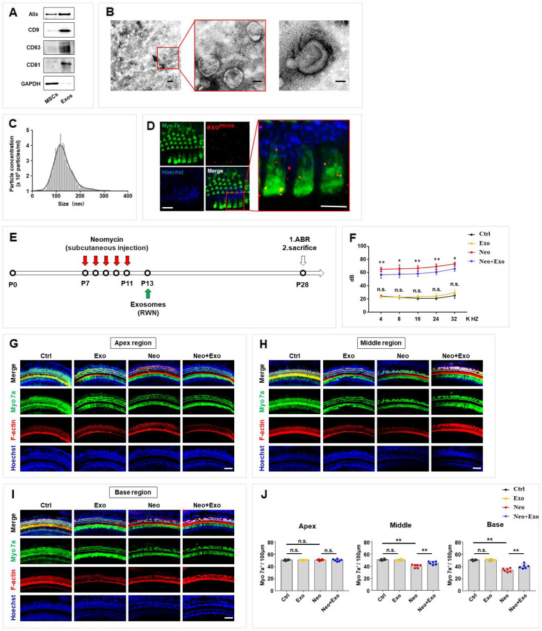
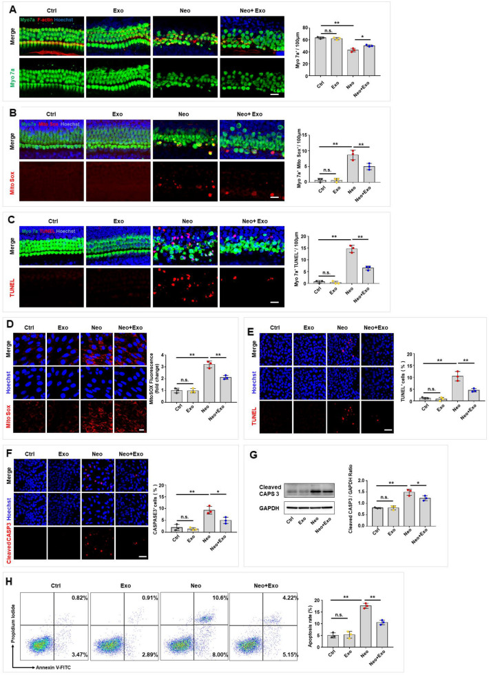
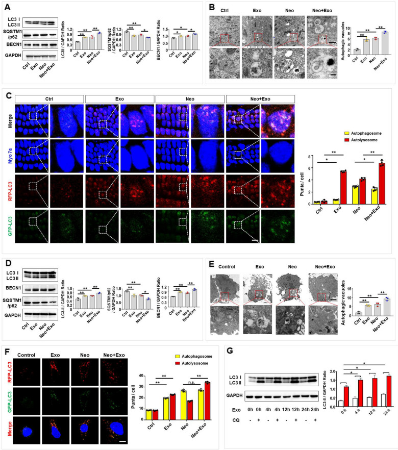
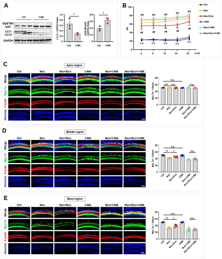
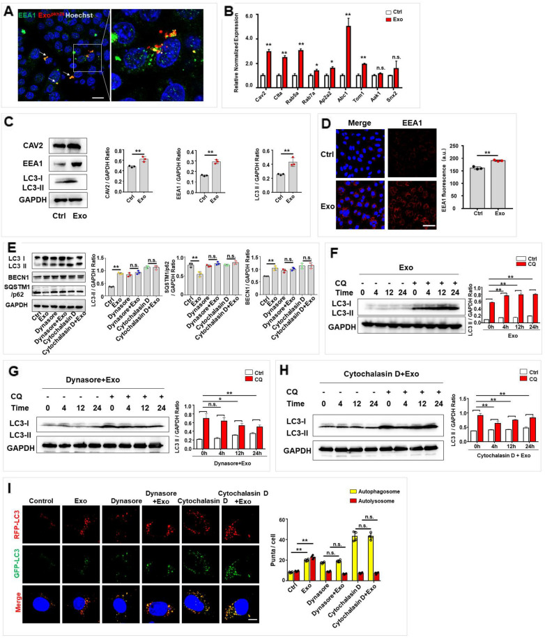
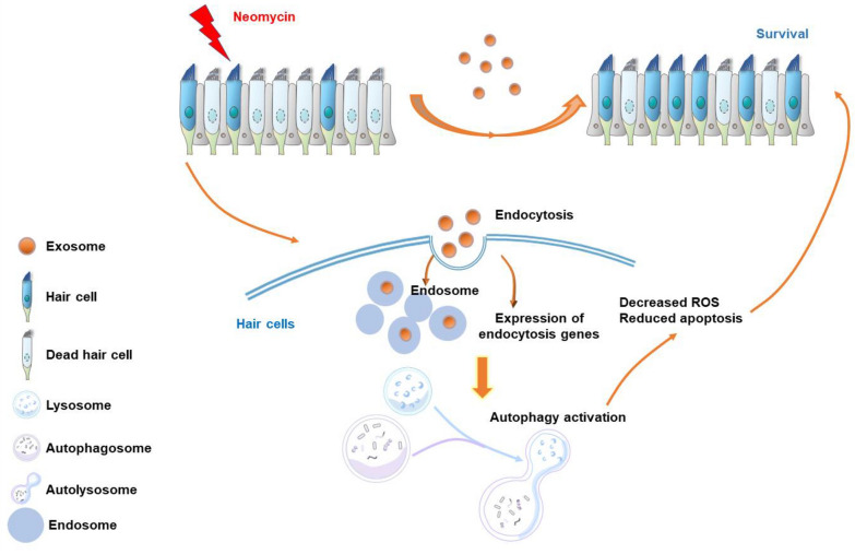
Results: Exosomes derived from MSCs reduced hearing and hair cell loss caused by neomycin-induced damage in models in vivo and in vitro. In addition, MSC-derived exosomes modulated autophagy in hair cells to exert a protective effect. Mechanistically, exogenously administered exosomes were internalized by hair cells and subsequently upregulated endocytic gene expression and endosome formation, ultimately leading to autophagy activation. This increased autophagic activity promoted cell survival, decreased the mitochondrial oxidative stress level and the apoptosis rate in hair cells, and ameliorated neomycin-induced ototoxicity.

Conclusions: In summary, our findings reveal the otoprotective capacity of exogenous exosome-mediated autophagy activation in hair cells in an endocytosis-dependent manner, suggesting possibilities for deafness treatment.

Keywords: Aminoglycoside; Autophagy; Endocytosis; Exosome; Hair cell; Mesenchymal stem cell; Neomycin.


Conflict of interest statement

The authors have no relevant financial or non-financial interests to disclose.

Figures

Fig. 1
Fig. 1
Characterization of exosomes and their capacity to reduce hearing loss and decrease hair cell loss after neomycin damage in mice. A Western blot analysis of exosome markers Alix, CD9, CD63, and CD81 in MSCs and MSC-derived exosomes, and the negative intracellular protein marker GAPDH. B Transmission electron microscope (TEM) analysis of MSC-derived exosomes. Scale bar, 200 nm (left), 100 nm (middle) and 50 nm (right). C Size distribution of exosomes measured by nanoparticle tracking analysis (NTA). D Confocal microscopy image showing exosomes internalization by hair cells in vitro. Scale bar, 20 μm (left) and 10 μm (right). (E) Schematic diagram of animal experiment workflow, round window niche (RWN). F Analysis of ABR thresholds in mice treated with neomycin (Neo, 200 mg/kg for five consecutive days) and/or exosomes (Exo, 20 μg in 10 μl PBS). n = 6 mice GI Immunofluorescence staining with Myo 7a (green), F-actin (red) and Hoechst (blue) in the apical (G), middle (H), and basal (I) turns of the cochleae from different groups. Scale bars, 50 μm. J Quantification of Myo 7a-positive hair cells in the apical, middle, and basal turns of the cochleae. The results were representative of the data generated in at least three independent experiments. The data were presented as mean ± s.d. n.s., not significant; *P < 0.05; **P < 0.01 by one-way ANOVA (F, J)
Fig. 2
Fig. 2
Exosomes protected hair cells against neomycin-induced damage in vitro. AC Immunofluorescence staining was performed after neomycin damage (0.5 mM, 24 h) then with/without exosomes treatments (30 μg/ml, 24 h) in the middle turn of cochleae. A Immunofluorescence staining with Myo 7a (green), F-actin (red) and Hoechst (blue) after different treatments. Quantification of Myo 7a-positive hair cells per 100 μm in the middle turn of different groups. Scale bar, 20 µm. B Immunofluorescence staining with Myo 7a (green), Mito-SOX (red) and Hoechst (blue) after different treatments. The numbers and proportions of Mito-SOX and Myo 7a double-positive cells were quantified. Scale bar, 20 µm. C Immunofluorescence staining with Myo 7a (green), TUNEL (red), and Hoechst (blue) after different treatments. The numbers of TUNEL and Myo 7a double-positive cells were quantified. Scale bar, 20 µm. DH HEI-OC1 cells were treated with neomycin (2 mM, 24 h), then with/ without exosomes (30 μg/ml, 24 h) treatment. D HEI-OC1 cells were labeled with Mito-SOX (red) and Hoechst (blue) and the relative fluorescence intensity was quantified. Scale bar, 20 µm. E TUNEL and Hoechst double staining and F Cleaved CASP3 and Hoechst double staining were performed to detect the percentage of apoptotic HEI-OC1 cells. Scale bar, 50 µm. G Cleaved CASP3 expression was detected by western blot in HEI-OC1 cells and was quantified by ImageJ software. H Analysis of apoptotic HEI-OC1 cells by flow cytometry. The results were representative of the data generated in at least three independent experiments. The data were presented as mean ± s.d. n.s., not significant; *P < 0.05; **P < 0.01 by one-way ANOVA (A-H)
Fig. 3
Fig. 3
Exosomes improved autophagy in cochlear hair cells and HEI-CO1 cells. A Expression of LC3, SQSTM1/p62 and BECN1 in cochlear explants treated with neomycin (0.5 mM, 24 h) and/or exosomes (30 μg/ml, 24 h) was evaluated by western blot and quantified by ImageJ software. B TEM analysis was used to evaluate autophagy in cochlear hair cells, and the numbers of autophagic vacuoles were quantified. Scale bar, 1 µm (up), 200 nm (down). C Immunofluorescence staining with Myo 7a (blue) in cochleae from CAG-RFP-EGFP-LC3 mice. The numbers of autophagosomes (yellow) and autolysosomes (red-only puncta) per cell were quantified. Scale bar, 10 µm. D Expression of LC3, SQSTM1/p62 and BECN1 in HEI-OC1 cells treated with neomycin and/or exosomes was evaluated by western blot and quantified by ImageJ software. E Autophagy in HEI-OC1 cells was detected by TEM, and the numbers of autophagic vacuoles were quantified. Scale bar, 2 µm (up), 500 nm (down). F HEI-OC1 cells were infected with mRFP-GFP-LC3 (tfLC3) and then treated with neomycin and/or exosomes. The numbers of autophagosomes (yellow) and autolysosomes (red-only puncta) per cell were quantified. Scale bar, 10 µm. G Autophagy flux was evaluated by western blotting for LC3 with or without CQ (20 μM). Autophagy flux assay was used in HEI-OC1 cells treated with exosomes at different time points, and LC3-II (CQ-Ctrl) was quantified by ImageJ software. The results were representative of the data generated in at least three independent experiments and presented as mean ± s.d. n.s., not significant; *P < 0.05; **P < 0.01 by one-way ANOVA (A-G)
Fig. 4
Fig. 4
Autophagy was required for exosome-mediated hearing functional recovery in the neomycin-induced SNHL mice model. A The expression levels of LC3 and SQSTM1/p62 in cochlear tissues were evaluated by western blot after 3-MA treatment and LC3-II and SQSTM1/p62 was quantified by ImageJ software. B ABR thresholds were analyzed in mice with different treatment. Untreated control (Ctrl, black), neomycin alone (Neo, yellow), neomycin and exosome treatment (Neo + Exo, red) 3-MA alone (3-MA, blue), neomycin and 3-MA (Neo + 3-MA, green), and neomycin, exosome and 3-MA (Neo + Exo + 3-MA, pink). n = 6 mice. ##p < 0.01 Ctrl vs. Neo; *p < 0.01; **p < 0.01 Neo vs. Neo + Exo; n.s., not significant Ctrl vs. 3-MA; NS, not significant Neo + 3-MA vs. Neo + Exo + 3-MA by one-way ANOVA. CE Immunofluorescence staining with Myo 7a (green), F-actin (red), and Hoechst (blue) in the apical (C), middle (D), and basal (E) turns of cochleae from different groups. Scale bars = 50 μm. Quantification of Myo 7a-positive hair cells in the apical (C), middle (D), and basal (E) turns of the cochleae from different groups. The results were representative of the data generated in at least three independent experiments and data were presented as mean ± s.d. n.s., not significant; *P < 0.05; **P < 0.01 by Student’s t-test (A) or by one-way ANOVA (BE)
Fig. 5
Fig. 5
Autophagy was necessary for exosome-mediated otoprotection. AC Cochlear explants were treated with 3-MA (5 mM) for 16 h. Cell survival, oxidative stress and apoptosis of hair cells were respectively detected by phalloidin staining (A), Mito-SOX Red (B), and TUNEL assay (C) following co-culture with exosomes after neomycin exposure. The number of F-actin and Myo 7a double-positive cells, Mito-SOX and Myo 7a double-positive cells and TUNEL and Myo 7a double-positive cells per 100 μm in the middle turn of different groups were quantified. Scale bar, 20 µm. DH HEI-OC1 cells were treated with 3-MA (5 mM) for 16 h to inhibit autophagy. D HEI-OC1 cells were labeled with Mito-SOX (red), and the relative fluorescence intensity was quantified after different treatments. Scale bar, 20 µm. E TUNEL and Hoechst double staining and F Cleaved CASP3 and Hoechst double staining were performed to detect the percentage of apoptotic HEI-OC1 cells after different treatments. Scale bar, 50 µm. G Cleaved CASP3 expression was detected by western blot in HEI-OC1 cells treated with exosomes and/or 3-MA following neomycin insults and was quantified by ImageJ software. H Analysis of apoptotic HEI-OC1 cells by flow cytometry after different treatments. The results were representative of the data generated in at least three independent experiments. The data were presented as mean ± s.d. n.s., not significant; *P < 0.05; **P < 0.01 by one-way ANOVA (AH)
Fig. 6
Fig. 6
Exosomes promoted endocytosis, which is required for exosomes to upregulate autophagy of hair cells. A PKH26-labeled exosomes co-localized with endosomes, which were immunofluorescence stained with EEA1 in HEI-OC1. Scale bar: 20 µm. B RT-qPCR was performed to detect the endocytosis-associated mRNA levels of HEI-OC1 cells after treatment with exosomes (30 μg/ml, 24 h). C The protein expression of CAV2, EEA1 and LC3 was detected by western blot and was quantified by ImageJ software. D The expression of EEA1 was detected by immunofluorescence staining, and the fluorescence intensity was quantified by ImageJ software. Scale bar, 50 μm. E Expression of LC3, SQSTM1/p62 and BECN1 were detected by western blot after exosome treatment with or without pre-treatment of dynasore (80 μM, 4 h) or cytochalasin D (2 μM, 30 min) and were quantified by ImageJ software. F Autophagy flux assay of HEI-OC1 cells treated with exosomes without endocytosis inhibition, and LC3-II (CQ-Ctrl) was quantified by ImageJ software. G, H Autophagy flux assay of HEI-OC1 cells treated with exosomes with endocytosis inhibition by either dynasore (G) or cytochalasin D (H), and LC3-II (CQ-Ctrl) was quantified by ImageJ software. I HEI-OC1 cells were infected with mRFP-GFP-LC3 (tfLC3) and then treated with exosomes with/without dynasore or cytochalasin D. The numbers of autophagosomes (yellow) and autolysosomes (red-only puncta) per cell were quantified. Scale bar, 10 µm. The results were representative of the data generated in at least three independent experiments. The data were presented as mean ± s.d. n.s., not significant; *P < 0.05; **P < 0.01 by Student’s t-test (BD) or by one-way ANOVA (EI)
Fig. 7
Fig. 7
Schema for MSC-derived exosomes protect auditory hair cells from neomycin-induced damage by regulating the autophagy of recipient hair cells. This work describes that exogenously exosomes were internalized by hair cells and subsequently upregulated endocytic gene expression and endosome formation, leading to autophagy activation, which ultimately protect against neomycin-induced damage

References

    1. Deafness and Hearing Loss. Available online at: WHO; https://www.who.int/news-room/fact-sheets/detail/deafness-and-hearing-loss
    1. Wang J, Puel JL. Toward cochlear therapies. Physiol Rev. 2018;98(4):2477–2522. doi: 10.1152/physrev.00053.2017. - DOI - PubMed
    1. Geleoc GS, Holt JR. Sound strategies for hearing restoration. Science. 2014;344(6184):1241062. doi: 10.1126/science.1241062. - DOI - PMC - PubMed
    1. Copeland BJ, Pillsbury HC., 3rd Cochlear implantation for the treatment of deafness. Annu Rev Med. 2004;55:157–167. doi: 10.1146/annurev.med.55.091902.105251. - DOI - PubMed
    1. Wolf BJ, Kusch K, Hunniford V, Vona B, Kuhler R, Keppeler D, et al. Is there an unmet medical need for improved hearing restoration? EMBO Mol Med. 2022;14(8):e15798. doi: 10.15252/emmm.202215798. - DOI - PMC - PubMed

LinkOut - more resources