

Spontaneous Pituitary Neoplasm in Two Female Geriatric Southern Giant Pouched Rats (Cricetomys ansorgei)
- PMID: 38217070
- PMCID: PMC10752360
- DOI: 10.30802/AALAS-CM-23-000051
Spontaneous Pituitary Neoplasm in Two Female Geriatric Southern Giant Pouched Rats (Cricetomys ansorgei)
Abstract
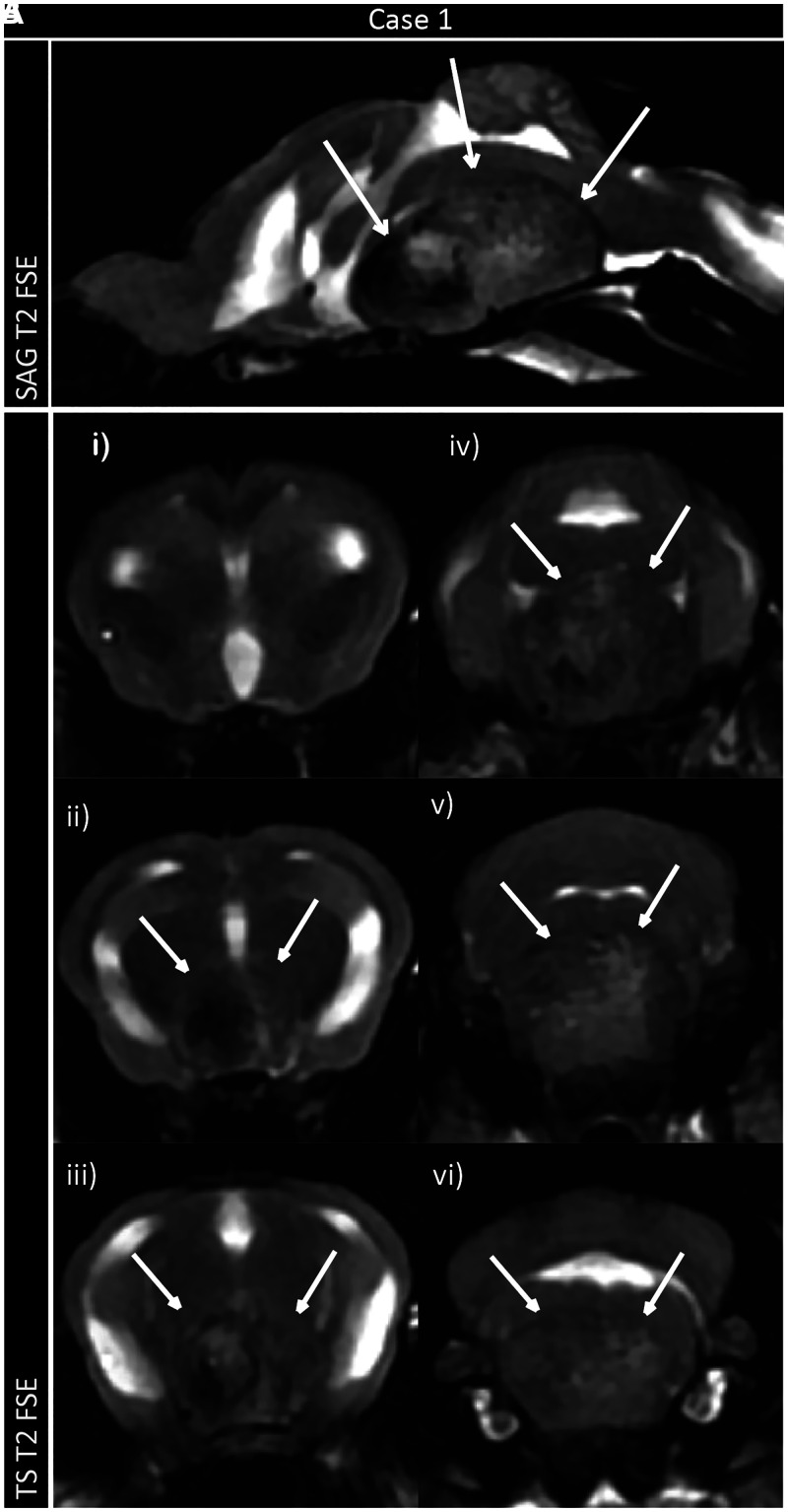
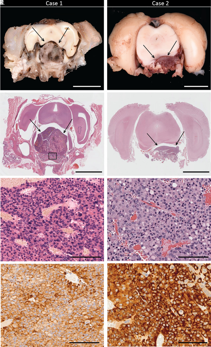
Southern giant pouched rats (Cricetomys ansorgei) are a small muroid species native to the sub-Saharan Africa. Their exceptionally developed olfactory system, trainability, and relatively small size makes them useful working animals for various applications in humanitarian work. At our institution, a breeding colony of Southern giant pouched rats is maintained to study their physiology and utility as scent detectors. This case report describes the occurrence of spontaneous pituitary neoplasms with distinct clinical presentations in 2 geriatric (approximately 7.5 y old) wild-caught female Southern giant pouched rats. The first pouched rat displayed vestibular deficits, including left-sided head tilt, ataxia, disorientation, and circling. MRI revealed a large, focal heterogeneous mass arising from the pituitary fossa. The second pouched rat presented with polyuria, polydipsia, and hyperglycemia but no neurologic signs. Examination after euthanasia revealed a prolactin (PRL)-expressing pituitary carcinoma and adenoma in the first and second pouched rat, respectively, associated with mammary hyperplasia in both animals. This is the first report of spontaneous PRL-producing pituitary tumors in Southern giant pouched rats.
Figures
Similar articles
-
The giant pouched rat (Cricetomys ansorgei) olfactory receptor repertoire.PLoS One. 2020 Apr 2;15(4):e0221981. doi: 10.1371/journal.pone.0221981. eCollection 2020. PLoS One. 2020. PMID: 32240170 Free PMC article.
-
Chromosome-scale genome assembly of the African giant pouched rat (Cricetomys ansorgei) and evolutionary analysis reveals evidence of olfactory specialization.Genomics. 2022 Nov;114(6):110521. doi: 10.1016/j.ygeno.2022.110521. Epub 2022 Nov 8. Genomics. 2022. PMID: 36351561
-
Radiologic Anatomy of the Thorax of the Southern Giant Pouched Rat (Cricetomys ansorgei).Anat Histol Embryol. 2025 Mar;54(2):e70023. doi: 10.1111/ahe.70023. Anat Histol Embryol. 2025. PMID: 39936551
-
[Unilateral exophthalmos caused by a prolactin producing ectopic pituitary adenoma: case report].No Shinkei Geka. 2002 Jun;30(6):623-8. No Shinkei Geka. 2002. PMID: 12094689 Review. Japanese.
-
Rats sniff out pulmonary tuberculosis from sputum: a diagnostic accuracy meta-analysis.Sci Rep. 2021 Jan 21;11(1):1877. doi: 10.1038/s41598-021-81086-x. Sci Rep. 2021. PMID: 33479276 Free PMC article.
References
-
- Barthold SW, Griffey SM, Percy DH. 2016. Pathology of laboratory rodents and rabbits 4th ed. West Sussex (England): Wiley-Blackwell. 10.1002/9781118924051. - DOI
Publication types
MeSH terms
LinkOut - more resources
Full Text Sources
Medical
