Review
. 2024 Feb;44(2):185-204.
doi: 10.1002/cac2.12519. Epub 2024 Jan 13.

Cellular metabolism: A key player in cancer ferroptosis

Affiliations
Review

Cellular metabolism: A key player in cancer ferroptosis

Xianjie Jiang et al. Cancer Commun (Lond). 2024 Feb.

Abstract

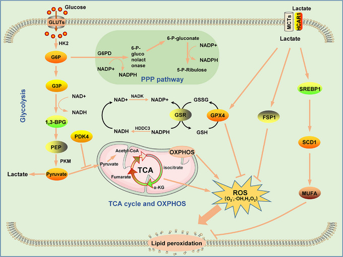
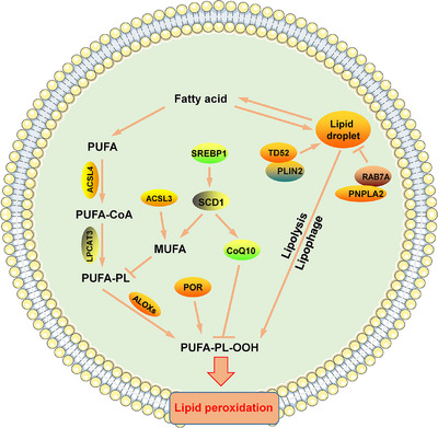
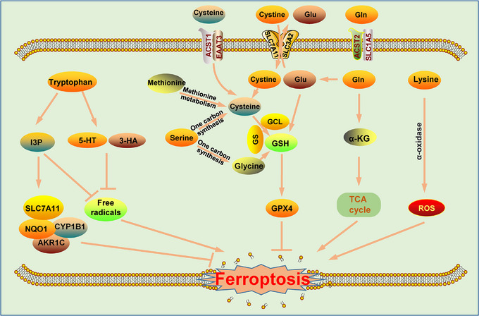
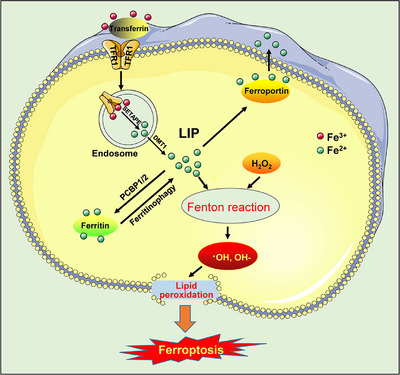
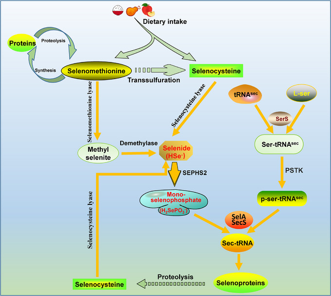
Cellular metabolism is the fundamental process by which cells maintain growth and self-renewal. It produces energy, furnishes raw materials, and intermediates for biomolecule synthesis, and modulates enzyme activity to sustain normal cellular functions. Cellular metabolism is the foundation of cellular life processes and plays a regulatory role in various biological functions, including programmed cell death. Ferroptosis is a recently discovered form of iron-dependent programmed cell death. The inhibition of ferroptosis plays a crucial role in tumorigenesis and tumor progression. However, the role of cellular metabolism, particularly glucose and amino acid metabolism, in cancer ferroptosis is not well understood. Here, we reviewed glucose, lipid, amino acid, iron and selenium metabolism involvement in cancer cell ferroptosis to elucidate the impact of different metabolic pathways on this process. Additionally, we provided a detailed overview of agents used to induce cancer ferroptosis. We explained that the metabolism of tumor cells plays a crucial role in maintaining intracellular redox homeostasis and that disrupting the normal metabolic processes in these cells renders them more susceptible to iron-induced cell death, resulting in enhanced tumor cell killing. The combination of ferroptosis inducers and cellular metabolism inhibitors may be a novel approach to future cancer therapy and an important strategy to advance the development of treatments.

Keywords: cancer therapy; cellular metabolism; ferroptosis; ferroptosis inducer.


Conflict of interest statement

The authors declare that they have no competing interests.

Figures

FIGURE 1
FIGURE 1
The role of glucose metabolism in cancer ferroptosis. Glycolysis: NADH and lactate are generated NADH is a reduction agent that eliminates ROS; lactate promotes the production of MUFAs and induces the expression of GPX4 and SLC7A11. The pentose phosphate pathway (PPP): This pathway produces large amounts of NADPH, which is involved in the reduction of GPX4 and inhibits lipid peroxidation. TCA cycle: the TCA cycle contributes to the accumulation of lipid ROS. OXPHOS ROS are produced and ferroptosis is promoted via the OXPHOS. Abbreviations: HK2, hexokinase 2; G6P, glucose‐6‐phosphate; G3P, glyceraldehyde 3‐phosphate; 1,3‐BPG, 1,3‐bisphosphoglycerate; PEP, phosphoenolpyruvate; PKM, pyruvate kinase, muscle; PDK4, pyruvate dehydrogenase kinase isoform 4; G6PD, glucose‐6‐phosphate 1‐dehydrogenase; FSP1, ferroptosis suppressor protein 1; SREBP1, sterol‐regulatory element binding protein 1; SCD1, stearoyl‐CoA desaturase 1; MUFA, monounsaturated fatty acid; ROS, reactive oxygen species; NADH, nicotinamide adenine dinucleotide; NADPH, nicotinamide adenine dinucleotide phosphate; GPX4, glutathione peroxidase 4; TCA, the tricarboxylic acid; OXPHOS, oxidative phosphorylation.
FIGURE 2
FIGURE 2
The role of lipid metabolism in cancer ferroptosis. Lipid peroxidation: free PUFAs are peroxidized by the action of the ACSL4/LPCAT3/ALOX axis, ultimately inducing ferroptosis. Storage and degradation: free fatty acids are stored in the form of lipid droplets, and TD52 and PLIN2 promote the formation of lipid droplets. Lipolysis and lipophagy are important contributors to the degradation of lipid droplets, resulting in lipid peroxidation. Moreover, RAB7A and PNPLA2 facilitate the degradation of lipid droplets. Regulation: ACSL3 and SREBP1/SCD1 regulate ferroptosis by promoting the synthesis of MUFAs and inhibiting the formation of PUFA‐PLs. In addition, SREBP1/SCD1 inhibits lipid peroxidation by promoting the production of CoQ10, and POR promotes lipid peroxidation. Abbreviations: PUFA, polyunsaturated fatty acid; ACSL4, acyl‐CoA synthetase long‐chain family 4; LPCAT3, lysophosphatidyl transferase 3; ALOXS, lipoxygenase; ACSL3, acetyl‐CoA synthetase family 3; MUFA, monounsaturated fatty acid; POR, cytochrome p450 oxidoreductase; TD52, tumor protein D52; PLIN2, perilipin2; PL, phospholipid.
FIGURE 3
FIGURE 3
Amino acid metabolism in cancer cell ferroptosis. Tryptophan mitigates intracellular oxidative stress by either forming I3P or metabolizing it to yield 5‐HT and 3‐HA. Moreover, tryptophan exerts a protective effect against ferroptosis through the upregulation of SLC7A11/NQO1/CYP1B1/AKR1C pathway activity by inducing I3P activity. After extracellular transport into a cell via the transport carrier SLC7A11/SLC3A2, cystine is rapidly reduced to cysteine (or extracellular cysteine is directly absorbed via ACST1/EAAT3) and synthesized into GSH with glutamate and glycine by the action of GCL and GS. As a cofactor of GPX4, GSH inhibits ferroptosis via enhanced GPX4 activity. methionine is converted into cysteine through methionine metabolism, which is involved in the regulation of ferroptosis; alternatively, serine can be converted into cysteine or glycine through a one‐carbon reaction, and this synthetic pathway is involved in the regulation of GSH. After extracellular Gln enters a cell through the ACST2/SLC1A5 transporter, it can be converted to glutamate and participate in GSH synthesis. Alternatively, glutamate can enter the mitochondria to form α‐KG and thus participates in the TCA cycle and regulates ferroptosis. L‐lysine α‐oxidase activates ferroptosis signaling by catalyzing the oxidative decarboxylation of lysine and ROS production. Abbreviations: I3P, indole‐3‐pyruvate; 5‐HT, serotonin; 3‐HA, 3‐hydroxyanthranilic acid; GCL, glutamate‐cysteine ligase; GS, glutathione synthetase; GSH, glutathione; GPX4, glutathione peroxidase 4; ROS, reactive oxygen species; TCA, tricarboxylic acid; SLC7A11, solute carrier family 7 member 11; NQO1, NAD(P)H quinone dehydrogenase 1; CYP1B1, cytochrome P450 family 1 subfamily B member 1; AKR1C, aldo‐keto reductase family 1 member C; SLC3A2, solute carrier family 3 member 2; ACST1, alanine/serine/cysteine/threonine transporter 1; EAAT3, excitatory amino acid transporter 3; ACST2, alanine/serine/cysteine/threonine transporter 2; SLC1A5, solute carrier family 1 member 5; α‐KG, α‐ketoglutarate
FIGURE 4
FIGURE 4
Iron metabolism in cancer cell ferroptosis. Import: Fe3+ forms a complex with transferrin and TFR1 to enter cells through endocytosis. Transport: Fe3+ entering a cell is reduced to Fe2+ by SETAPE and is then transported to the cytoplasm via DMT1. Storage: Free Fe2+ in the cytoplasm is bound to ferritin vis the action of PCBP1/2 and stored in the cytoplasm. Export: Free Fe2+ in the cytoplasm is transported out of a cell by ferroportin. Abbreviations: TFR1, membrane protein TF receptor 1; SETAPE, six‐transmembrane epithelial antigens of the prostate 3; DMT1, divalent metal transporter 1; PCBP1/2, poly‐(rC)‐binding protein 1/2; LIP, labile iron pool.
FIGURE 5
FIGURE 5
Schematic illustration of selenium metabolism. Metabolic pathways of selenomethionine from plant food sources: (1) protein synthesis; (2) transfer to selenocysteine via transsulfuration; and (3) conversion to selenide. Metabolic pathways of selenocysteine ingested from animal food sources: selenocysteine is converted to selenide by selenocysteine lyase, which is recognized and synthesized as selenoprotein after conversion to sec‐tRNA. Abbreviations: SEPHS2, selenophosphate synthetase 2; HSe, selenide; PSTK, phosphoseryl‐tRNA kinase; L‐ser, L‐serine; SerS, seryl‐tRNA synthetase; SelA, selenocysteine synthase; SecS, selenocysteine tRNA synthase; tRNAsec, selenocysteine‐specific transfer RNA; ser‐tRNAsec, serly‐tRNAsec; p‐ser‐tRNAsec, phosphorylation of serly‐tRNAsec; sec‐tRNA, selenocysteine.

Similar articles

Cited by

References

    1. Qiu H, Cao S, Xu R. Cancer incidence, mortality, and burden in China: a time‐trend analysis and comparison with the United States and United Kingdom based on the global epidemiological data released in 2020. Cancer Commun (Lond). 2021;41(10):1037–1048. - PMC - PubMed
    1. Sung H, Ferlay J, Siegel RL, Laversanne M, Soerjomataram I, Jemal A, et al. Global Cancer Statistics 2020: GLOBOCAN Estimates of Incidence and Mortality Worldwide for 36 Cancers in 185 Countries. CA Cancer J Clin. 2021;71(3):209–249. - PubMed
    1. Dixon SJ, Lemberg KM, Lamprecht MR, Skouta R, Zaitsev EM, Gleason CE, et al. Ferroptosis: an iron‐dependent form of nonapoptotic cell death. Cell. 2012;149(5):1060–1072. - PMC - PubMed
    1. Jiang X, Stockwell BR, Conrad M. Ferroptosis: mechanisms, biology and role in disease. Nat Rev Mol Cell Biol. 2021;22(4):266–282. - PMC - PubMed
    1. Chen X, Kang R, Kroemer G, Tang D. Broadening horizons: the role of ferroptosis in cancer. Nat Rev Clin Oncol. 2021;18(5):280–296. - PubMed

Publication types

Grants and funding