. 2024 Mar 6;226(3):iyae004.
doi: 10.1093/genetics/iyae004.

Natural variation in the zinc-finger-encoding exon of Prdm9 affects hybrid sterility phenotypes in mice

Affiliations

Natural variation in the zinc-finger-encoding exon of Prdm9 affects hybrid sterility phenotypes in mice

Khawla F N AbuAlia et al. Genetics. .

Abstract

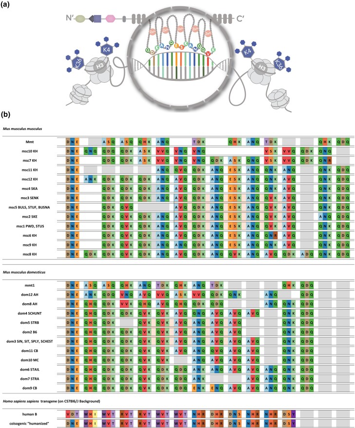
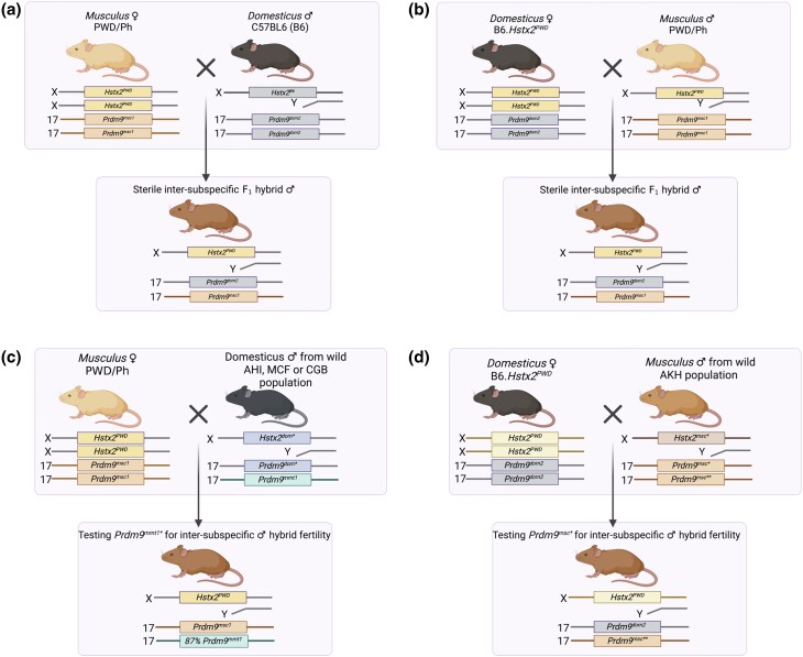
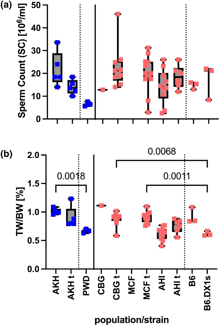
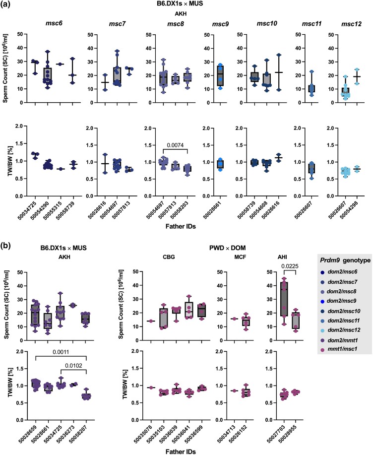
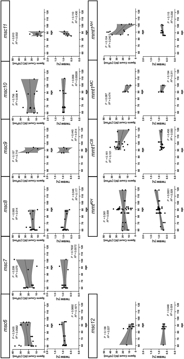
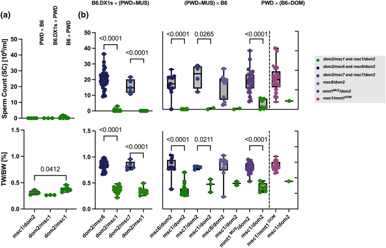
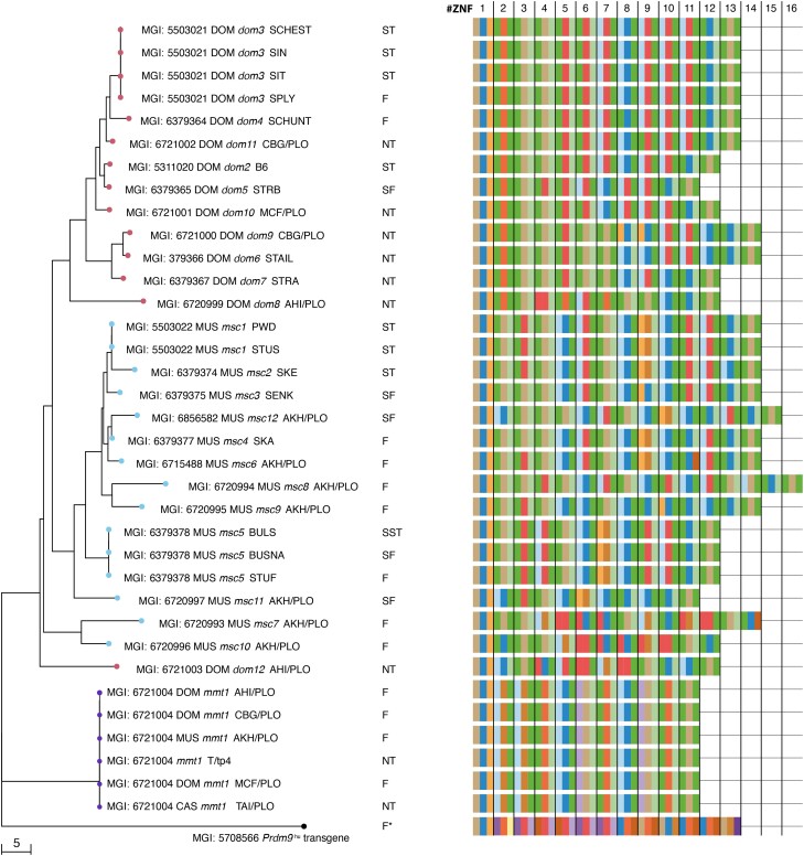
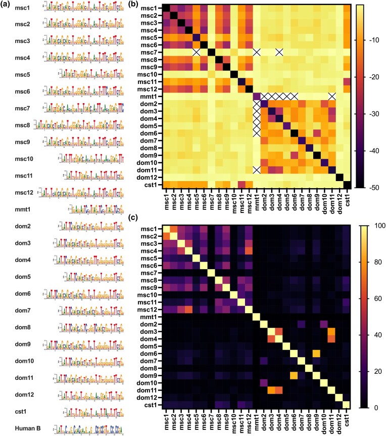
PRDM9-mediated reproductive isolation was first described in the progeny of Mus musculus musculus (MUS) PWD/Ph and Mus musculus domesticus (DOM) C57BL/6J inbred strains. These male F1 hybrids fail to complete chromosome synapsis and arrest meiosis at prophase I, due to incompatibilities between the Prdm9 gene and hybrid sterility locus Hstx2. We identified 14 alleles of Prdm9 in exon 12, encoding the DNA-binding domain of the PRDM9 protein in outcrossed wild mouse populations from Europe, Asia, and the Middle East, 8 of which are novel. The same allele was found in all mice bearing introgressed t-haplotypes encompassing Prdm9. We asked whether 7 novel Prdm9 alleles in MUS populations and the t-haplotype allele in 1 MUS and 3 DOM populations induce Prdm9-mediated reproductive isolation. The results show that only combinations of the dom2 allele of DOM origin and the MUS msc1 allele ensure complete infertility of intersubspecific hybrids in outcrossed wild populations and inbred mouse strains examined so far. The results further indicate that MUS mice may share the erasure of PRDM9msc1 binding motifs in populations with different Prdm9 alleles, which implies that erased PRDM9 binding motifs may be uncoupled from their corresponding Prdm9 alleles at the population level. Our data corroborate the model of Prdm9-mediated hybrid sterility beyond inbred strains of mice and suggest that sterility alleles of Prdm9 may be rare.

Keywords: Hstx2; Mus musculus; Prdm9; t-haplotype; asynapsis; fertility; reproductive isolation.


Conflict of interest statement

Conflicts of interest The author(s) declare no conflicts of interest.

Figures

Fig. 1.
Fig. 1.
Types of C2H2 zinc-finger arrays studied for hybrid sterility phenotypes. a) Cartoon depicting the amino acids in positions −1, 3, and 6 of the alpha-helix of each C2H2 ZNFs that are responsible for DNA binding. b) Representation of all C2H2 zinc-finger arrays using only acronyms of the amino-acid positions responsible for DNA-binding (as in Supplementary Figs. 2 and 3) zinc-finger arrays of Prdm9 alleles msc2, msc3, msc4, msc5, dom3, dom4, dom5, dom6, and dom7 with alleles from Mukaj et al. (2020).
Fig. 2.
Fig. 2.
Scheme of intersubspecific crosses and genotypes. a) Classical model for Prdm9-driven hybrid male sterility (PWD × B6) and b) reciprocal HS model of B6.Hstx2PWD female crossed to B6 male. a, b) Hybrid male sterility depends on 3 main factors: the incompatibility of the PWD and B6 alleles of the Prdm9 gene (Prdm9msc1 and Prdm9dom2), the presence of the PWD (musculus) allele of the X-linked Hstx2 locus, and the MUS/DOM heterozygosity of the F1 genetic background. c) By substituting the B6 male for the wild DOM male, a change in hybrid sterility can be attributed to the wild DOM Prdm9 allele. Prdm9mmt1 was over-transmitted with the t-haplotype as all wild DOM were heterozygous for t-haplotype. d) By substituting the PWD male for the wild MUS male, a change in hybrid sterility can be attributed to the tester wild MUS Prdm9 alleles with an unknown effect on hybrid male fertility, Prdm9msc*, or Prdm9mmt1 (not shown again in MUS for simplicity). Image created with BioRender.
Fig. 3.
Fig. 3.
Fertility parameters of wild mouse populations in Ploen, Germany. MUS: AKH, Almaty, Kazakhstan; DOM: AHI, Ahvaz, Iran; CBG, Cologne-Bonn Germany; MCF, Massif-Central France; t, tt/wt-haplotype genotype. a) Represents the sperm count for each population. b) Represents the values of the normalized testes weight (TW/BW) ratio for the same populations—we compared fertility parameters between populations using pairwise ANOVA with Bonferroni correction.
Fig. 4.
Fig. 4.
Fertility phenotypes of hybrid offspring grouped by Prdm9 genotype and sire ID (a) mice with different MUS Prdm9 from Kazakhstan without t-haplotypes. Each graph represents offspring sorted by Prdm9 genotype, with the father (sire) ID on the X-axis. Only the second sire (50054290) was homozygous for Prdm9, while all of the others were heterozygous for different Prdm9 alleles. b) Offspring with t-haplotypes is grouped by the source population of the fathers and sire IDs on the X-axis. (Left) MUS from Kazakhstan (right), DOM from Cologne-Bonn, Germany (CBG), Massif-Central, France (MCF), and Ahvaz, Iran (AHI). Data pairs were compared using Welch's t-test, and multiple comparisons were performed using ANOVA with Kruskal–Wallis’ test and corrected for multiple comparisons using Dunn's test. Only significant values are shown on the graph.
Fig. 5.
Fig. 5.
Age correlation with fertility parameters grouped by paternal Prdm9 allele of F1 hybrids. Each vertical graph represents correlation analyses between age (in days) and fertility parameters for F1-hybrid offspring arranged based on the fathers’ allele, sperm count on the top graphs, and normalized testes weight (TW/BW) ratio on the bottom. The same data, as in Fig. 1, were analyzed for correlation with age using simple linear regression with 95% confidence intervals shown.
Fig. 6.
Fig. 6.
Fertility parameters of intersubspecific hybrids were grouped by Prdm9 genotype with (a) sperm count and (b) paired testes weights normalized by body weight (TW/BW) tested separately for intersubspecific offspring of (B6.DX1s × wild MUS) or (PWD × wild DOM) intersubspecific crosses (depicted in Fig. 2). We compared their fertility parameters between hybrids grouped by Prdm9 genotype and also to offspring of (B6.DX1s × PWD) and (PWD × B6), with known hybrid sterility phenotypes using pairwise ANOVA with Bonferroni correction (additional statistics shown in Supplementary Tables 3 and 4). All hybrid males carry the Hstx2PWD allele on Chr X. c) The panels show spermatocyte spreads of intersubspecific B6.DX1s × wild MUS hybrids, with differing Prdm9 genotypes. The defects in chromosome asynapsis were assessed by antibody staining for HORMAD2 protein (green), which marks asynapsed autosomal chromosomes in addition to the nonhomologous parts of XY sex chromosomes that are physiologically observed in normally progressing meiocytes. DNA is counterstained with DAPI (blue). Synaptonemal complex assembly was evaluated by SYCP3 protein immunostaining (red) and the presence of yH2AX (gray). At the zygotene/pachytene transition, clouds of yH2AX mark chromatin associated with asynapsed axes. The localized gray dots represent CEN-labeled centromeres. d) The percentages of asynaptic cells on the Y-axis were grouped by the Prdm9 genotype on the X-axis. The percentage of asynaptic cells correlated with fertility parameters of intersubspecific F1 hybrids, namely e) sperm count and f) normalized testes weight (TW/BW) ratio, with dotted lines representing 95% confidence intervals, P-values, and Pearson R2 values.
Fig. 7.
Fig. 7.
Fertility phenotypes segregate with parental Prdm9 alleles in reciprocal intersubspecific hybrids. a) Fertility parameters of control cross with the Prdm9 allelic combination dom2/msc1 and Hstx2 allele from PWD or B6. b) Intersubspecific F1 male offspring of (left) B6.DX1s females crossed to intrasubspecific MUS males (middle), intrasubspecific MUS hybrid females crossed to B6 males (right), and PWD females crossed to intrasubspecific DOM males (as shown in Fig. 2). Data were pooled from parents with the same Prdm9 genotype and compared using pairwise ANOVA with Bonferroni correction, as in Figs. 4 and 5.
Fig. 8.
Fig. 8.
Neighbor-joining tree of the Prdm9 exon12 minisatellite, which encodes the DNA-binding domain of PRDM9 calculated on the nucleotide sequences of all Prdm9 alleles in this study and Mukaj et al. (2020) that code for the C2H2 ZNFs array, with red nodes for DOM and blue nodes for MUS alleles, and with purple nodes depicting the t-haplotype allele found in MUS and DOM. To the right of the tree, a table depicts the C2H2 ZNF array encoded by each allele, with boxes (colored as in Fig. 1, Supplementary Figs. 2 and 3) representing only the amino acids responsible for the DNA contacting of each ZNF.
Fig. 9.
Fig. 9.
In silico predicted PRDM9 DNA binding. (a) PRDM9 DNA-binding motifs are represented as sequence logos of the underlying positional weight matrices, which were predicted using the polynomial kernel method by Persikov et al. (2009) and Persikov and Singh (2014) on translated nucleotide sequences of alleles in this study, and Mukaj et al. (2020). (b) Motifs were compared using TomTom, within the MEME suite, which computes the probabilities that a random motif would be better matched than the input motif. TomTom output P-values were log-transformed and are shown in a heatmap matrix, such that darker colors represent better matching of sequence motifs, and lighter colors represent weaker similarities of motifs, with crossed-out values representing incidences where no similarity was found. When the motif is compared to itself, the probability of another motif binding better than the motif itself is zero in most cases; however, as values of zero cannot be log-transformed, these incidences are represented by black boxes. c) Overlap of genome-wide binding sites, predicted for each PRDM9 binding motif, with brighter colors showing more extensive genomic binding site overlap between different DNA-binding motifs.

Similar articles

Cited by

References

    1. Altemose N, Noor N, Bitoun E, Tumian A, Imbeault M, Chapman JR, Aricescu AR, Myers SR. 2017a. A map of human PRDM9 binding provides evidence for novel behaviors of PRDM9 and other zinc-finger proteins in meiosis. Elife. 6:e28383. doi:10.7554/eLife.28383. - DOI - PMC - PubMed
    1. Altemose N, Noor N, Bitoun E, Tumian A, Imbeault M, Chapman JR, Aricescu AR, Myers SR. 2017b. Human PRDM9 can bind and activate promoters, and other zinc-finger proteins associate with reduced recombination in cis. bioRxiv. 10.1101/144295, preprint: not peer reviewed. - DOI
    1. Ambrosini G, Groux R, Bucher P. 2018. PWMScan: a fast tool for scanning entire genomes with a position-specific weight matrix. Bioinformatics. 34(14):2483–2484. doi:10.1093/bioinformatics/bty127. - DOI - PMC - PubMed
    1. Anderson LK, Reeves A, Webb LM, Ashley T. 1999. Distribution of crossing over on mouse synaptonemal complexes using immunofluorescent localization of MLH1 protein. Genetics. 151(4):1569–1579. doi:10.1093/genetics/151.4.1569. - DOI - PMC - PubMed
    1. Arora UP, Dumont BL. 2022. Meiotic drive in house mice: mechanisms, consequences, and insights for human biology. Chromosome Res. 30(2–3):165–186. doi:10.1007/s10577-022-09697-2. - DOI - PMC - PubMed

Publication types