. 2024 Jan 13;15(1):48.
doi: 10.1038/s41419-024-06434-x.

Akt enhances the vulnerability of cancer cells to VCP/p97 inhibition-mediated paraptosis

Affiliations

Akt enhances the vulnerability of cancer cells to VCP/p97 inhibition-mediated paraptosis

Dong Min Lee et al. Cell Death Dis. .

Abstract

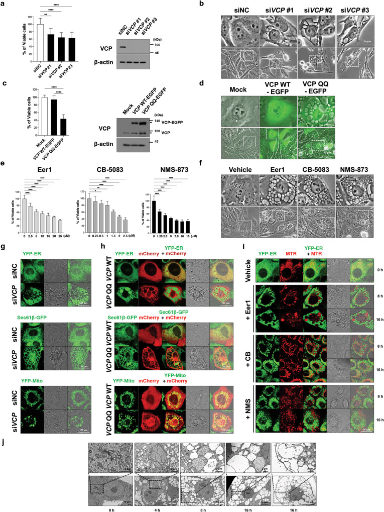
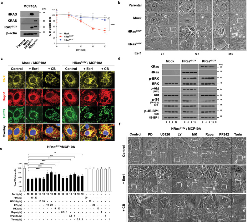
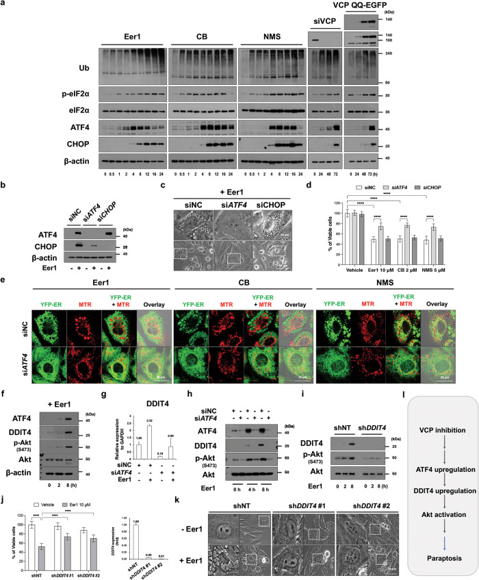
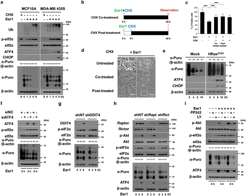
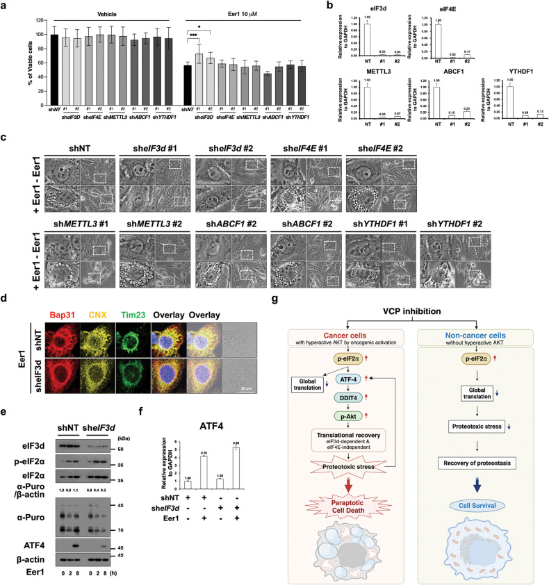
Valosin-containing protein (VCP)/p97, an AAA+ ATPase critical for maintaining proteostasis, emerges as a promising target for cancer therapy. This study reveals that targeting VCP selectively eliminates breast cancer cells while sparing non-transformed cells by inducing paraptosis, a non-apoptotic cell death mechanism characterized by endoplasmic reticulum and mitochondria dilation. Intriguingly, oncogenic HRas sensitizes non-transformed cells to VCP inhibition-mediated paraptosis. The susceptibility of cancer cells to VCP inhibition is attributed to the non-attenuation and recovery of protein synthesis under proteotoxic stress. Mechanistically, mTORC2/Akt activation and eIF3d-dependent translation contribute to translational rebound and amplification of proteotoxic stress. Furthermore, the ATF4/DDIT4 axis augments VCP inhibition-mediated paraptosis by activating Akt. Given that hyperactive Akt counteracts chemotherapeutic-induced apoptosis, VCP inhibition presents a promising therapeutic avenue to exploit Akt-associated vulnerabilities in cancer cells by triggering paraptosis while safeguarding normal cells.


Conflict of interest statement

The authors declare no competing interest.

Figures

Fig. 1
Fig. 1. VCP impairment induces paraptotic cell death in MDA-MB 435 S cells.
a, b MDA-MB 435 S cells were transfected with either a negative control siRNA (siNC) or a VCP-targeting siRNA (siVCP) and incubated with fresh medium for 48 h. c, d MDA-MB 435 S cells infected with adenovirus encoding VCP WT-EGFP or VCP QQ-EGFP for 72 h. e, f MDA-MB 435 S cells were treated with the indicated concentrations of VCP inhibitors for 24 h e or with 10 μM Eer1, 2 μM CB-5083, or 5 μM NMS-873 for 12 h f. g, h YFP-ER, Sec61β-GFP, and YFP-Mito cells were transfected with siNC or siVCP for 48 h g or with VCP WT-mCherry or VCP QQ-mCherry for 36 h h. i YFP-ER cells treated with 10 μM Eer1, 2 μM CB-5083, and 5 μM NMS were stained with MTR. a (left), c (left), e Cell viability was assessed using an IncuCyte system, as described in the Materials and Methods. The percentage of live cells was normalized to that of untreated cells (100%). Cell viability data are presented as the means ± SD of three independent experiments. n = 10. The p-values in panels a, c, and e were calculated by one-way ANOVA. *p < 0.05, **p < 0.01, **p < 0.001, ****p < 0.0001. ns, not significant. a (right), c (right) Western blotting of VCP using β-actin as a loading control. b, f Representative phase-contrast microscopic images. d Phase-contrast/fluorescence microscopy. gi Confocal microscopy. j Electron microscopy of cells treated with 10 μM Eer1.
Fig. 2
Fig. 2. Apoptosis, necroptosis, ferroptosis, or autophagy may not critically contribute to the anticancer effect of VCP inhibition.
a Immunocytochemistry of cytochrome c (cyto. c) and Tom20 in MDA-MB 435 S cells treated with 200 ng/ml TRAIL, 10 μM Eer1, or 2 μM CB-5083 for the indicated time durations, transfected with siNC or siVCP for 48 h, or infected with adenoviruses encoding VCP WT-EGF or VCP QQ-EGF for 48 h. b Western blotting of Caspase-3 and PARP in MDA-MB 435 S cells treated with 10 μM Eer1, 2 μM CB-5083, or 200 ng/μl TRAIL using β-actin as a loading control. The representative blots of two independent experiments are shown. c, d MDA-MB 435 S cells pretreated with 20 μM z-VAD-fmk were further treated with Eer1 or TRAIL for 24 h. c Cellular viability assay. d Phase-contrast microscopy. e, f MDA-MB 435 S cells pretreated with the indicated death inhibitors were further treated with 10 μM Eer1, 2 μM CB-5083, or 5 μM NMS for 24 h e or 12 h f. e Cell viability assay. f Representative phase-contrast microscopic images. g, h MDA-MB 435 S cells pretreated with CHX were further treated with 10 μM Eer1, 2 μM CB-5083, and 5 μM NMS for 24 h g or 12 h h. g Cell viability assay. h Phase-contrast microscopy. i Confocal microscopy in YFP-ER cells pretreated with 1 μM CHX, further treated with Eer1, CB-5083, or NMS for 8 h, and stained with MTR. Cell viability data b, e, g represent the means ± SD of three independent experiments. n = 10. The p-value was calculated by one-way ANOVA. *p < 0.05, **p < 0.01, **p < 0.001, ****p < 0.0001. ns, not significant.
Fig. 3
Fig. 3. VCP inhibition is preferentially cytotoxic to cancer cells compared to non-malignant cells and induces paraptosis in vitro and in vivo.
ac Cells were treated with the indicated concentrations of Eer1 or CB-5083 for 24 h a, 10 μM Eer1 or 2 μM CB-5083 for 12 h b, c. a Cell viability assay. b Phase-contrast microscopy. c Confocal microscopy of the immunocytochemical staining of Bap31, calnexin (CNX), and Tim23. dh Xenograft-bearing mice were treated with the indicated amounts of CB-5083 as described in the Material and Methods. The tumor volume d and body weight e were measured twice a week for 15 days, and the growth curve was plotted. On the 15th day, tumors were isolated, photographed f, and weighed g. h H&E staining. Yellow arrows indicate the cellular vacuoles in the tumor tissues of CB-5083-treated mice. Data a, d, e, g are presented as the means ± SD of three independent experiments. The p-values in panels a, d, and e were calculated by two-way ANOVA, and the p-values in panel g were calculated by one-way ANOVA. *p < 0.05, **p < 0.01, ***p < 0.001, ****p < 0.0001. ns, not significant.
Fig. 4
Fig. 4. PI3K/Akt/mTOR signals may be required for the oncogenic Ras-mediated cell vulnerability to VCP inhibition.
ad Parental MCF10A cells, mock vector-transfected, HRasG12V-, or KRasG12V-expressing MCF10A cells were treated with Eer1 for 24 h a or for the indicated time durations b, d, or treated with 10 μM Eer1 or 2 μM CB-5083 for 12 h c. e, f HRasG12V/MCF10A cells pretreated with the indicated inhibitors were further treated with 10 μM Eer1 for 24 h e, or treated with 10 μM Eer1 or 2 μM CB-5083 for 12 h f. a (right), e Cell viability assay. a (left) Western blotting to confirm the overexpression of HRasG12V or KRasG12V using β-actin as a loading control. b, f Phase-contrast microscopy. c Immunocytochemistry of Bap31, calnexin (CNX), and Tim23. d Western blotting. Cell viability data a, e represent the means ± SD of three independent experiments. n = 10. The p-values in panels a and e were calculated by two-way ANOVA and one-way ANOVA, respectively. *p < 0.05, **p < 0.01, ***p < 0.001, ****p < 0.0001. ns not significant.
Fig. 5
Fig. 5. mTORC2/Akt activation is critical for VCP inhibition-induced paraptosis.
a Western blotting of the proteins associated with mTOR signals in 10 μM Eer1- or 2 μM CB-5083-treated MDA-MB 435 S cells for the indicated time durations. Representative blots of two independent experiments are shown. b, c MDA-MB 435 S cells transduced with the lentivirus encoding shRNA against Raptor or Rictor gene were treated with 10 μM Eer1 or 2 μM CB-5083 12 h b (left) and c or for 24 h b (right). b (left) qRT-PCR using GAPDH as a reference gene. b (right) Cell viability assay. c Phase-contrast microscopy. d, e MDA-MB 435 S cells transfected with Mock vector or the plasmid encoding mTOR or Myr-Akt were treated with 10 μM Eer1 or 2 μM CB-5083 for 24 h d or 12 h e. d (left) Western blotting. d (middle) Cell viability assay. d (right) IC50 of Eer1 or CB-5083 in the respective cells was assessed. e Phase-contrast microscopy. f, g MDA-MB 435 S cells pretreated with the indicated inhibitors were further treated with 10 μM Eer1 or 2 μM CB-5083 for 24 h f or 12 h g. f Cell viability assay. g Phase-contrast microscopy. h Confocal microscopy in YFP-ER cells pretreated with 1 μM PP242 or 10 μM LY294002, further treated with 10 μM Eer1 or 2 μM CB-5083 for 8 h, and stained with MTR. Cell viability data (b (right), d (middle), f are presented as the means ± SD of three independent experiments. n = 10. The p-values in panel b (right) and f were calculated by one-way ANOVA. The p-values in panel d (middle) were calculated by two-way ANOVA. **p < 0.01, ****p < 0.0001. ns not significant.
Fig. 6
Fig. 6. The ATF4/DDIT4 axis is crucial for VCP inhibition-mediated paraptosis, affecting Akt activation.
a Western blotting of the ISR-associated proteins in MDA-MB 435 S cells treated with 10 μM Eer1, 2 μM CB, or 5 μM NMS-873, transfected with siVCP, or infected with VCP QQ-EGFP. b, c MDA-MB 435 S cells transfected with siATF4 or siCHOP were treated with 10 μM Eer1 for 24 h b or 12 h c. b Western blotting. The representative blots of two independent experiments are shown. c Phase-contrast microscopy. d Cell viability assay in MDA-MB 435 S cells transfected with siATF4 or siCHOP were treated with the indicated VCP inhibitors for 24 h. e Confocal microscopy in YFP-ER cells transfected with siNC or siATF4, treated with the VCP inhibitors for 16 h, and stained with MTR. f Western blotting in MDA-MB 435 S cells treated with 10 μM Eer1. gk MDA-MB 435 S cells transfected with siNC or siATF4 g, h, or those transduced with the lentivirus encoding shNT or shDDIT4 ik were further treated with 10 μM Eer1 for 12 h g, j (right), k, indicated time points h, i, or 24 h j (left). g, j (right) qRT-PCR of DDIT4 using GAPDH as a reference. h, i Western blotting. j (left) Cell viability assay. k Phase-contrast microscopy. l Hypothetical upstream signals for VCP inhibition-mediated paraptosis. Cell viability data d, j are presented as the means ± SD of three independent experiments. n = 10. The p-values were calculated by two-way ANOVA. ****p < 0.0001.
Fig. 7
Fig. 7. The ATF4/DDIT4 axis and mTORC2/Akt are critically involved in translational recovery during VCP inhibition-mediated paraptosis.
a Western blotting of the ISR-associated proteins and newly synthesized puromycinylated peptides was performed in MCF10A and MDA-MB 435 S cells treated with 1 μM CHX or 10 μM Eer1. Representative blots of two independent experiments are shown. The band intensity of puromycinylated proteins was analyzed using ImageJ. bd Eer1-treated MDA-MB 435 S cells were co-treated or post-treated (after Eer1 treatment for 4 h) with 1 μM CHX and further incubated for 24 h. b The treatment schedule. c Cell viability assay. Data are presented as the means ± SD of three independent experiments. n = 10. The p-value was calculated by one-way ANOVA. ****p < 0.0001. d Phase-contrast microscopy. e Mock vector-transfected or HRasG12V-expressing MCF10A cells were treated with 10 μM Eer1. fh MDA-MB 435 S cells transfected with siNC or siATF4 f or those transduced with the lentivirus encoding the indicated shRNA g, h were further treated with Eer1. i MDA-MB 435 S cells pretreated with 1 μM PP242 or 10 μM LY294002 were further treated with Eer1. ei Western blotting of the indicated proteins was performed with β-actin as a positive control. The representative blots of two independent experiments are shown.
Fig. 8
Fig. 8. eIF3d critically contributes to VCP inhibition-mediated translational recovery and subsequent paraptosis.
af MDA-MB 435 S cells transduced with the lentivirus encoding shRNAs against the indicated genes were treated with 10 μM Eer1 for 24 h a, 12 h bd, indicated time duration e or 8 h f. a Cell viability. Data are presented as the means ± SD of three independent experiments. n = 10. The p-value was calculated by two-way ANOVA. *p < 0.05, ***p < 0.001. b, f qRT-PCR of the indicated genes using GAPDH as a reference gene. c Phase-contrast microscopy. d Immunocytochemistry. e Western blotting. g Hypothetical model for the cause of the preferential cytotoxicity of VCP inhibition to cancer cells with hyperactive Akt. While normal cells with low Akt activity are less sensitive to VCP inhibition due to translational suppression, cancer cells with hyperactive Akt due to oncogenic activation are more vulnerable to VCP inhibition-mediated paraptosis via enhanced proteotoxic stress triggered by ATF4/DDIT4/p-Akt-and eIF3d-mediated translational recovery. Therefore, cellular fates in response to VCP inhibition may depend on their Akt activity.

Similar articles

Cited by

References

    1. Ohashi A, Ohori M, Iwai K, Nakayama Y, Nambu T, Morishita D, et al. Aneuploidy generates proteotoxic stress and DNA damage concurrently with p53-mediated post-mitotic apoptosis in SAC-impaired cells. Nat Commun. 2015;6:7668. doi: 10.1038/ncomms8668. - DOI - PMC - PubMed
    1. Guang MHZ, Kavanagh EL, Dunne LP, Dowling P, Zhang L, Lindsay S, et al. Targeting proteotoxic stress in cancer: a review of the role that protein quality control pathways play in oncogenesis. Cancers (Basel). 2019;11. - PMC - PubMed
    1. Brancolini C, Iuliano L. Proteotoxic Stress and Cell Death in Cancer Cells. Cancers (Basel). 2020;12. - PMC - PubMed
    1. van den Boom J, Meyer H. VCP/p97-mediated unfolding as a principle in protein homeostasis and signaling. Mol Cell. 2018;69:182–94. doi: 10.1016/j.molcel.2017.10.028. - DOI - PubMed
    1. Ye Y, Meyer HH, Rapoport TA. The AAA ATPase Cdc48/p97 and its partners transport proteins from the ER into the cytosol. Nature. 2001;414:652–6. doi: 10.1038/414652a. - DOI - PubMed

Publication types

MeSH terms

Substances