. 2024 Apr:70:103021.
doi: 10.1016/j.redox.2023.103021. Epub 2024 Jan 5.

Inhibition of xanthine oxidase by allopurinol suppresses HMGB1 secretion and ameliorates experimental asthma

Affiliations

Inhibition of xanthine oxidase by allopurinol suppresses HMGB1 secretion and ameliorates experimental asthma

Yanhong Wang et al. Redox Biol. 2024 Apr.

Abstract

Background: Extracellular high mobility group box 1 (HMGB1) is a key mediator in driving allergic airway inflammation and contributes to asthma. Yet, mechanism of HMGB1 secretion in asthma is poorly defined. Pulmonary metabolic dysfunction is recently recognized as a driver of respiratory pathology. However, the altered metabolic signatures and the roles of metabolic to allergic airway inflammation remain unclear.

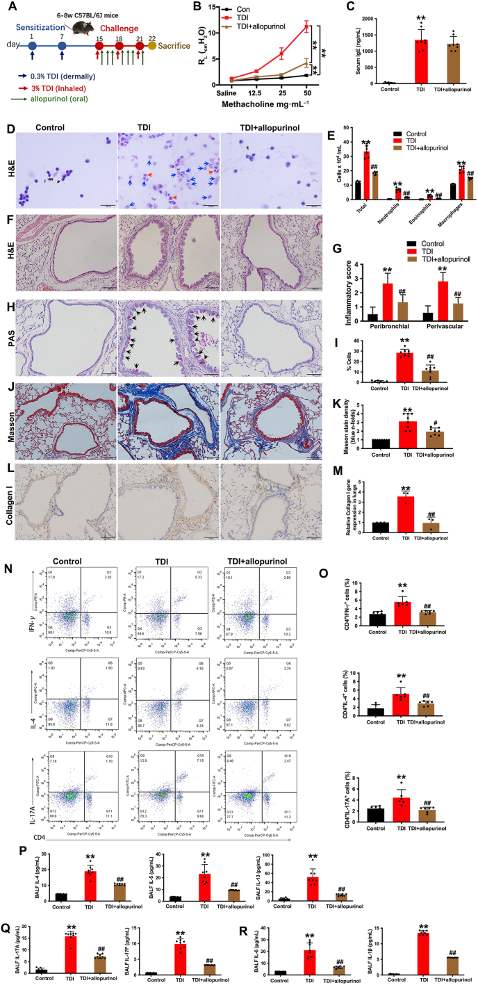
Methods: Male C57BL/6 J mice were sensitized and challenged with toluene diisocyanate (TDI) to generate a chemically induced asthma model. Pulmonary untargeted metabolomics was employed. According to results, mice were orally administered allopurinol, a xanthine oxidase (XO) inhibitor. Human bronchial epithelial cells (16HBE) were stimulated by TDI-human serum albumin (HSA).

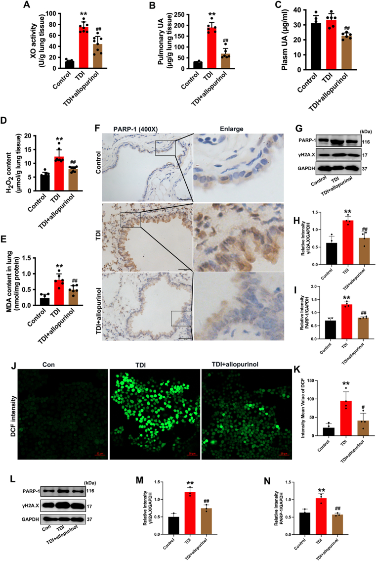
Results: We identified the purine metabolism was the most enriched pathway in TDI-exposed lungs, corresponding to the increase of xanthine and uric acid, products of purine degradation mediated by XO. Inhibition of XO by allopurinol ameliorates TDI-induced oxidative stress and DNA damage, mixed granulocytic airway inflammation and Th1, Th2 and Th17 immunology as well as HMGB1 acetylation and secretion. Mechanistically, HMGB1 acetylation was caused by decreased activation of the NAD+-sirtuin 1 (SIRT1) axis triggered by hyperactivation of the DNA damage sensor poly (ADP-ribose)-polymerase 1 (PARP-1). This was rescued by allopurinol, PARP-1 inhibitor or supplementation with NAD+ precursor in a SIRT1-dependent manner. Meanwhile, allopurinol attenuated Nrf2 defect due to SIRT1 inactivation to help ROS scavenge.

Conclusions: We demonstrated a novel regulation of HMGB1 acetylation and secretion by purine metabolism that is critical for asthma onset. Allopurinol may have therapeutic potential in patients with asthma.

Keywords: Allopurinol; Asthma; HMGB1; Purine metabolism; SIRT1.


Conflict of interest statement

Declaration of competing interest The authors declare no conflict of interest in relation to this work.

Figures

Image 1
Graphical abstract
Fig. 1
Fig. 1
Metabolomic profiles and functional alterations in toluene diisocyanate (TDI)-induced asthma model. (a) The principle component analysis (PCA) plots of Control and TDI groups. (b) The partial least squares-discriminant analysis (PLS-DA) plots of Control and TDI groups. (c) Importance of altered metabolites analyzed by PLS-DA. The Y axis is FDR-corrected P value and X axis is variable importance in the projection (VIP). (d–e) Heatmap (d) or mean-heatmap (e) of the top 30 altered metabolites in lungs of TDI-induced mice. The degree of change is marked with different colors. (f–g) Enrichment analysis of altered metabolites. (h) Heatmap of purine metabolism products in lungs of Control and TDI mice. (h) Summary of metabolites related to purine metabolism in lungs of TDI-sensitization and challenged mice. R-5-P, Ribose-5-phosphate; PRPP, Phosphoribosyl pyrophosphate; AMP, Adenosine 5′-monophosphate; GMP, Guanosine monophosphate; IMP, Inosine 5‘-monophosphate; XMP, xanthine monophosphate; XO, xanthine oxidase; n = 5 per group. (For interpretation of the references to color in this figure legend, the reader is referred to the Web version of this article.)
Fig. 2
Fig. 2
Inhibition of xanthine oxidase (XO) by allopurinol ameliorates toluene diisocyanate (TDI)-induced airway hyperresponsiveness and airway inflammation. (A) Schematic of TDI sensitization and challenge and allopurinol treatment model. The XO inhibitor, allopurinol (25 mg/kg/day), or the same volume of vehicle was administered by gavage every day since the first day of TDI challenge. (B) Airway hyperresponsiveness was measured by RL (lung resistance). Results were expressed as RL value for each concentration of methacholine. n = 5. (C) Level of total IgE in serum. n = 8. (D) Representative hematoxylin and eosin (H&E) staining of cell smears from bronchoalveolar lavage fluid (BALF). Scale bar = 50 μm. Blue arrows indicated neutrophils and red arrows indicated eosinophils. (E) Total and differential inflammatory cell counts in BALF. n = 6. (F–G) Representative H&E staining of lung sections (Scale bar = 100 μm) with semiquantitative analysis. n = 6. (H–I) Representative Periodic acid–Schiff (PAS)-stained lung sections of different groups (Scale bar = 100 μm) with semiquantitative analysis. n = 8. Black arrows showed the PAS-positive cells. (J–K) Representative Masson's trichrome-stained lung sections of different groups (Scale bar = 100 μm) with semiquantitative analysis. n = 8. (L) Collagen I expression was detected by Immunohistochemistry in lung sections of different groups. Scale bar = 100 μm. (M) Collagen I gene expression in lungs was detected by qPCR, n = 3. (N–O) The percentages of Th1 (CD4+IFN-γ+), Th2 (CD4+IL-4+) and Th17 (CD4+IL-17 A+) cell subsets in lungs analyzed by Flow cytometry. n = 6. (P–Q) Representative the levels of Th2- (IL-4, IL-5 and IL-13) and Th17- (IL-17 A and IL-17F) related cytokines in BALF. n = 8. (R) Representative the levels of IL-6 and IL-1β in BALF. n = 8. Data were presented as mean ± SD. *Denotes statistical significance compared with Control group, and #denotes statistical significance compared with TDI group. */#P < 0.05 and **/##P < 0.01. (For interpretation of the references to color in this figure legend, the reader is referred to the Web version of this article.)
Fig. 3
Fig. 3
Inhibition of xanthine oxidase (XO) by allopurinol downregulated HMGB1 acetylation and secretion and activated SIRT1 in toluene diisocyanate (TDI) asthma. (A) Pulmonary HMGB1 expression and distribution was detected by Immunohistochemistry (IHC). Scale bar = 50 μm. (B) HMGB1 secretion in serum and bronchoalveolar lavage fluid (BALF) was detected by ELISA. n = 6. (C) Expression and distribution of acetylation (Ac)-HMGB1 (K12) was detected by Immunofluorescent in lung sections of different groups. Scale bar = 20 μm. (D) Pulmonary SIRT1 expression was detected by IHC. Scale bar = 50 μm. (E–J) Expression of Ac-HMGB1 (K12), Ac-HMGB1 (K29), SIRT1, Ac-p53 (Lys379) and H4K16ac were detected by Western blot analysis with densitometric quantification in lung tissues. n = 4. (K) The cell viability of TDI-HSA-treated 16HBE cells was determined by CCK-8 assay. n = 4. (L–Q) Expression of Ac-HMGB1 (K12), Ac-HMGB1 (K29), SIRT1, Ac-p53 (Lys379) and H4K16ac were detected by Western blot analysis with densitometric quantification in 16HBE cells treated by TDI-HSA (100 μg/ml, 12 h) with or without allopurinol pretreatment (50 μg/ml). n = 3. (R) HMGB1 expression and translocation in 16HBE cells was detected by Immunofluorescent. Scale bar = 20 μm. Data were presented as mean ± SD. *Denotes statistical significance compared with Control group, and #denotes statistical significance compared with TDI group. */#P < 0.05 and **/##P < 0.01.
Fig. 4
Fig. 4
Inhibition of xanthine oxidase (XO) by allopurinol reduced oxidative stress and DNA damage induced by toluene diisocyanate (TDI). (A) Quantification of the pulmonary XO activity. n = 8. (B–C) Level of uric acid (UA) in lung tissue and plasm. n = 6. (D) Quantification of H2O2 content in lung tissue. n = 8. (E) Lipid peroxidation was quantified by using malondialdehyde (MDA) assay kit. n = 6. (F) The DNA damage sensor poly (ADP-ribose)-polymerase 1 (PARP-1) expression was detected by Immunohistochemistry (IHC) in lung sections of different groups. Scale bar = 50 μm. (G–I) DNA damage was measured by immunoblotting of γH2A.X and PARP-1 in lung tissue with densitometric analysis. n = 4. (J–K) Representative microphotographs and quantification of fluorescence intensity of DCF staining (reactive oxygen species, ROS) in 16HBE cells treated by 100 μg/ml TDI-HSA for 8 h with or without allopurinol (50 μg/ml) pretreatment. Scale bar = 50 μm. n = 4. (L–N) 16HBE cells were treated by 100 μg/ml TDI-HSA for 12 h with or without allopurinol (50 μg/ml) pretreatment, then expressions of γH2A.X and PARP-1 were detected by Western blot analysis with densitometric quantification. n = 3. Data were presented as mean ± SD. *Denotes statistical significance compared with Control group, and #denotes statistical significance compared with TDI group. */#P < 0.05 and **/##P < 0.01.
Fig. 5
Fig. 5
PARP-1 upregulation induced by toluene diisocyanate (TDI) impedes SIRT1 activity by competitively consuming NAD+. (A–B) The pulmonary NAD+, NADH content and the ratio of NAD+/NADH were detected in different groups. n = 6. (C–D) 16HBE cells pretreated with or without allopurinol (50 μg/ml) were stimulated with TDI-HSA (100 μg/ml, 12 h). The cellular NAD+, NADH content and the ratio of NAD+/NADH were detected. n = 3. (E) 16HBE cells were stimulated by TDI-HSA (100 μg/ml, 12 h) with or without NAD+ precursor NMN (0.5 mM) or PARP-1 inhibitor Olaparib (OLA, 50 nM) pretreatment, then cellular NAD+/NADH ratio was detected. n = 3. (F–G) Expression of H4K16ac was detected by Western blot analysis with densitometric quantification. n = 3. (H–J) 16HBE cells transfected with NC-siRNA or si-sirt1 were treated by TDI-HSA with or without allopurinol or OLA pretreatment, then expression of Ac-HMGB1 (K12) and Ac-HMGB1 (K29) in 16HBE cells of different groups was detected by Western blot analysis with densitometric quantification. n = 3. $P < 0.05 versus to Control group in si-sirt1-transfected cells. (H–J) 16HBE cells transfected with NC-siRNA or si-sirt1 were treated by TDI-HSA with or without allopurinol or OLA pretreatment, then expression of Ac-HMGB1 (K12) and Ac-HMGB1 (K29) in 16HBE cells of different groups was detected by Western blot analysis with densitometric quantification. n = 3. $P < 0.05 versus to Control group in si-sirt1-transfected cells. (K–L) 16HBE cells were stimulated by TDI-HSA (100 μg/ml, 12 h) with or without Resveratrol (10 μM) or Ex-527 (10 μM) pretreatment, then protein-protein direct interaction between SIRT1 and HMGB1 in bronchial epithelial cells was demonstrated by co-Immunoprecipitation with densitometric quantification. n = 3. Data were presented as mean ± SD. *Denotes statistical significance compared with Control group, and #denotes statistical significance compared with TDI group. */#P < 0.05 and **/##P < 0.01.
Fig. 6
Fig. 6
Inhibition of xanthine oxidase (XO) by allopurinol improves Nrf2 defect induced by toluene diisocyanate (TDI) in a SIRT1-dependent manner. (A) Nrf2 expression and distribution was detected by Immunohistochemistry (IHC) in lung sections of different groups. Scale bar = 50 μm. Red arrows showed the high-expression of Nrf2 and blue arrows showed the faint staining in the nucleus of bronchial epithelial cells. (B) Expression of Ho-1 and Nqo1 mRNA in lungs of mice were quantified by qPCR. n = 4. (C–E) 16HBE cells transfected with NC-siRNA or si-sirt1 were treated by 100 μg/ml TDI-HSA with or without allopurinol (50 μg/ml) pretreatment, then expression of Nrf2 in subcellular fractions was analyzed by Western blot with densitometric quantification. Purity of cytoplasmic extracts (CE) and nuclear extracts (NE) was confirmed by immunoblotting with antibodies against GAPDH and Histone H3. n = 3. (F–G) 16HBE cells transfected with NC-siRNA or si-sirt1 were treated by TDI-HSA with or without allopurinol pretreatment, then expression of Ho-1 and Nqo1 mRNA were quantified by qPCR. n = 3. (H) The cell viability was determined by CCK-8 assay. n = 5. (I–L) The expression of HMGB1, Ac-HMGB1 (K12) and Ac-HMGB1 (K29) in 16HBE cells treated by uric acid (UA) at indicated concentration for 24 h was detected by Western blot analysis with densitometric quantification. n = 3. (M − N) Expression of Ho-1 and Nqo1 mRNA in 16HBE cells treated by UA at indicated concentration for 6 h were quantified by qPCR. n = 3. Data were presented as mean ± SD. *Denotes statistical significance compared with Control group, and #denotes statistical significance compared with TDI group, and $denotes statistical significance compared with Control group in si-sirt1 transfected cells. */#/$P < 0.05 and **/##/$$P < 0.01. (For interpretation of the references to color in this figure legend, the reader is referred to the Web version of this article.)
figs1
figs1
figs2
figs2

References

    1. Israel E., Reddel H.K. Severe and difficult-to-treat asthma in adults. N. Engl. J. Med. 2017;377(10):965–976. - PubMed
    1. Agache I., Akdis C.A., Akdis M., et al. EAACI biologicals guidelines—recommendations for severe asthma. Allergy. 2021;76(1):14–44. - PubMed
    1. Chen R., Zhang Q., Chen S., et al. IL-17F, rather than IL-17A, underlies airway inflammation in a steroid-insensitive toluene diisocyanate-induced asthma model. Eur. Respir. J. 2019;53(4) - PubMed
    1. Wang Y., Le Y., Zhao W., et al. Short thymic stromal lymphopoietin attenuates toluene diisocyanate-induced airway inflammation and inhibits high mobility group box 1-receptor for advanced glycation end products and long thymic stromal lymphopoietin expression. Toxicol. Sci. 2017;157(2):276–290. - PubMed
    1. Yao L., Zhao H., Tang H., et al. Blockade of β-catenin signaling attenuates toluene diisocyanate-induced experimental asthma. Allergy. 2017;72(4):579–589. - PubMed