. 2024 Dec:66:267-284.
doi: 10.1016/j.jare.2024.01.007. Epub 2024 Jan 12.

The circUbqln1, regulated by XBP1s, interplays with 14-3-3ζ to inhibit collagen synthesis and promote osteoarthritis by controlling PRODH activity and proline metabolism

Affiliations

The circUbqln1, regulated by XBP1s, interplays with 14-3-3ζ to inhibit collagen synthesis and promote osteoarthritis by controlling PRODH activity and proline metabolism

Naibo Feng et al. J Adv Res. 2024 Dec.

Abstract

Introduction: Osteoarthritis (OA) is a degenerative bone disease associated with ageing, characterized by joint pain, stiffness, swelling and deformation. Currently, pharmaceutical options for the clinical treatment of OA are very limited. Circular RNAs(cirRNAs) have garnered significant attention in OA and related drug development due to their unique RNA sequence characteristics.Therefore,exploring the role of cirRNAs in the occurrence and development of OA is of paramount importance for the development of effective medications for OA.

Objectives: To identify a novel circRNA, circUbqln1, for treating osteoarthritis and elucidate its pathophysiological role and mechanisms in the treatment of OA.

Methods: The circUbqln1 expression and distribution were determined by qRT-PCR and FISH. XBP1 gene knockout(XBP1 cKO) spontaneous OA and DMM model and WT mouse CIOA model were used to explore the role of XBP1 and circUbqln1 in OA.Overexpression or knockdown of circUbqln1 lentivirus was used to observe the impacts of circUbqln1 on primary chondrocytes,C28/I2 and mice in vitro and in vivo.Chromatin immunoprecipitation,luciferase reporter assay,RNA pulldown,mass spectrometry,RNA immunoprecipitation,fluorescence in situ hybridization,and flow cytometry to explore the molecular mechanisms of circUbqln1.

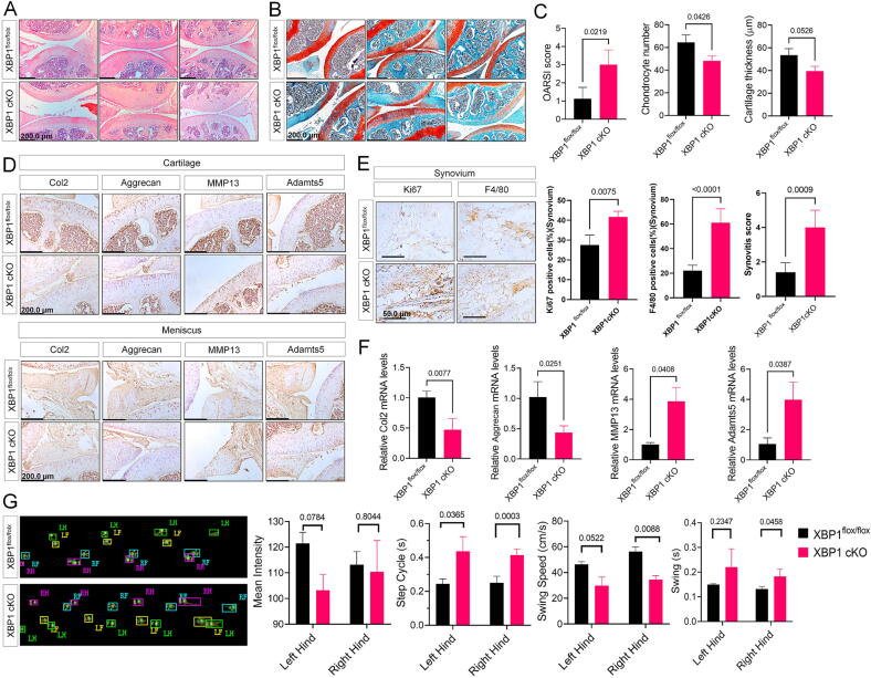
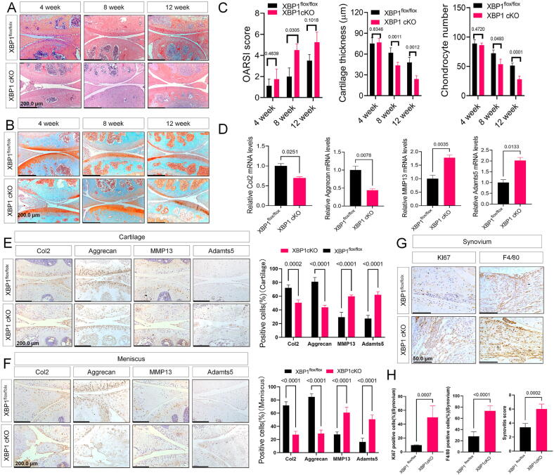
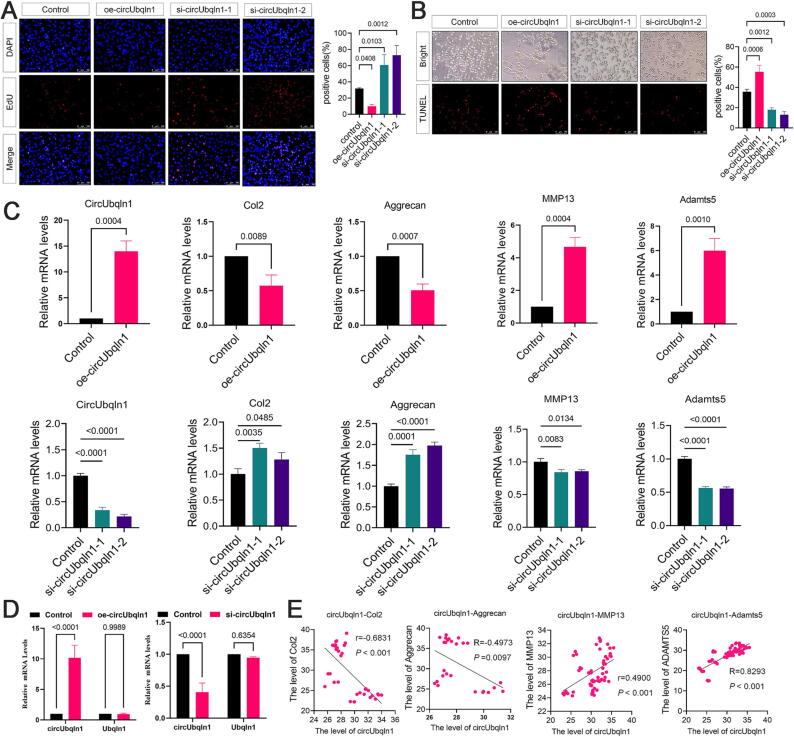
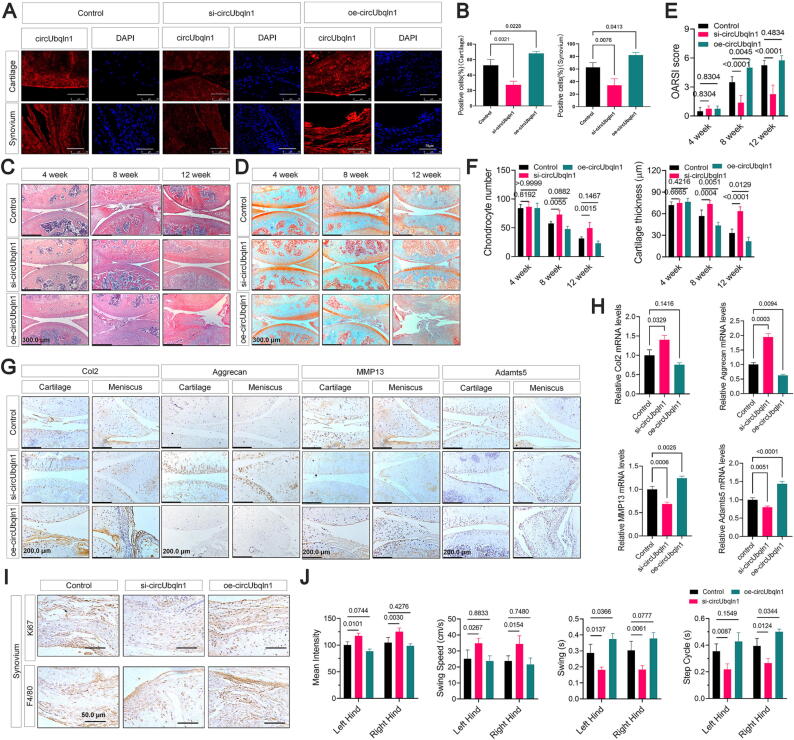
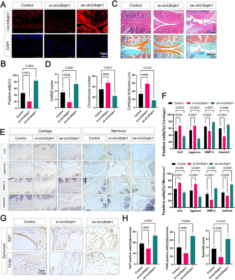
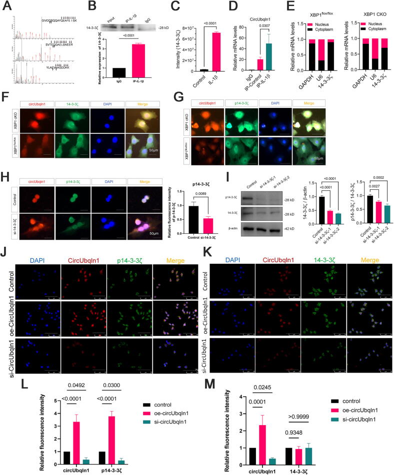
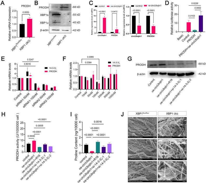
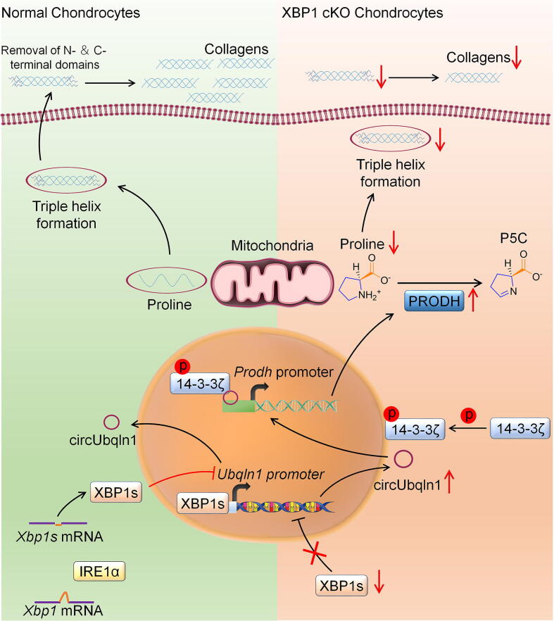
Results: It was found that cartilage-specific XBP1 cKO mice exhibited a faster OA progression compared to normal's.Importantly,transcript factor XBP1s has the capacity to impede the biogenesis of circUbqln1,derived from Ubqln1. The circUbqln1 promotes cartilage catabolism and inhibits anabolism, therefore accelerates the occurrence of OA.Mechanismly,circUbqln1 can translocate to the chondrocyte nucleus with the assistance of phosphorylated 14-3-3ζ, upregulate the transcriptional activity of the proline dehydrogenase(Prodh) promoter and PRODH enzyme activity. Consequently, this leads to the promotion of proline degradation and the inhibition of collagen synthesis,ultimately culminating in the impairment of cartilage and its structural integrity.

Conclusion: CircUbqln1 plays a crucial role in the occurrence and development of OA, indicating that the inhibition of circUbqln1 holds promise as a significant approach for treating OA in the future.

Keywords: Cartilage; Circubqln1; Osteoarthritis; PRODH; Proline; XBP1s.


Conflict of interest statement

Declaration of competing interest The authors declare that they have no known competing financial interests or personal relationships that could have appeared to influence the work reported in this paper.

Figures

None
Graphical abstract
Fig. 1
Fig. 1
XBP1 cKO mice exhibited more severe cartilage damage compared to XBP1flox/flox mice. (A) Representative images of HE staining sections, and (B) Saffron-O and Fast Green staining sections of knee joints from the 12-month old XBP1 cKO mice and XBP1flox/flox control mice, n = 6. Scale bar: 200 µm. (C) The severity of articular cartilage damage in XBP1 cKO mice and XBP1flox/flox control mice were analyzed using the OARSI score, quantification assay of number of chondrocytes, quantification assay of articular cartilage thickness. (D) Immunohistochemistry of Col2, Aggrecan, MMP13 and ADAMTS5 in the articular cartilage and meniscus of knee joint from the 12-month old XBP1 cKO mice and XBP1flox/flox control mice, n = 6. Scale bar: 200 µm. (E) Immunohistochemistry of Ki67, F4/80, and statistical analysis of synovitis scores and quantification of Ki67-, F4/80-positive cells in the synovium of knee joint from the 12-month old XBP1 cKO mice and XBP1flox/flox control mice, n = 6. Scale bar:50 µm. (F) QPCR assay for mRNA levels of Col2, Aggrecan, MMP13 and ADAMTS5 in the articular cartilage of knee joint from the 12-month old XBP1 cKO mice and XBP1flox/flox control mice, n = 3. (G) The posture changes and gait parameters assay on the 12-month old XBP1 cKO mice and XBP1flox/flox control mice by the animal gait analysis system. n = 3. Data represent means ± SEM using Student’s t test in chondrocytes number, articular cartilage thickness, positive cells and F), Mann-Whitney U test in (OARSI score and synovitis score).
Fig. 2
Fig. 2
The OA phenotype was more severe in the surgically induced arthritis model in XBP1 cKO mice. (A) Representative images of HE staining sections, and (B) Saffron-O and Fast Green staining sections of knee joints from the DMM-induced OA model at 4-, 8-, and 12-week in XBP1 cKO mice and XBP1flox/flox control mice respectively. n = 6. Scale bar: 200 µm. (C) The severity of articular cartilage damage in XBP1 cKO mice and XBP1flox/flox mice were analyzed using the OARSI score, quantification assay of number of chondrocytes, quantification assay of articular cartilage thickness. (D-F) QPCR assay for mRNA levels of Col2, Aggrecan, MMP13 and ADAMTS5 in the articular cartilage (D), immunohistochemistry of Col2, Aggrecan, MMP13 and ADAMTS5 in the articular cartilage (E) and meniscus (F) of the knee joint from the DMM-induced OA model at 12-week in XBP1 cKO mice and XBP1flox/flox control mice respectively. n = 6 per group. Scale bar: 200 µm. (G, H) Immunohistochemistry of Ki67, F4/80 (G) and statistical analysis of synovitis scores and quantification of Ki67-, F4/80-positive cells (H) in the synovium of knee joint from the DMM-induced OA model at 12-week in XBP1 cKO mice and XBP1flox/flox control mice respectively. n = 8 per group. Scale bar:50 µm. Data represent means ± SEM using Student’s t test (chondrocytes number, articular cartilage thickness, positive cells, and D), Mann-Whitney U test in (OARSI score and synovitis score).
Fig. 3
Fig. 3
Identification of the circUbqln1 and XBP1s repressed circUbqln1production in chondrocytes. (A) Heat map of differential circRNAs in XBP1cKO and XBP1flox/flox cartilage. (B) Schematic illustration of circUbqln1 formation via the circularization from exons 2 and exon 6 in Ubqln1 gene. The back-splice junction sequences were confirmed by Sanger sequencing. (C) The localization of circUbqln1 was investigated in primary chondrocytes with or without IL-1β treatment. (D) The localization of circUbqln1 was investigated in articular cartilage. (E) The expression of circUbqln1 in the cytoplasm and nucleus of the primary chondrocytes of the two groups of mice were detected by RT-qPCR. (F) Luciferase activity detected the regulation of the transcriptional activity of Ubqln1 gene by overexpression (OE) or knockdown (KD) of XBP1s. (G) The Ubqln1 protein expression after OE-XBP1s or si-XBP1s were measured by Western blot and quantification assay. (H) JASPAR database predicted that there are four binding sites between XBP1s and Ubqln1 promoter region. (I) CHIP assay showed that the binding site between XBP1s and Ubqln1 promoter was located within the range of the first pair of primers. (J) Overexpression (OE) of XBP1s could inhibit the expression of Ubqln1 mRNA and circUbqln1, and the expressions of Ubqln1 and circUbqln1 mRNA were upregulated when XBP1s was knocked down (KD). Data represent means ± SEM using ANOVA followed by post hoc test in (F, G, and J), and Student’s t test in (I).
Fig. 4
Fig. 4
CircUbqln1 augments chondrocyte apoptosis and modulates chondrocyte catabolism and anabolism. (A) EdU Assay and quantification results showed that overexpression (OE) circUbqln1 inhibited chondrocyte proliferation, while knock down (KD) circUbqln1 expression by siRNA promoted chondrocyte proliferation. Two-way ANOVA was used. (B) TUNEL staining showed that OE circUbqln1 induced the increased of chondrocyte apoptosis, while KD of circUbqln1 inhibited chondrocyte apoptosis. Two-way ANOVA was used. (C) The expression of cartilage anabolic markers Col2 and Aggrecan, and the expression of cartilage catabolic markers MMP13 and Adamts5 were measured by RT-qPCR after OE or KD circUbqln1 in primary chondrocytes. (D) The Ubqln1 mRNA expression has no significant change after overexpression (OE) or knockdown (KD) of CircUbqln1 in chondrocytes by qPCR detection. (E) Knee joint samples of XBP1 cKO mice at different ages were collected, and the expressions of circUbqln1 and cartilage anabolic and catabolic markers in cartilage tissue were detected by RT-qPCR. Pearson correlation analysis was used. Data represent means ± SEM using ANOVA followed by post hoc test in (A, B, C(down)), and Student’s t test in (C(up), D), Pearson correlation analyses were performed using IBM SPSS Statistics 23 and were used in (E).
Fig. 5
Fig. 5
The effect of circUbqln1 was verified in the DMM-induced OA model in XBP1 cKO mice. DMM model was constructed in XBP1 cKO mice at 4-, 8-, and 12-week, respectively, then the mice were intra-articularly injected with either phosphate buffered saline (PBS), circUbqln1 overexpression or si-circUbqln1 knockdown lentivirus once a week, and the samples were collected after 4 weeks of injection . (A) The expression and positioning of circUbqln1 was detected by the cy5-fluorescent probe of circUbqln1 in the joint cartilage and synovium of DMM-induced OA model in XBP1 cKO mice at 12-week. n = 6 per group. Scale bar: 75 µm. (B) Quantitative analysis of circUbqln1 positive cells in the cartilage and synovium. (C-F) Intra-articular injection of si-circUbqln1 lentivirus dramatically protected cartilage from degeneration following surgically induced OA model, assayed by HE staining sections (C) and Saffron-O and Fast Green staining sections (D) and the OARSI score (E), quantification assay of number of chondrocytes, quantification assay of articular cartilage thickness (F). n = 6 per group. Scale bar: 300 µm. (G-I) Immunohistochemistry of Col2, Aggrecan, MMP13 and ADAMTS5 in the articular cartilage and meniscus (G), QPCR assay for mRNA levels of Col2, Aggrecan, MMP13 and ADAMTS5 in the articular cartilage (H), and immunohistochemistry of Ki67, F4/80 in the synovium (I) of the knee joint from the DMM-induced OA model in XBP1 cKO mice at 12-week . n = 6 per group. Scale bar: 200 µm, 50 µm. (J) The posture changes and gait behaviors parameters assay on the DMM-induced OA model in XBP1 cKO mice at 12-week . n = 3 per group. Data represent means ± SEM using ANOVA followed by post hoc test in (B), (H), and (J), for (E, F), Kruskal-Wallis test followed by the Mann-Whitney U test were used in OARSI score, ANOVA followed by post hoc test in chondrocytes number and articular cartilage thickness.
Fig. 6
Fig. 6
Local delivery of si-circUbqln1 lentivirus attenuated osteoarthritis (OA) development in the CIOA mice model. CIOA model was constructed in C57 wild-type mice, then circUbqln1 overexpression or si-circUbqln1 knockdown lentivirus was injected into the joint cavity once a week, and the knee joints were collected 4 weeks later. (A) The expression and positioning of circUbqln1 was detected by the cy5-fluorescent probe of circUbqln1 in the joint cartilage. (B) Quantification assay of circUbqln1 positive cells in the cartilage. (C, D) Intra-articular injection of si-circUbqln1 lentivirus obviously protected cartilage damage in CIOA model, assayed by HE staining sections and Saffron-O and Fast Green staining sections(C), the OARSI score, quantification assay of number of chondrocytes, quantification assay of articular cartilage thickness (D). n = 6 per group. Scale bar: 250 µm, (E-H) Immunohistochemistry of Col2, Aggrecan, MMP13 and ADAMTS5 in the articular cartilage and meniscus (E), and quantification assay (F), immunohistochemistry of Ki67, F4/80 in the synovium (G), statistical analysis of synovitis score and quantification of Ki67-, F4/80-positive cells in the synovium of the knee joint from the CIOA mice model respectively (H). n = 6 per group. Scale bar: 75 µm,50 µm. Data represent means ± SEM using ANOVA followed by post hoc test in (B), and (F). For (D, H), Kruskal-Wallis test followed by the Mann-Whitney U test were used in OARSI score and synovitis score, ANOVA followed by post hoc test in chondrocytes number, articular cartilage thickness, and in positive cells.
Fig. 7
Fig. 7
CircUbqln1 binds to 143-3ζ and influences the nuclear translocation of circUbqln1 in XBP1 KO chondrocytes. (A) MS/MS spectrum of 14–3-3ζ. (B) RNA pull-down experiment was performed using the specific biotin-labeled CircUbqln1 probe in ATDC5 cell lysates, and the protein bands were analyzed by mass spectrometry and western blot. Unpaired t test was used. (C) Protein profiling analysis results showed that after chondrocytes were treated with IL-1β, the binding amount of 14–3-3ζ with circUbqln1 were increased. Unpaired t test was used. (D) RIP assay was executed in ATDC5 cell lysates using anti-14–3-3ζ or anti-IgG, then the enrichment of circUbqln1 was detected by RT-qPCR. Unpaired t test was used. (E) Nuclear-cytoplasmic fractionation assay displayed that 14–3-3ζ was mostly distributed in the cytoplasm of XBP1flox/flox primary chondrocytes, while mostly distributed less in the cytoplasm of XBP1 cKO primary chondrocytes. U6 was considered as a nuclear control and GAPDH was used as a cytoplasmic protein control. (F) Co-localized of CircUbqln1 and 14–3-3ζ and (G) Co-localized of circUbqln1 and p14-3-3ζ detected by FISH-IF assay in primary chondrocytes isolated from the cartilage of XBP1 cKO and XBP1flox/flox mice. (H) Co-localized of circUbqln1 and p14-3-3ζ detected by FISH-IF assay in ATDC5 cells after si-14–3-3ζ infection and quantification assay. (I) Western blot and quantification results of 14–3-3ζ total protein and p14-3-3ζ in ATDC5 cells after si-14–3-3ζ. Two-way ANOVA were used, n = 5. (J) Co-localized of CircUbqln1 and p14-3-3ζ and (K) Co-localized of CircUbqln1 and 14–3-3ζ detected by FISH-IF assay in ATDC5 cells infected by si-CircUbqln1 or oe-CircUbqln1. (L) Quantification of CircUbqln1 and p14-3-3ζ. (M) Quantification of CircUbqln1 and 14–3-3ζ. Data represent means ± SEM using Student’s t test in (B) to (D), (H), ANOVA followed by post hoc test in (I), and (L, M).
Fig. 8
Fig. 8
CircUbqln1 enhances the activity of PRODH and promotes collagen degradation. (A) The mRNA level of PRODH mRNA in XBP1 cKO and XBP1flox/flox mice. (B) Western blot results of PRODH in XBP1 cKO and XBP1flox/flox mice. Paired t test was used. (C) The mRNA expression of PRODH and circUbqln1 after overexpressing (OE) or knock down (KD) circUbqln1 in primary chondrocytes. Two- way ANOVA were used. (D) OE circUbqln1 enhanced the promoter activity of PRODH by dual luciferase reporter assay. Two-way ANOVA were used. (E) The mRNA levels of PRODH and 143-3ζ after treated with difference dose 143-3ζ siRNA in C28/I2 cells. (F) The mRNA expressions of PRODH and 143-3ζ after treated with difference dose R18 (14–3-3ζ competitive inhibitor) in C28/I2 cells. Paired t test was used. (G-I) C28/I2 chondrocytes treated with oe-CircUbqln1, oe-CircUbqln1 + R18(100 nM), oe-CircUbqln1 + 14–3-3ζ siRNA(100 nM), then detected the protein expression of PRODH (G), the PRODH activity (H), and the proline content (I). Two-way ANOVA was used. (J) Representative TEM images of collagen fibers from the cartilage of XBP1 cKO and XBP1flox/flox mice. Data represent means ± SEM using ANOVA followed by post hoc test in (A), and (C), ANOVA followed by post hoc test in (D), (E), (F), (H), and (I).
Fig. 9
Fig. 9
Schematic diagram of the regulation of CircUbqln1 in the process of osteoarthritis.XBP1s inhibits the generation of circUbqln1. With assistance from phosphorylated 14–3–3ζ, circUbqln1 enters the nucleus and upregulates the transcriptional activity of Prodh, leading to abnormal proline metabolism, consequently disrupting the balance of cartilage synthesis and degradation, then accelerating the progression of OA.

Similar articles

Cited by

  • IRE1α pathway: A potential bone metabolism mediator.
    Yu C, Zhang Z, Xiao L, Ai M, Qing Y, Zhang Z, Xu L, Yu OY, Cao Y, Liu Y, Song K. Yu C, et al. Cell Prolif. 2024 Oct;57(10):e13654. doi: 10.1111/cpr.13654. Epub 2024 May 12. Cell Prolif. 2024. PMID: 38736291 Free PMC article. Review.

References

    1. Bernabei I., So A., Busso N., Nasi S. Cartilage calcification in osteoarthritis: mechanisms and clinical relevance. Nat Rev Rheumatol. 2023;19(1):10–27. doi: 10.1038/s41584-022-00875-4. - DOI - PubMed
    1. Favero M., Belluzzi E., Ortolan A., Lorenzin M., Oliviero F., Doria A., et al. Erosive hand osteoarthritis: latest findings and outlook. Nat Rev Rheumatol. 2022;18(3):171–183. - PubMed
    1. Barnett R. Osteoarthritis. Osteoarthritis Lancet. 2018;391(10134):1985. - PubMed
    1. Yao Q., Wu X., Tao C., Gong W., Chen M., Qu M., et al. Osteoarthritis: pathogenic signaling pathways and therapeutic targets. Signal Transduct Target Ther. 2023;8(1) doi: 10.1038/s41392-023-01330-w. - DOI - PMC - PubMed
    1. Arden N.K., Perry T.A., Bannuru R.R., Bruyère O., Cooper C., Haugen I.K., et al. Non-surgical management of knee osteoarthritis: comparison of ESCEO and OARSI 2019 guidelines. Nat Rev Rheumatol. 2021;17(1):59–66. - PubMed

LinkOut - more resources