. 2024 May;13(13):e2303785.
doi: 10.1002/adhm.202303785. Epub 2024 Feb 9.

Pump-Less, Recirculating Organ-on-Chip (rOoC) Platform to Model the Metabolic Crosstalk between Islets and Liver

Affiliations

Pump-Less, Recirculating Organ-on-Chip (rOoC) Platform to Model the Metabolic Crosstalk between Islets and Liver

Aleksandra Aizenshtadt et al. Adv Healthc Mater. 2024 May.

Abstract

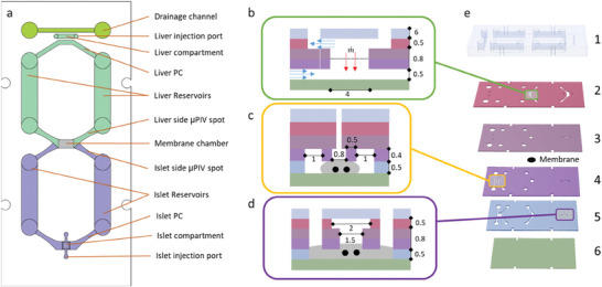
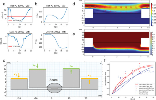
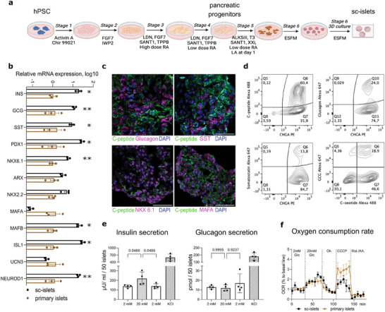
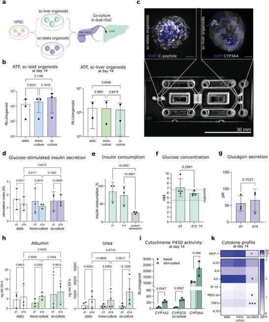
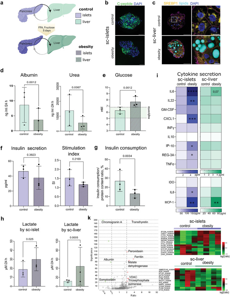
Type 2 diabetes mellitus (T2DM), obesity, and metabolic dysfunction-associated steatotic liver disease (MASLD) are epidemiologically correlated disorders with a worldwide growing prevalence. While the mechanisms leading to the onset and development of these conditions are not fully understood, predictive tissue representations for studying the coordinated interactions between central organs that regulate energy metabolism, particularly the liver and pancreatic islets, are needed. Here, a dual pump-less recirculating organ-on-chip platform that combines human pluripotent stem cell (sc)-derived sc-liver and sc-islet organoids is presented. The platform reproduces key aspects of the metabolic cross-talk between both organs, including glucose levels and selected hormones, and supports the viability and functionality of both sc-islet and sc-liver organoids while preserving a reduced release of pro-inflammatory cytokines. In a model of metabolic disruption in response to treatment with high lipids and fructose, sc-liver organoids exhibit hallmarks of steatosis and insulin resistance, while sc-islets produce pro-inflammatory cytokines on-chip. Finally, the platform reproduces known effects of anti-diabetic drugs on-chip. Taken together, the platform provides a basis for functional studies of obesity, T2DM, and MASLD on-chip, as well as for testing potential therapeutic interventions.

Keywords: Type 2 diabetes (T2DM); drug testing; energy metabolism; metabolic dysfunction‐associated steatotic liver disease (MASLD); obesity; organ‐on‐chip; sc‐islet organoids; sc‐liver organoids.


Conflict of interest statement

A.A., M.B., and S.J.K.K. have applied for a patent covering the main principle of fluid actuation and application and are planning to commercialize the technology.

Figures

Figure 1
Figure 1
Principle layout of the dual‐rOoC. a) Different circuits and elements of the dual‐rOoC as top‐view; b) cross‐section of the membrane chamber with the separated top and bottom flow (blue arrows) and red arrows indicating trans‐membrane flow; c,d) cross‐section of the liver and islet compartment with organoids indicated as black dots and the ECM marked in grey; e) explosion‐view of the full device consisting of 6 layers. All dimensions are given in mm.
Figure 2
Figure 2
Modeling and technical characterization of the dual‐rOoC. a) Measured velocity–time curves obtained at the positions indicated in Figure 1a with a tilt of 18°, a speed of 3 rpm, and a total volume of 300 µL. The red line indicates the mean flow rate; b) transported volume obtained by integrating (a); c) model description (cross‐section) indicating the two reservoirs and the exchange membrane in the middle. The green lines indicate the fixed concentration boundary condition at the donor side and the orange line indicates the continuous boundary condition with all molecules leaving the system to the right and reentering the reservoir to the left; d) flow profile at the sc‐islet‐PC (top) and the sc‐liver‐PC (bottom) (zoomed area of (c). The red arrows indicate the flow velocity field; e) color plots of the insulin concentration ratio c 2/c 0 distribution for the islet‐PC (top) as donor and the liver‐PC (btm) as acceptor channel; f) insulin concentration ratio c 2/c 0 for the transport from the islet to the liver circuit (red) and vice versa (blue). The crosses indicate measured values (donor concentration: 2 µg mL−1, n = 2 replicates), the dashed line is the result obtained via the Comsol model and the continuous line is the result obtained via regression and solving the ODE from Equation (6).
Figure 3
Figure 3
Generation and characterization of sc‐islet organoids from human pluripotent stem cells (hPSC). a) Scheme of the differentiation protocol; b) comparison of gene expression between sc‐islet organoids (n = 3 independent differentiations) and primary adult islets (n = 3 donors). INS: insulin, GCG: glucagon. Significance was calculated using multiple unpaired t‐tests, * p < 0.05, ** p < 0.01; c) representative confocal images of sc‐islet organoid cryosections, labeled with antibodies against human insulin, glucagon, somatostatin (SST), NKX6.1, MAFA and nuclei (DAPI); d) representative flow cytometry density plots showing the percentage of chromogranin A (CHGA), C‐peptide, somatostatin and glucagon‐positive cells, as well as C‐peptide and glucagon double‐positive cells in sc‐islet organoids; e) insulin and glucagon secretion by sc‐islets in response to glucose stimulation and potassium chloride (KCl) (n = 3 independent differentiations). Significance was calculated using one‐way ANOVA; f) glucose‐stimulated mitochondrial capacity in sc‐islet organoids and primary islets (n = 4 replicates for each group).
Figure 4
Figure 4
Generation and characterization of sc‐liver organoids. a) Scheme of differentiation protocol; b) comparison of gene expression levels between sc‐islet organoids (n = 3 independent differentiation) and PHH spheroids (n = 3 donors), primary human LSEC (n = 1 donor) and HSC (n = 2 donors), 0 h culture. Significance was calculated using multiple unpaired t‐tests, * p < 0.05, ** p < 0.01; c) representative confocal images of sc‐liver organoids labeled with antibodies against HNF4α, albumin, CYP3A4, ZO1, α1‐antitrypsin (A1AT), E‐cadherin, nuclei (DAPI) and PDGFR‐β. Scale bar 50 µm; d) albumin and urea secretion by sc‐liver organoids (sc‐liver organoids from n = 3 independent differentiation) in comparison to primary human hepatic spheroids, 5 days of culture (PHH from n = 4 donors for albumin secretion and PHH from n = 2 donors for the urea secretion assay). Significance was calculated using an unpaired t‐test and Mann–Whitney test correspondingly; cytochrome P450 activity was measured in basal conditions and after 24 h of stimulation with omeprazole (50 µM, for CYP1A2) or rifampicin (10 µM, for CYP2C9 and CYP3A4). Significance was calculated using multiple unpaired t‐tests. e) Hepatic steatosis model using sc‐liver organoids in response to treatment with oleic and palmitic acids (250 µM each, 1:1) in combination with fructose (10 mM). Confocal imaging shows lipids accumulation in steatotic organoids (white—lipid droplets stained with Nile red, green—E‐cadherin), scale bar 50 µm; f) basal and insulin‐stimulated glucose uptake and accumulation in sc‐liver organoids under control and steatosis‐inducing conditions using substrate oxidation assay with D‐[14C U]glucose (0.5 µCi mL−1, 200 µM). Significance was calculated using one‐way ANOVA on ranks (n = 5 replicates).
Figure 5
Figure 5
Coupling of sc‐islets and sc‐liver organoids on‐chip. a) Scheme of the experimental setup; b) viability of sc‐islet (left) and sc‐liver organoids after 2 weeks of mono‐ and co‐culture on the dual‐rOoC, analyzed by ATP content (n = 3 and 2 independent experiments with N > 3 replicates in each). Significance was calculated using paired one‐way ANOVA test; c) confocal on‐chip imaging of sc‐islet (left) and sc‐liver organoids (right) stained with antibodies against C‐peptide and CYP3A4 respectively. Scale bar = 50 µm; d) insulin secretion by sc‐islets measured during GSIS after 1 and 14 days of culture (n = 3 independent experiments, N > 3 replicates each). Significance was calculated using paired two‐way ANOVA test; e) insulin consumption rate (R) by sc‐liver organoids measured during GSIS after 1 and 14 days of culture (n = 3 independent experiments, N > 3 replicates each). Significance was calculated using a paired two‐way ANOVA test; f) glucose concentration in the background media after 24 h incubation at the beginning and end of islets‐liver co‐culture in the dual‐rOoC (n = 4 independent experiments, N > 3 replicates each). Significance was calculated using paired two‐way ANOVA test; g) glucagon secretion by sc‐islet organoids measured at 1 and 14 days of culture (n = 3 independent experiments, N > 3 replicates each). Significance was calculated using paired two‐way ANOVA test; h) albumin and urea secretion by sc‐liver organoids in static conditions, mono‐ and co‐culture on the dual‐rOoC (n = 3 independent experiments, N > 3 replicates each). Significance was calculated using paired two‐way ANOVA test; i) cytochrome P450 (CYP) activity of sc‐liver organoids after 2 weeks of co‐culture on the dual‐rOoC, represented as relative luminescence units (RLU), normalized to organoids number (n = 3 replicates). Significance is calculated by one‐way ANOVA test; j) Heat map showing the absolute concentration of islets‐relevant cytokines in the background media in the sc‐islets compartment per 24 h after 14 days of sc‐islet organoids culture under static conditions, in mono‐culture and co‐culture on the dual rOoC. (N = 5 replicates). Significance was calculated using an unpaired one‐way ANOVA test. On‐chip groups were compared to static condition, * p < 0.05, ** p < 0.01, *** p < 0.005.
Figure 6
Figure 6
Sc‐islet and sc‐liver organoids co‐cultured on‐chip in control and obesity medium. a) Scheme of experimental setup; b) representative confocal on‐chip imaging of sc‐islet organoids stained with antibodies against C‐peptide and glucagon. Scale bar = 50 µm; c) representative confocal on‐chip imaging of sc‐liver organoids stained with antibodies against SREBP1 and BODIPY 493/503. Scale bar = 50 µm; d) albumin and urea secretion by sc‐liver organoids (n = 3 independent experiments, N > 3 replicates in each). Significance was calculated by two‐way ANOVA test; e) glucose concentration in control and obesity groups after having been shifted to a 24‐h culture in basic medium (n = 3 independent experiments, N > 3 replicates in each). Significance was calculated by two‐way ANOVA test; f) insulin secretion by sc‐islet organoids represented as GSIS index (left) and insulin concentration in high‐glucose buffer (right) during GSIS assay, (n = 3 independent experiments, N > 3 replicates in each). Significance was calculated by two‐way ANOVA test; g) insulin consumption during GSIS by sc‐liver organoids (n = 3 independent experiments, N > 3 replicates in each). Significance was calculated by two‐way ANOVA test; h) lactate production by sc‐islets (left) and sc‐liver organoids (right) (n = 3 independent experiments, N > 3 replicates in each). Significance was calculated by two‐way ANOVA test; i) heat map showing absolute concentration of cytokines in the sc‐islet and sc‐liver organoid compartments 24 h after shifting from control and obesity conditions to basic media conditions (N = 5 replicates). Significance was calculated by unpaired one‐way ANOVA test, * p < 0.05, ** p < 0.01, *** p < 0.005; j) Proteomics analysis of sc‐islet and sc‐liver organoids in control and obesity groups. Left: volcano plot showing the changes in protein expression in sc‐islets, represented as the ratio between the obesity and the low glucose group; right the most significantly up‐ and downregulated proteins in sc‐islets and sc‐liver organoids.
Figure 7
Figure 7
Anti‐diabetic drug testing on‐chip. a) Albumin and urea secretion by sc‐liver organoids in control and obesity groups after having been shifted to a 24‐h culture in control medium without drugs (ObC group) and with drugs (Metf, Tolb, PK‐act groups) (N > 3 replicates). Significance was calculated by one‐way ANOVA test; b) glucose concentration in control and obesity groups after having been shifted to a 24 h culture in control medium (ObC group) (N > 3 replicates). Significance was calculated by one‐way ANOVA test; c) insulin secretion by sc‐islets represented as insulin concentration in the high‐glucose buffer during GSIS assay (left) and SI (right), (N > 3 replicates). Significance was calculated by one‐way ANOVA test; d) insulin consumption during GSIS by sc‐liver organoids (N > 3 replicates in each). Significance was calculated by one‐way ANOVA test; e) heat map showing an absolute concentration of cytokines in the sc‐islet and sc‐liver organoid compartments 24 h after shifting from control and obesity conditions to ObC media conditions (N > 3 replicates). Drug‐treated groups were compared to the ObC group. Significance was calculated by unpaired one‐way ANOVA test, *** p < 0005. f) viability of organoids, measured by ATP content (N > 3 replicates in each). Significance was calculated by one‐way ANOVA test; Abbreviations: Metf: metformin‐treated group, Tolb: tolbutamide‐treated group, PK‐act: PK‐act‐treated group.

References

    1. Rinella M. E., Lazarus J. V., Ratziu V., Francque S. M., Sanyal A. J., Kanwal F., Romero D., Abdelmalek M. F., Anstee Q. M., Arab J. P., Arrese M., Bataller R., Beuers U., Boursier J., Bugianesi E., Byrne C. D., Castro Narro G. E., Chowdhury A., Cortez‐Pinto H., Cryer D. R., Cusi K., El‐Kassas M., Klein S., Eskridge W., Fan J., Gawrieh S., Guy C. D., Harrison S. A., Kim S. U., Koot B. G., et al., J. Hepatol. 2023, 79, 1542. - PubMed
    1. Paik J. M., Golabi P., Younossi Y., Srishord M., Mishra A., Younossi Z. M., Hepatol. Commun. 2020, 4, 1769. - PMC - PubMed
    1. Magliano D. J., Boyko E. J., IDF Diabetes Atlas, 10th ed., International Diabetes Federation, Brussels: 2021. - PubMed
    1. Cotter T. G., Rinella M., Gastroenterology 2020, 158, 1851. - PubMed
    1. Lobstein T., Brinsden H., Neveux M., 2022. https://policycommons.net/artifacts/2266990/world_obesity_atlas_2022_web....

Publication types

MeSH terms

LinkOut - more resources