. 2024 Jan 15;16(1):3.
doi: 10.1038/s41368-023-00268-7.

Force-induced Caspase-1-dependent pyroptosis regulates orthodontic tooth movement

Affiliations

Force-induced Caspase-1-dependent pyroptosis regulates orthodontic tooth movement

Liyuan Chen et al. Int J Oral Sci. .

Abstract

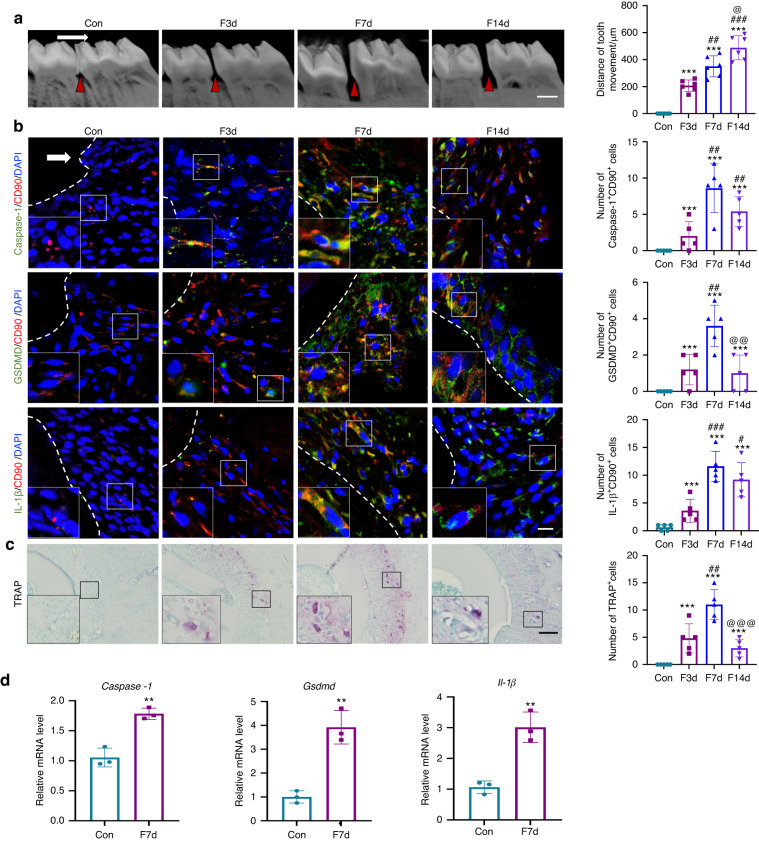
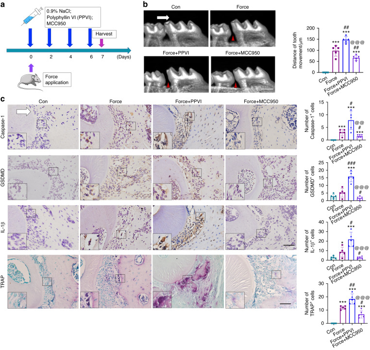
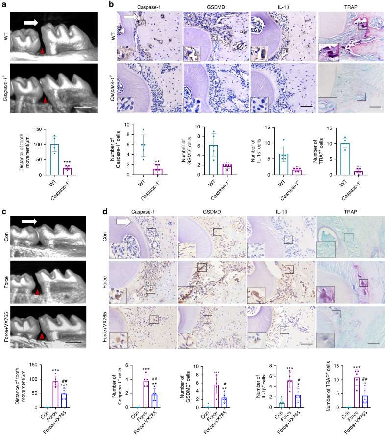
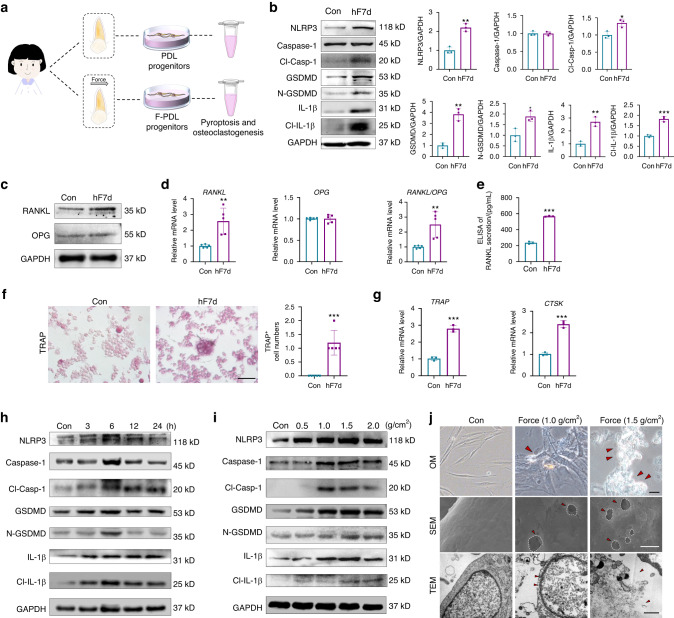
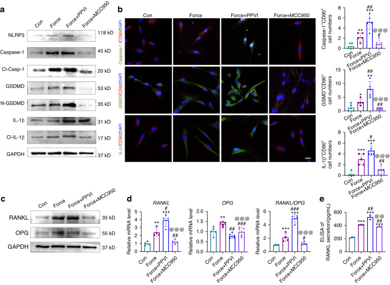
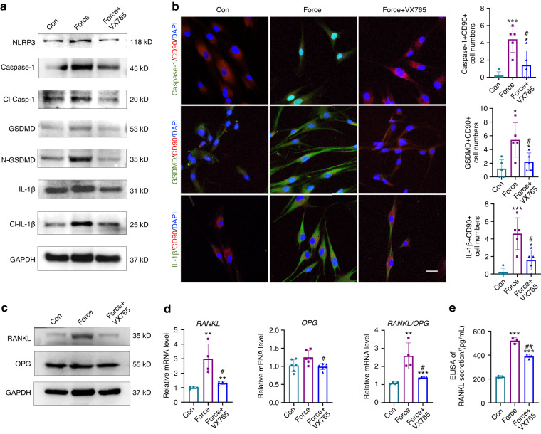
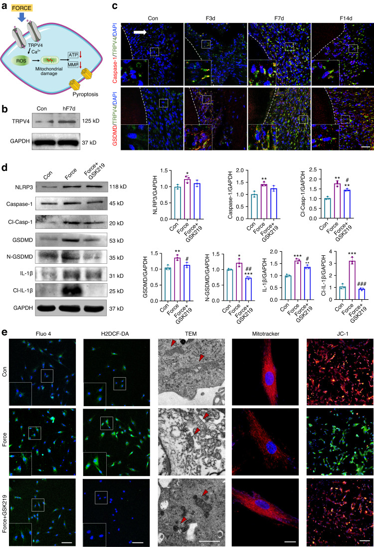
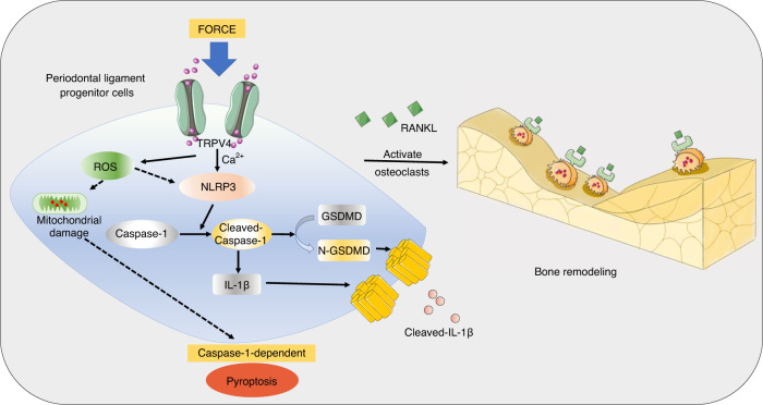
Pyroptosis, an inflammatory caspase-dependent programmed cell death, plays a vital role in maintaining tissue homeostasis and activating inflammatory responses. Orthodontic tooth movement (OTM) is an aseptic force-induced inflammatory bone remodeling process mediated by the activation of periodontal ligament (PDL) progenitor cells. However, whether and how force induces PDL progenitor cell pyroptosis, thereby influencing OTM and alveolar bone remodeling remains unknown. In this study, we found that mechanical force induced the expression of pyroptosis-related markers in rat OTM and alveolar bone remodeling process. Blocking or enhancing pyroptosis level could suppress or promote OTM and alveolar bone remodeling respectively. Using Caspase-1-/- mice, we further demonstrated that the functional role of the force-induced pyroptosis in PDL progenitor cells depended on Caspase-1. Moreover, mechanical force could also induce pyroptosis in human ex-vivo force-treated PDL progenitor cells and in compressive force-loaded PDL progenitor cells in vitro, which influenced osteoclastogenesis. Mechanistically, transient receptor potential subfamily V member 4 signaling was involved in force-induced Caspase-1-dependent pyroptosis in PDL progenitor cells. Overall, this study suggested a novel mechanism contributing to the modulation of osteoclastogenesis and alveolar bone remodeling under mechanical stimuli, indicating a promising approach to accelerate OTM by targeting Caspase-1.


Conflict of interest statement

The authors declare no competing interests.

Figures

Fig. 1
Fig. 1
Mechanical force induces pyroptosis during OTM and alveolar bone remodeling in vivo. a Representative image of micro-CT and semiquantification analysis of force-induced OTM distance in rats. Scale bar: 1 mm. b Representative immunofluorescence images on the compression side of distobuccal roots and semiquantification analysis of double-labeled cells. Dashed lines mark the outline of distobuccal roots. Scale bar: 50 µm. c Representative tartrate-resistant acid phosphatase (TRAP) staining images of distobuccal roots. Scale bar: 200 µm. Results were presented as mean ± SD. n = 5 biologically independent samples. d Real time-PCR of Caspase-1, Il-1β, and Gsdmd in periodontal tissues. Results were presented as mean ± SD. n = 3 biologically independent samples. **P < 0.01, ***P < 0.001 versus Con; #P < 0.05, ##P < 0.01, ###P < 0.001 versus F3d; @P < 0.05, @@P < 0.01, @@@P < 0.001 versus F7d. The white arrow represents the direction of the force application. Large boxed areas show high magnification views of the small boxed areas
Fig. 2
Fig. 2
Modulating pyroptosis level influences OTM and alveolar bone remodeling. a Schematic of the in vivo study. b Representative micro-CT images of OTM in mice. Con: mice without force stimuli in vivo; Force: mice receiving force stimuli for 7 d; Force+Polyphyllin VI (PPVI): mice receiving force stimuli for 7 d and the application of pyroptosis activator PPVI treatment. Force+MCC950: mice receiving force stimuli for 7 d and the application of pyroptosis inhibitor MCC950 treatment. Scale bar: 500 µm. c Representative immunohistochemical staining images and TRAP staining images of the compression side of distobuccal roots. The expressions of pyroptosis-related proteins Caspase-1, GSDMD, IL-1β, and the TRAP positive cells were detected. Large boxed areas show high magnification views of the small boxed areas. Scale bar: 100 µm. *P < 0.05, ***P < 0.001 versus Con; #P < 0.05, ##P < 0.01, ###P < 0.001 versus Force; @@P < 0.01, @@@P < 0.001 versus Force+PPVI. Results were presented as mean ± SD. n = 5 biologically independent samples. The white arrow represents the direction of the force application
Fig. 3
Fig. 3
Force-induced pyroptosis modulates OTM and alveolar bone remodeling in a Caspase-1-depended manner. a Representative micro-CT images of force-induced tooth movement distance in wild type (WT) or Caspase-1−/− mice. Scale bar: 500 µm. b Representative immunohistochemical staining images and TRAP staining images of the compression side of distobuccal roots. Large boxed areas show high magnification views of the small boxed areas. Scale bars: 100 µm. **P < 0.01, ***P < 0.001 versus WT. c Representative images of micro-CT of force-induced tooth movement distance in mice with or without Caspase-1 inhibitor Belnacasan (VX765) application. Scale bar: 500 µm. d Representative immunohistochemically stained images and TRAP staining images of the compression sides of distobuccal roots. Large boxed areas show high magnification views of the small boxed areas. Scale bars: 100 µm. *P < 0.05, **P < 0.01, ***P < 0.001 versus Con; #P < 0.05, ##P < 0.01 versus Force. The white arrow represents the direction of the force application. Results were presented as mean ± SD. n = 5 biologically independent samples
Fig. 4
Fig. 4
Mechanical force induces pyroptosis in human PDL progenitor cells and influences osteoclastic activity. a Schematic of the isolation of ex vivo human PDL (h-PDL) progenitor cells. b Western blotting of pyroptosis-related proteins in ex-vivo h-PDL progenitor cells under clinical force for 7 d. c, d Western blotting and Real time-PCR of RANKL and OPG expressions in ex-vivo h-PDL progenitor cells. e ELISA of RANKL secretion in ex-vivo h-PDL progenitor cells. f Representative images of TRAP staining of osteoclasts in peripheral blood mononuclear cells (PBMCs) co-cultured with ex-vivo h-PDL progenitor cells. Scale bar: 50 µm. g Real time-PCR of TRAP and Cathepsin K (CTSK)expressions in PBMCs. *P < 0.05, **P < 0.01, ***P < 0.001 versus Con. h, i Western blotting of pyroptosis-related proteins in PDL progenitor cells under 1.5 g/cm2 compressive force at different time points and under different force loading for 6 h in vitro. j Representative images of optical microscope (OM), scanning electron microscope (SEM) and transmission electron microscope (TEM) of PDL progenitor cells under 1.0 g/cm2 and 1.5 g/cm2 compressive force loading for 6 h in vitro. The red arrows in OM show the bubbles. Scale bar: 20 µm. The white circles in SEM show multiple pores in the membranes, and the red arrows in TEM show membrane disruption, cell swelling, and lysis. Scale bar: 1 µm. Results were presented as mean ± SD. n = 3–5 biologically independent samples
Fig. 5
Fig. 5
Regulation of PDL progenitor pyroptosis influences osteoclastic activity. a Western blotting of pyroptosis-related proteins in PDL progenitor cells under 1.5 g/cm2 force loading for 6 h with or without the appliance of PPVI and MCC950 in vitro. Con: PDL progenitor cells without force loading and drug application; Force: PDL progenitor cells under 1.5 g/cm2 force loading for 6 h; Force + PPVI: PDL progenitor cells with force loading and pyroptosis activator PPVI treatment. Force + MCC950: PDL progenitor cells with force loading and pyroptosis inhibitor MCC950 treatment. b Representative immunocytofluorescense images of PDL progenitor cells of Con, Force, Force + PPVI and Force + MCC950. Scale bar: 10 µm. n = 5 independent experiments. c Western blotting of RANKL and OPG in PDL progenitor cells of Con, Force, Force + PPVI and Force + MCC950. d Real time-PCR of RANKL, OPG, and the ratio of RANKL/OPG in PDL progenitor cells of Con, Force, Force + PPVI and Force + MCC950. e ELISA of RANKL secretion in PDL progenitor cells of Con, Force, Force + PPVI and Force + MCC950. *P < 0.05, **P < 0.01, ***P < 0.001 versus Con; #P < 0.05, ##P < 0.01, ###P < 0.001 versus Force; @P < 0.05, @@P < 0.01, @@@P < 0.001 versus Force+PPVI. Results were presented as mean ± SD. n = 3–5 biologically independent samples
Fig. 6
Fig. 6
Regulation of Caspase-1 influences RANKL/OPG expression in PDL progenitor cells in vitro. a Western blotting of pyroptosis-related proteins in PDL progenitor cells under 1.5 g/cm2 force loading for 6 h with or without the appliance of the Caspase-1 inhibitor VX765. Con: PDL progenitor cells without force loading and drug application; Force: PDL progenitor cells under 1.5 g/cm2 force loading for 6 h; Force + VX765: PDL progenitor cells with force loading and VX765 treatment. b Representative immunocytofluorescense images of PDL progenitor cells under force loading with or without VX765 application. Scale bar:10 µm. n = 5 independent experiments. c Western blotting of RANKL and OPG in PDL progenitor cells under force loading with or without VX765 application. d Real time-PCR of RANKL, OPG, and the ratio of RANKL/OPG under 1.5 g/cm2 force loading for 6 h. e ELISA of RANKL secretion in PDL progenitor cells of Con, Force, Force+VX765. *P < 0.05, **P < 0.01, ***P < 0.001 versus Con; #P < 0.05, ##P < 0.01, ###P < 0.001 versus Force. Results were presented as mean ± SD. n = 3–5 biologically independent samples
Fig. 7
Fig. 7
TRPV4 signaling is involved in force-induced pyroptosis in PDL progenitor cells. a Schematic of the experiment. b Western blotting of TRPV4 in ex-vivo h-PDL progenitor cells with or without clinical orthodontic force stimulated for 7 d. c Representative immunofluorescence images on the compression side of distobuccal roots. Dashed lines mark the outline of roots. Arrow represents the direction of the force. Scale bar: 50 µm. d Western blotting of pyroptosis-related proteins and semiquantification analysis in PDL progenitor cells under 1.5 g/cm2 force loading for 6 h. *P < 0.05, **P < 0.01, ***P < 0.001 versus Con, #P < 0.05, ##P < 0.01, ###P < 0.001 versus Force. e Representative immunofluorescence images of Fluo-4 (green) in PDL progenitor cells. Scale bar: 100 µm. Representative photomicrographs of intracellular reactive oxygen species (ROS) shown by H2DCF-DA (green) in PDL progenitor cells. Scale bar: 50 µm. Representative images of mitochondrial morphology detected by TEM (Scale bar: 1 µm). Representative images of mitochondrial morphology detected by MitoTracker Red (Scale bar: 5 µm). Representative images of PDL progenitor cells mitochondrial membrane potential detected by JC-1. Scale bar: 100 µm
Fig. 8
Fig. 8
Schematic showing force-induced Caspase-1-dependent pyroptosis in PDL progenitor cells via TRPV4 signaling, ultimately contributing to the activation of osteoclastogenesis and alveolar bone remodeling

Similar articles

Cited by

References

    1. Shi J, Gao W, Shao F. Pyroptosis: gasdermin-mediated programmed necrotic cell death. Trends Biochem. Sci. 2017;42:245–254. doi: 10.1016/j.tibs.2016.10.004. - DOI - PubMed
    1. Shi JJ, et al. Cleavage of GSDMD by inflammatory caspases determines pyroptotic cell death. Nature. 2015;526:660–665. doi: 10.1038/nature15514. - DOI - PubMed
    1. Chen T, et al. Vector analysis of cytoskeletal structural tension and the mechanisms that underpin spectrin-related forces in pyroptosis. Antioxid. Redox. Signal. 2019;30:1503–1520. doi: 10.1089/ars.2017.7366. - DOI - PubMed
    1. Wu, J. et al. The key role of NLRP3 and STING in APOL1-associated podocytopathy. J. Clin. Invest.131, 10.1172/JCI136329 (2021). - PMC - PubMed
    1. Qi, S., Wang, Q., Zhang, J., Liu, Q. & Li, C. Pyroptosis and its role in the modulation of cancer progression and antitumor immunity. Int. J. Mol. Sci.23, 10.3390/ijms231810494 (2022). - PMC - PubMed

Publication types

LinkOut - more resources