

Scaffold-based 3D cell culture models in cancer research
- PMID: 38221607
- PMCID: PMC10789053
- DOI: 10.1186/s12929-024-00994-y
Scaffold-based 3D cell culture models in cancer research
Abstract
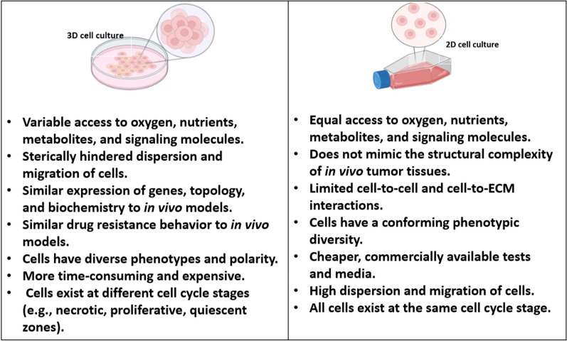
Three-dimensional (3D) cell cultures have emerged as valuable tools in cancer research, offering significant advantages over traditional two-dimensional (2D) cell culture systems. In 3D cell cultures, cancer cells are grown in an environment that more closely mimics the 3D architecture and complexity of in vivo tumors. This approach has revolutionized cancer research by providing a more accurate representation of the tumor microenvironment (TME) and enabling the study of tumor behavior and response to therapies in a more physiologically relevant context. One of the key benefits of 3D cell culture in cancer research is the ability to recapitulate the complex interactions between cancer cells and their surrounding stroma. Tumors consist not only of cancer cells but also various other cell types, including stromal cells, immune cells, and blood vessels. These models bridge traditional 2D cell cultures and animal models, offering a cost-effective, scalable, and ethical alternative for preclinical research. As the field advances, 3D cell cultures are poised to play a pivotal role in understanding cancer biology and accelerating the development of effective anticancer therapies. This review article highlights the key advantages of 3D cell cultures, progress in the most common scaffold-based culturing techniques, pertinent literature on their applications in cancer research, and the ongoing challenges.
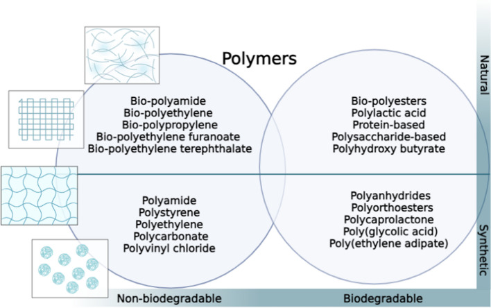
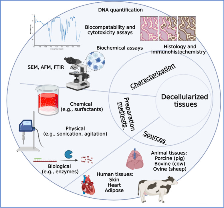
Keywords: Decellularized tissues; Extracellular matrix (ECM); Hydrogels; Microfluidics; Scaffolds; Three-dimensional (3D) cell culture.
© 2024. The Author(s).
Conflict of interest statement
The authors declare no competing interest.
Figures
References
-
- Grønning T. History of cancer. In: Colditz GA, editor. The sage encyclopedia of cancer and society. 2. CA: SAGE Publications, Inc.; 2015. pp. 549–554.
-
- Guimarães* I dos S, Daltoé* RD, Herlinger AL, Madeira KP, Ladislau T, Valadão IC, Junior PCML, Fernandes Teixeira S, Amorim GM, Santos DZ dos, Demuth KR, Rangel LBA. Conventional Cancer Treatment. Cancer Treatment—Conventional and Innovative Approaches. 2013; Available from: https://www.intechopen.com/chapters/42057.
-
- De Vita VT. The evolution of therapeutic research in cancer. N Engl J Med. 2010;298(16):907–910. - PubMed
Publication types
MeSH terms
LinkOut - more resources
Full Text Sources
Other Literature Sources
Medical
Research Materials
