Case Reports
. 2023 Dec 1;70(4):178-183.
doi: 10.2344/anpr-70-03-03.

Management of an Ingested Foreign Body in a COVID-Positive Patient

Case Reports

Management of an Ingested Foreign Body in a COVID-Positive Patient

Tiffany Smith et al. Anesth Prog. .

Abstract

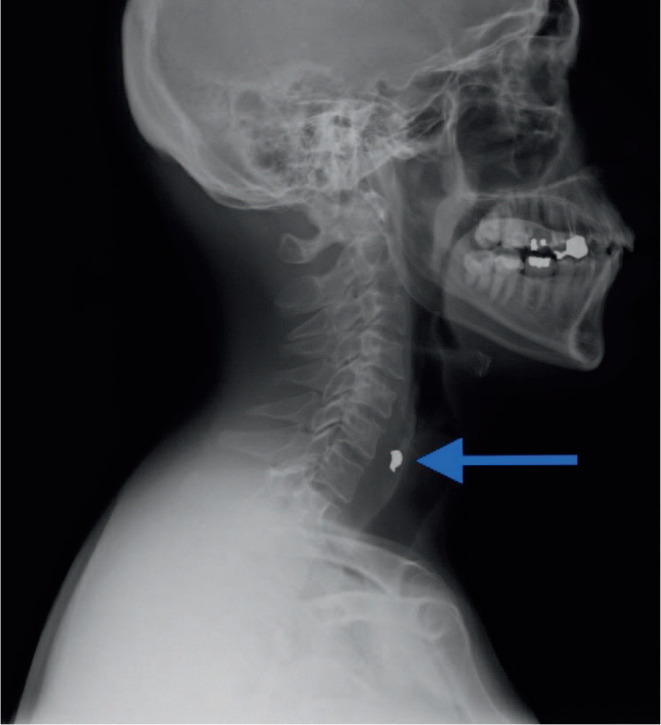
This case report describes a 51-year-old man who swallowed an amalgam fragment dislodged during dental treatment performed without a throat screen. The patient was transferred to the emergency department, where the foreign body was confirmed to be in the esophagus following radiographic imaging. Foreign body removal from the esophagus is routinely achieved via esophagogastroduodenoscopy (EGD). However, this incident occurred in September 2020, at the height of the COVID-19 pandemic. Because of the patient's preoperative positive COVID-19 test, the option for EGD retrieval was eliminated per hospital protocol. Instead, a noninvasive approach with serial radiographic monitoring was deemed mandatory to observe the fragment as it passed through the gastrointestinal tract, warranted by the small size of the foreign body and the patient's lack of signs and symptoms of respiratory distress. This case report reinforces the importance of using airway protection during every dental procedure. Furthermore, reevaluation of EGD as the gold standard for treatment of ingested small materials may be warranted.

Keywords: Amalgam; Aspiration; COVID; Case report; Foreign body; Ingestion; Isolation.


Figures

Figure 1.
Figure 1.
Initial Anteroposterior Radiograph Containing an Amalgam Fragment Anteroposterior radiograph obtained upon patient's arrival at the emergency department. The amalgam fragment was located lateral to the midline at the level of C6.
Figure 2.
Figure 2.
Initial Lateral Radiograph Containing an Amalgam Fragment Lateral radiograph obtained upon patient's arrival at the emergency department. The amalgam fragment was located within the esophagus at the level of C6.
Figure 3.
Figure 3.
Anteroposterior Radiograph With an Outline of the Esophagus and a Potential Zenker's Diverticulum The esophagus and likely position of the Zenker's diverticulum on the anteroposterior radiograph have been outlined in red. This provides an explanation for the lateral positioning of the amalgam fragment seen in Figure 1.
Figure 4.
Figure 4.
Lateral Radiograph With an Outline of the Esophagus and a Potential Zenker's Diverticulum The esophagus and likely position of the Zenker's diverticulum on the lateral radiograph have been outlined in red. This outline has been provided to explain the lateral positioning of the amalgam fragment seen in Figure 1.
Figure 5.
Figure 5.
Anteroposterior Radiograph After Passing the Amalgam Fragment Anteroposterior radiograph obtained approximately 4 hours after the initial anteroposterior radiograph showing the amalgam fragment no longer present and presumed to have passed into the stomach.
Figure 6.
Figure 6.
Lateral Radiograph After Passing the Amalgam Fragment Lateral radiograph obtained approximately 4 hours after the initial lateral radiograph. The amalgam fragment is no longer present and presumed to have passed into the stomach. Extracorporeal radiopacities represent hospital gown buttons (green arrowheads).

References

    1. Susini G,, Pommel L,, Camps J. Accidental ingestion and aspiration of root canal instruments and other dental foreign bodies in a French population Int Endod J 2007. 40 (8) 585–589 10.1111/j.1365-2591.2007.01249.x - DOI - PubMed
    1. Drobyazgin EA,, Chikinev YV,, Arkhipov DA. Inorodnye tela verkhnikh otdelov zheludochno-kishechnogo trakta. Diagnostic and treatment of foreign bodies of the upper digestive tract. Khirurgiia (Mosk). 2021. (6) 38–44 10.17116/hirurgia2021061387 - DOI - PubMed
    1. Law R,, Katzka DA,, Baron TH. Zenker's diverticulum Clin Gastroenterol Hepatol 2014. 12 (11) 1773–e112 10.1016/j.cgh.2013.09.016 - DOI - PubMed
    1. Zenker's diverticulum. Mount Sinai Health System. Accessed January 11, 2023. https://www.mountsinai.org/locations/grabscheid-voice-swallowing-center/...
    1. Malamed SF,, Orr DL,, Orr TM. Foreign body airway obstruction In: Medical Emergencies in the Dental Office. Elsevier; 2015: 186–204

Publication types