

Indocyanine green fluorescence in gastrointestinal surgery: Appraisal of current evidence
- PMID: 38222003
- PMCID: PMC10784830
- DOI: 10.4240/wjgs.v15.i12.2693
Indocyanine green fluorescence in gastrointestinal surgery: Appraisal of current evidence
Abstract
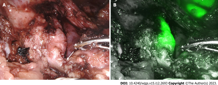
Applying indocyanine green (ICG) fluorescence in surgery has created a new dimension of navigation surgery to advance in various disciplines. The research in this field is nascent and fragmented, necessitating academic efforts to gain a comprehensive understanding. The present review aims to integrate diverse perspectives and recent advances in its application in gastrointestinal surgery. The relevant articles were selected by using the appropriate keyword search in PubMed. The angiography and cholangiography property of ICG fluorescence is helpful in various hepatobiliary disorders. In gastroesophageal and colorectal surgery, the lymphangiography and angiography property of ICG is applied to evaluate bowel vascularity and guide lymphadenectomy. The lack of objective parameters to assess ICG fluorescence has been the primary limitation when ICG is used to evaluate bowel perfusion. The optimum dose and timing of ICG administration need to be standardized in some new application areas in gastrointestinal surgery. Binding tumor-specific ligands with fluorophores can potentially widen the fluorescence application to detect primary and metastatic gastrointestinal tumors. The narrative review outlines prior contributions, limitations, and research opportunities for future studies across gastrointestinal sub-specialty. The findings of the present review would be helpful for scholars and practitioners to explore and progress in this exciting domain of gastrointestinal surgery.
Keywords: Angiography; Cholangiography; Fluorescence; Indocyanine green; Lymphangiography; Navigation surgery.
©The Author(s) 2023. Published by Baishideng Publishing Group Inc. All rights reserved.
Conflict of interest statement
Conflict-of-interest statement: All the authors report no relevant conflicts of interest for this article.
Figures


References
-
- Renz M. Fluorescence microscopy-a historical and technical perspective. Cytometry A. 2013;83:767–779. - PubMed
-
- Benson RC, Kues HA. Fluorescence properties of indocyanine green as related to angiography. Phys Med Biol. 1978;23:159–163. - PubMed
-
- Reinhart MB, Huntington CR, Blair LJ, Heniford BT, Augenstein VA. Indocyanine Green: Historical Context, Current Applications, and Future Considerations. Surg Innov. 2016;23:166–175. - PubMed
-
- Kogure K, David NJ, Yamanouchi U, Choromokos E. Infrared absorption angiography of the fundus circulation. Arch Ophthalmol. 1970;83:209–214. - PubMed
Publication types
LinkOut - more resources
Full Text Sources
