

Unraveling a rare splenic pathology: a case report of a benign hemorrhagic spleen with primary congenital splenic epidermoid cysts
- PMID: 38222723
- PMCID: PMC10783293
- DOI: 10.1097/MS9.0000000000001587
Unraveling a rare splenic pathology: a case report of a benign hemorrhagic spleen with primary congenital splenic epidermoid cysts
Abstract
Introduction and importance: A primary congenital splenic epidermoid cyst is an immensely rare pathology with mostly unknown epidemiological parameters. Misdiagnosis can easily happen and this results in life-threatening ramifications for patients. Considering this pathology as a potential differential diagnosis allows for the required surgical intervention to be timely accomplished. In this case, the authors are documenting this pathology and presenting how it was successfully managed via proper and informed preoperative analysis and meticulous intraoperative technique.
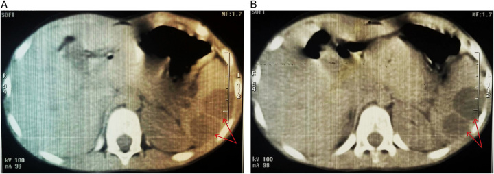
Case presentation: Hereby, we portray the exceptionally rare case of a 7-year-old male who presented to our surgical clinic complaining of a sudden left hypochondriac pain with early satiety for 1 month's duration. The preoperative radiological assessment displayed numerous splenic cystic lesions throughout the splenic parenchyma.
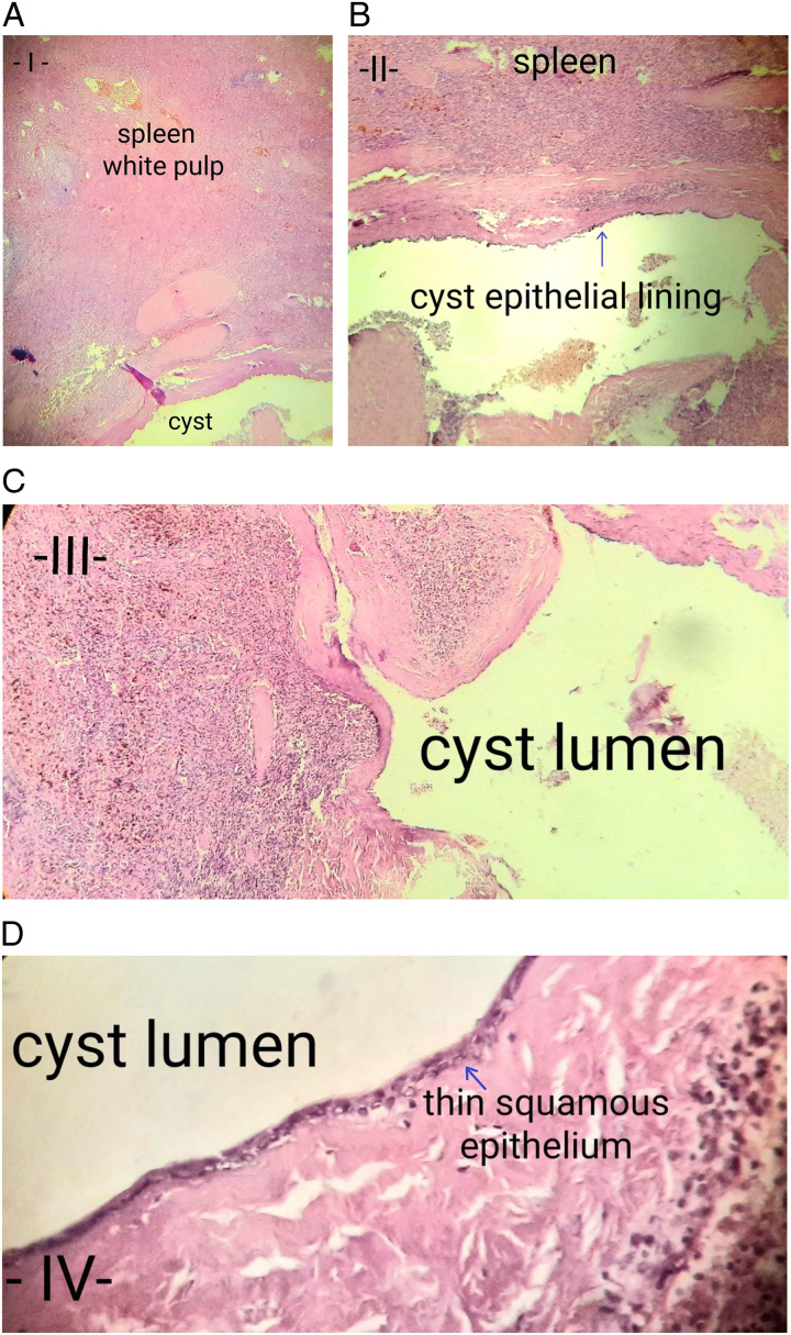
Clinical discussion: Resection of the cysts was accomplished via total splenectomy. The ensuing histopathological analysis via Hematoxylin and Eosin of the resected specimens established the diagnosis of a congested hemorrhagic spleen with multiple primary congenital splenic epidermoid cysts.
Conclusion: Primary congenital splenic epidermoid cysts are an extremely rare type of splenic pathology. There is profound scarcity amidst the published literature regarding it. This merits in-depth study and apt documentation to raise awareness regarding this pathology as a potential differential diagnosis in cases of abdominal pain. Documentation allows us to set up proper and innovative clinical and surgical protocols for these patients. Based on our conclusive review of the published literature, the authors conclude that ours is the first ever documented case from our country of a primary congenital splenic epidermoid cyst.
Keywords: abdominal surgery; case report; epidermoid cyst; primary congenital splenic epidermoid cyst; splenic cyst; total splenectomy.
Copyright © 2023 The Author(s). Published by Wolters Kluwer Health, Inc.
Conflict of interest statement
The authors declare that there are no conflicts of interest.Sponsorships or competing interests that may be relevant to content are disclosed at the end of this article.
Figures
References
-
- Khan Z, Chetty R. A review of the cysts of the spleen. Diagnostic Histopathol 2016;22:479–484.
-
- Reddi VR, Reddy MK, Srinivas B, et al. . Mesothelial splenic cyst--a case report. Ann Acad Med Singap 1998;27:880–882. - PubMed
-
- Verma M, Vashist MG, Dalal S, et al. . Epidermoid cyst of the spleen. IJRRMS 2013;3:47–48.
Publication types
LinkOut - more resources
Full Text Sources