Vertex Epidural Hematomas: Discussion of a Rare Traumatic Injury Through a Mini Series of 3 Cases
- PMID: 38222837
- PMCID: PMC10782103
- DOI: 10.13004/kjnt.2023.19.e45
Vertex Epidural Hematomas: Discussion of a Rare Traumatic Injury Through a Mini Series of 3 Cases
Abstract
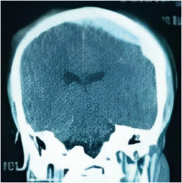
Vertex epidural hematomas are very uncommon complications of traumatic head injury. Besides the volume of the epidural bleeding, compression of the superior sagittal sinus may be source for added elevated intracranial pressure. Clinical presentation of such lesions is heterogenous and symptoms can develop in an acute to a chronic frame. Radiological diagnosis can sometimes be challenging. Due to its rarity, such lesions have been only reported on case reports and small series and the management remain controversial. Hereby we report 3 cases of surgically managed post traumatic acute epidural hematomas of the vertex. Wen also went through a literature-based discussion of clinical, radiological and therapeutic features related to this condition.
Keywords: Brain trauma; Cranial epidural hematoma; Neurosurgery; Vertex.
Copyright © 2023 Korean Neurotraumatology Society.
Conflict of interest statement
Conflict of Interest: The authors have no financial conflicts of interest.
Figures


References
-
- Bonilha L, Mattos JP, Borges WA, Fernandes YB, Andrioli MS, Borges G. Chronic epidural hematoma of the vertex. Clin Neurol Neurosurg. 2003;106:69–73. - PubMed
-
- Fernandes-Cabral DT, Kooshkabadi A, Panesar SS, Celtikci E, Borghei-Razavi H, Celtikci P, et al. Surgical management of vertex epidural hematoma: technical case report and literature review. World Neurosurg. 2017;103:475–483. - PubMed
-
- Harbury OL, Provenzale JM, Barboriak DP. Vertex epidural hematomas: imaging findings and diagnostic pitfalls. Eur J Radiol. 2000;36:150–157. - PubMed
-
- Klepinowski T, Kawalec P, Larysz M, Sagan L. Acute-on-chronic vertex epidural hematoma with diastasis of the sagittal suture in an adult. World Neurosurg. 2020;139:245–249. - PubMed
Publication types
LinkOut - more resources
Full Text Sources