

Point-of-Care Echocardiography in the Difficult-to-Image Patient in the ICU: A Narrative Review
- PMID: 38222871
- PMCID: PMC10786596
- DOI: 10.1097/CCE.0000000000001035
Point-of-Care Echocardiography in the Difficult-to-Image Patient in the ICU: A Narrative Review
Abstract
Objectives: The objective of this narrative review was to address common obstacles encountered in the ICU to acquiring quality and interpretable images using point-of-care echocardiography.
Data sources: Detailed searches were performed using PubMed and Ovid Medline using medical subject headings and keywords on topics related to patient positioning, IV echo contrast, alternative subcostal views, right ventricular outflow tract (RVOT) hemodynamics, and point-of-care transesophageal echocardiography. Articles known to the authors were also selected based on expert opinion.
Study selection: Articles specific to patient positioning, IV echo contrast, alternative subcostal views, RVOT hemodynamics, and point-of-care transesophageal echocardiography were considered.
Data extraction: One author screened titles and extracted relevant data while two separate authors independently reviewed selected articles.
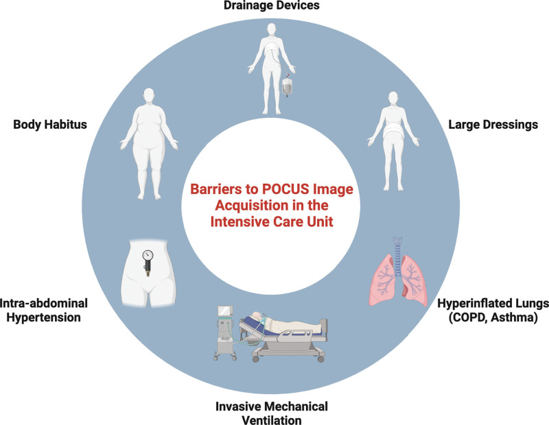
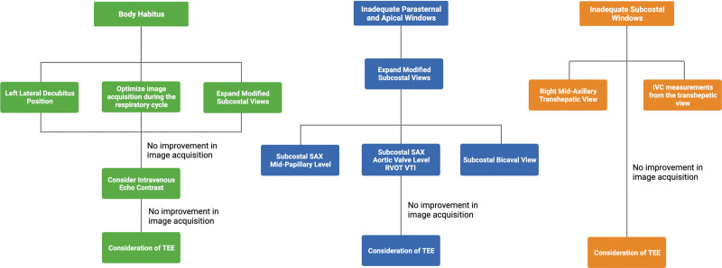
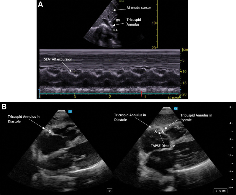
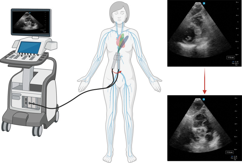
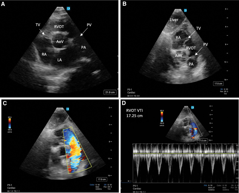
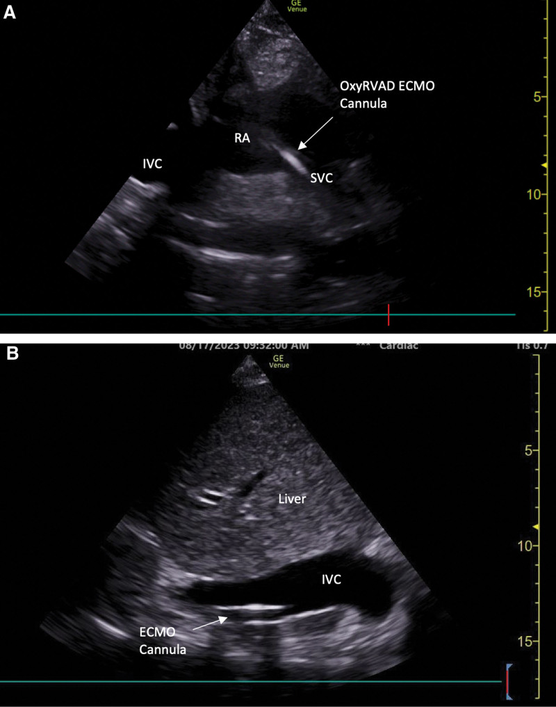
Data synthesis: Impediments to acquiring quality and interpretable images in critically ill patients are common. Notably, body habitus, intra-abdominal hypertension, dressings or drainage tubes, postoperative sternotomies, invasive mechanical ventilation, and the presence of subcutaneous emphysema or lung hyperinflation are commonly encountered obstacles in transthoracic image acquisition in the ICU. Despite these obstacles, the bedside clinician may use obstacle-specific maneuvers to enhance image acquisition. These may include altering patient positioning, respiratory cycle timing, expanding the subcostal window to include multilevel short-axis views for use in the assessment of RV systolic function and hemodynamics, coronal transhepatic view of the inferior vena cava, and finally point-of-care transesophageal echocardiography.
Conclusions: Despite common obstacles to point-of-care echocardiography in critically ill patients, the beside sonographer may take an obstacle-specific stepwise approach to enhance image acquisition in difficult-to-image patients.
Keywords: critical care; hemodynamics; transesophageal echocardiography; transthoracic echocardiography; ultrasound.
Copyright © 2024 The Authors. Published by Wolters Kluwer Health, Inc. on behalf of the Society of Critical Care Medicine.
Figures
References
-
- Moore CL, Copel JA: Point-of-care ultrasonography. N Engl J Med 2011; 364:749–757 - PubMed
-
- Via G, Hussain A, Wells M, et al. ; International Liaison Committee on Focused Cardiac UltraSound (ILC-FoCUS): International evidence-based recommendations for focused cardiac ultrasound. J Am Soc Echocardiogr 2014; 27:683.e1–683.e33 - PubMed
-
- Frankel HL, Kirkpatrick AW, Elbarbary M, et al. : Guidelines for the appropriate use of bedside general and cardiac ultrasonography in the evaluation of critically ill patients-part I: General ultrasonography. Crit Care Med 2015; 43:2479–2502 - PubMed
-
- Levitov A, Frankel HL, Blaivas M, et al. : Guidelines for the appropriate use of bedside general and cardiac ultrasonography in the evaluation of critically ill patients—part II: Cardiac ultrasonography. Crit Care Med 2016; 44:1206–1227 - PubMed
-
- Cosyns B, El Haddad P, Lignian H, et al. : Contrast harmonic imaging improves the evaluation of left ventricular function in ventilated patients: Comparison with transesophageal echocardiography. Eur J Echocardiogr 2004; 5:118–122 - PubMed
Publication types
Grants and funding
LinkOut - more resources
Full Text Sources
