. 2024 Jan 3;14(1):662-683.
doi: 10.21037/qims-23-847. Epub 2024 Jan 2.

U-fiber analysis: a toolbox for automated quantification of U-fibers and white matter hyperintensities

Affiliations

U-fiber analysis: a toolbox for automated quantification of U-fibers and white matter hyperintensities

Gaoxing Zheng et al. Quant Imaging Med Surg. .

Abstract

Background: Whether white matter hyperintensities (WMHs) involve U-fibers is of great value in understanding the different etiologies of cerebral white matter (WM) lesions. However, clinical practice currently relies only on the naked eye to determine whether WMHs are in the vicinity of U-fibers, and there is a lack of good neuroimaging tools to quantify WMHs and U-fibers.

Methods: Here, we developed a multimodal neuroimaging toolbox named U-fiber analysis (UFA) that can automatically extract WMHs and quantitatively characterize the volume and number of WMHs in different brain regions. In addition, we proposed an anatomically constrained U-fiber tracking scheme and quantitatively characterized the microstructure diffusion properties, fiber length, and number of U-fibers in different brain regions to help clinicians to quantitatively determine whether WMHs in the proximal cortex disrupt the microstructure of U-fibers. To validate the utility of the UFA toolbox, we analyzed the neuroimaging data from 246 patients with cerebral small vessel disease (cSVD) enrolled at Zhongshan Hospital between March 2018 and November 2019 in a cross-sectional study.

Results: According to the manual judgment of the clinician, the patients with cSVD were divided into a WMHs involved U-fiber group (U-fiber-involved group, 51 cases) and WMHs not involved U-fiber group (U-fiber-spared group, 163 cases). There were no significant differences between the U-fiber-spared group and the U-fiber-involved group in terms of age (P=0.143), gender (P=0.462), education (P=0.151), Mini-Mental State Examination (MMSE) scores (P=0.151), and Montreal Cognitive Assessment (MoCA) scores (P=0.411). However, patients in the U-fiber-involved group had higher Fazekas scores (P<0.001) and significantly higher whole brain WMHs (P=0.046) and deep WMH volumes (P<0.001) compared to patients in the U-fiber-spared group. Moreover, the U-fiber-involved group had higher WMH volumes in the bilateral frontal [P(left) <0.001, P(right) <0.001] and parietal lobes [P(left) <0.001, P(right) <0.001]. On the other hand, patients in the U-fiber-involved group had higher mean diffusivity (MD) and axial diffusivity (AD) in the bilateral parietal [P(left, MD) =0.048, P(right, MD) =0.045, P(left, AD) =0.015, P(right, AD) =0.015] and right frontal-parietal regions [P(MD) =0.048, P(AD) =0.027], and had significantly reduced mean fiber length and number in the right parietal [P(length) =0.013, P(number) =0.028] and right frontal-parietal regions [P(length) =0.048] compared to patients in the U-fiber-spared group.

Conclusions: Our results suggest that WMHs in the proximal cortex may disrupt the microstructure of U-fibers. Our tool may provide new insights into the understanding of WM lesions of different etiologies in the brain.

Keywords: MATLAB toolbox; U-fiber tractography; cerebral small vessel disease (cSVD); diffusion magnetic resonance imaging (dMRI); white matter hyperintensities (WMHs).


Conflict of interest statement

Conflicts of Interest: All authors have completed the ICMJE uniform disclosure form (available at https://qims.amegroups.com/article/view/10.21037/qims-23-847/coif). The authors have no conflicts of interest to declare.

Figures
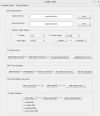
Figure 1
Figure 1
The GUI of the UFA toolbox. GUI, Graphical User Interface; UFA, U-fiber analysis; T1W, T1-weighted image; FLAIR, fluid-attenuated inversion recovery; DTI, diffusion tensor imaging; AP, anterior-posterior; DWI, diffusion-weighted imaging; MR, magnetic resonance; WMH, white matter hyperintensity.
Figure 2
Figure 2
Multimodal MRI data preprocessing and post-processing pipeline provided by the UFA toolbox. cSVD patient’s (A) T2 FLAIR image; (B) T1 image; and (C) DWI image with two phase encoding directions (DWI_AP, DWI_PA), AP indicates the anterior-posterior phase encoding direction and PA indicates the posterior-anterior phase encoding direction; (D) WMHs image after extraction by UBO Detector open source tool, white dots indicate WMHs extracted by the algorithm; (E) preprocessed T1 image; (F) schematic diagram of whole brain/periventricular/deep WMHs volume; (G) preprocessed DWI image; (H) diffusion tensor image obtained by diffusion tensor modeling of DWI image; (I) four diffusion metrics (FA/MD/AD/RD); (J) response function diagram of three tissues (gray matter/white matter/cerebrospinal fluid) in DWI image; (K) schematic of local fiber orientation distribution; (L) schematic of whole brain white matter fiber bundles; (M) schematic of subcortical gray/white matter ROI for exclusion of deep white matter fiber bundles passing through ROI; (N) schematic of U fibers; (O) schematic of mean fiber length (26) and fiber number (right) of 16 U fibers; (P) schematic diagrams of WMHs not involving U fibers (26) and involving U fibers (right); red circle represents WMHs with U-fiber involvement. cSVD, cerebral small vessel disease; T2 FLAIR, T2-weighted fluid-attenuated inversion recovery; DWI, diffusion-weighted imaging; AP, anterior-posterior; PA, posterior-anterior; WMHs, white matter hyperintensities; PV, periventricular; UBO, unidentified bright objects; FA, fractional anisotropy; MD, mean diffusivity; AD, axial diffusivity; RD, radial diffusivity; ROI, region of interest; ACT, anatomically-constrained tractography; Sup-F, superficial-frontal; Sup-FP, superficial-frontal-parietal; Sup-O, superficial-occipital; Sup-OT, superficial-occipital-temporal; Sup-P, superficial-parietal; Sup-PO, superficial-parietal-occipital; Sup-PT, superficial-parietal-temporal; Sup-T, superficial-temporal.
Video 1
Video 1
Video demonstration of how to use the UFA toolbox. UFA, U-fiber analysis.
Figure 3
Figure 3
Visualization examples of WMHs in different brain lobes and different arterial territories provided by the UFA toolbox. (A) WMH volume (left panel) and WMH clusters (right panel) in different brain lobes; (B) WMH volume (left panel) and WMH clusters (right panel) in different arteries; (C) four types of WMH clusters (punctuate, focal, medium, confluent) in different brain lobes (left lobes in the left panel, right lobes in the right panel); (D) four types of WMH clusters (punctuate, focal, medium, confluent) in different arteries (left arteries in the left panel, right arteries in the right panel). WMHs, white matter hyperintensities; UFA, U-fiber analysis; AAH, anterior artery hemisphere; AAC, anterior artery callosal; AAML, anterior artery medial lenticulostriate; MAH, middle artery hemisphere; MALL, middle artery lateral lenticulostriate, PAH, posterior artery hemisphere; PAC, posterior artery callosal; PATMP, posterior artery thalamic and midbrain perforators.
Figure 4
Figure 4
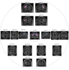
The 16 U-fiber clusters provided in the ORG atlas in the WMA tool. ORG, O’Donnell Research Group; WMA, white matter analysis; L, left hemisphere; R, right hemisphere; Sup-F, superficial-frontal; Sup-FP, superficial-frontal-parietal; Sup-O, superficial-occipital; Sup-OT, superficial-occipital-temporal; Sup-P, superficial-parietal; Sup-PO, superficial-parietal-occipital; Sup-PT, superficial-parietal-temporal; Sup-T, superficial-temporal.
Figure 5
Figure 5
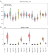
Visualization of diffusion microstructure parameters (FA/MD/AD/RD) in 16 superficial white matter tracts. FA, fractional anisotropy; MD, mean diffusivity; AD, axial diffusivity; RD, radial diffusivity; L, left hemisphere; R, right hemisphere; Sup-F, superficial-frontal; Sup-P, superficial-parietal; Sup-T, superficial-temporal; Sup-O, superficial-occipital; Sup-FP, superficial-frontal-parietal; Sup-PT, superficial-parietal-temporal; Sup-PO, superficial-parietal-occipital; Sup-OT, superficial-occipital-temporal.
Figure 6
Figure 6
Inclusion and exclusion flowchart of cSVD participants. Finally, 51 subjects with U-fibers involved and 163 subjects with U-fibers spared were analyzed by our UFA toolbox. MRI, magnetic resonance imaging; WMHs, white matter hyperintensities; cSVD, cerebral small vessel disease; UFA, U-fiber analysis.
Figure 7
Figure 7
Differences in the volume of WMHs in different brain regions between the U-fiber-spared group and the U-fiber-involved group. Significant differences were marked by black asterisks (*) at the alpha significance level of 0.05. The double asterisks (**) represent a significant P value less than 0.01. (A) WMH volume comparison in whole white matter (Whole), periventricular (PV) and deep white matter (15). (B) WMH volume comparison in different lobes (bilateral frontal, temporal, parietal, occipital lobes) and cerebellum. (C) WMH volume comparison in different arterial territories. WMHs, white matter hyperintensities; L, left hemisphere; R, right hemisphere; AAH, anterior artery hemisphere; AAC, anterior artery callosal; AAML, anterior artery medial lenticulostriate; MAH, middle artery hemisphere; MALL, middle artery lateral lenticulostriate, PAH, posterior artery hemisphere; PAC, posterior artery callosal; PATMP, posterior artery thalamic and midbrain perforators.
Figure 8
Figure 8
Differences in the U-fiber diffusion measures in different brain regions between the U-fiber-spared group and the U-fiber-involved group. Significant differences are marked by black asterisks (*) at the alpha significance level of 0.05. (A) FA; (B) MD; (C) AD; (D) RD. FA, fractional anisotropy; MD, mean diffusivity; AD, axial diffusivity; RD, radial diffusivity; L, left hemisphere; R, right hemisphere; Sup-FP, superficial-frontal-parietal; Sup-F, superficial-frontal; Sup-OT, superficial-occipital-temporal; Sup-O, superficial-occipital; Sup-PO, superficial-parietal-occipital; Sup-PT, superficial-parietal-temporal; Sup-P, superficial-parietal; Sup-T, superficial-temporal.
Figure 9
Figure 9
Differences in the macrostructural characteristics in different brain regions between the U-fiber spared group and the U-fiber involved group. (A) Mean fiber length of U-fibers; (B) number of U-fibers. Significant differences are marked by black asterisks (*) at the alpha significance level of 0.05. L, left hemisphere; R, right hemisphere; Sup-FP, superficial-frontal-parietal; Sup-F, superficial-frontal; Sup-OT, superficial-occipital-temporal; Sup-O, superficial-occipital; Sup-PO, superficial-parietal-occipital; Sup-PT, superficial-parietal-temporal; Sup-P, superficial-parietal; Sup-T, superficial-temporal.

Similar articles

Cited by

References

    1. Shastin D, Genc S, Parker GD, Koller K, Tax CM, Evans J, Hamandi K, Gray WP, Jones DK, Chamberland M. Short Association Fibre Tractography. bioRxiv 2021. doi: . - PMC - PubMed
    1. Schüz A, Miller R. Cortical areas: unity and diversity. London: CRC press; 2002.
    1. Kirilina E, Helbling S, Morawski M, Pine K, Reimann K, Jankuhn S, Dinse J, Deistung A, Reichenbach JR, Trampel R, Geyer S, Müller L, Jakubowski N, Arendt T, Bazin PL, Weiskopf N. Superficial white matter imaging: Contrast mechanisms and whole-brain in vivo mapping. Sci Adv 2020;6:eaaz9281. 10.1126/sciadv.aaz9281 - DOI - PMC - PubMed
    1. Gahm JK, Shi Y. Surface-based Tracking of U-fibers in the Superficial White Matter. Med Image Comput Comput Assist Interv 2019;11766:538-46. - PMC - PubMed
    1. Movahedian Attar F, Kirilina E, Haenelt D, Pine KJ, Trampel R, Edwards LJ, Weiskopf N. Mapping Short Association Fibers in the Early Cortical Visual Processing Stream Using In Vivo Diffusion Tractography. Cereb Cortex 2020;30:4496-514. 10.1093/cercor/bhaa049 - DOI - PMC - PubMed