. 2024;39(1):137-149.
doi: 10.1557/s43578-023-01163-x. Epub 2023 Oct 25.

Conductive 3D nano-biohybrid systems based on densified carbon nanotube forests and living cells

Affiliations

Conductive 3D nano-biohybrid systems based on densified carbon nanotube forests and living cells

Roya Bagheri et al. J Mater Res. 2024.

Abstract

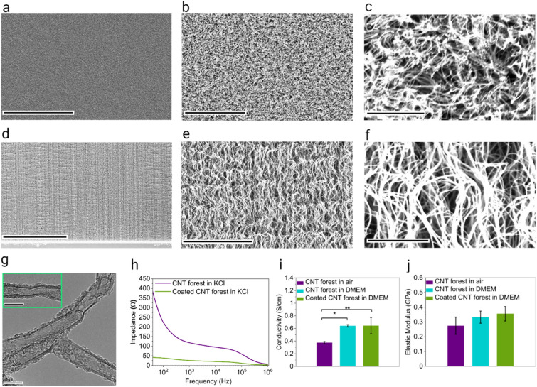
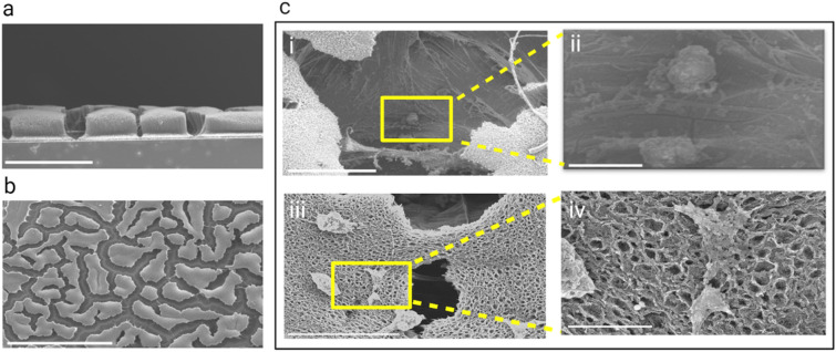
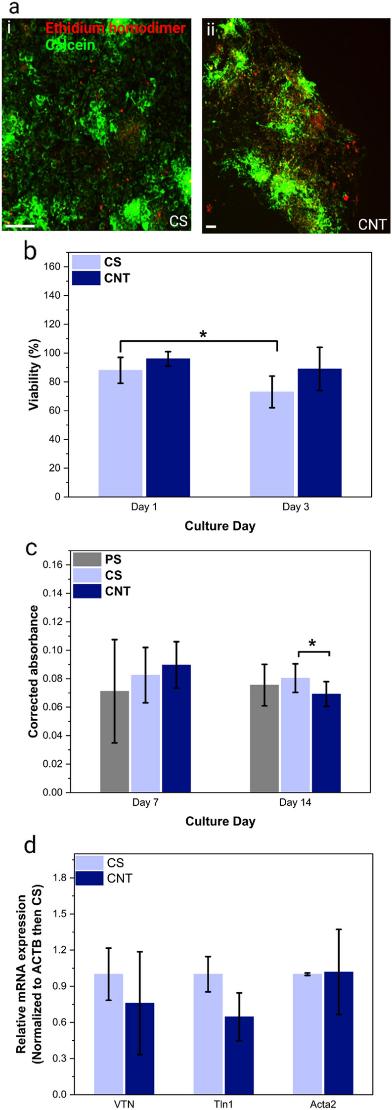
Conductive biohybrid cell-material systems have applications in bioelectronics and biorobotics. To date, conductive scaffolds are limited to those with low electrical conductivity or 2D sheets. Here, 3D biohybrid conductive systems are developed using fibroblasts or cardiomyocytes integrated with carbon nanotube (CNT) forests that are densified due to interactions with a gelatin coating. CNT forest scaffolds with a height range of 120-240 µm and an average electrical conductivity of 0.6 S/cm are developed and shown to be cytocompatible as evidenced from greater than 89% viability measured by live-dead assay on both cells on day 1. The cells spread on top and along the height of the CNT forest scaffolds. Finally, the scaffolds have no adverse effects on the expression of genes related to cardiomyocyte maturation and functionality, or fibroblast migration, adhesion, and spreading. The results show that the scaffold could be used in applications ranging from organ-on-a-chip systems to muscle actuators.

Keywords: Carbon nanotube forest; Cardiomyocytes; Cell scaffold; Conductive nano-biohybrid systems; Densification; Fibroblasts; Gelatin.


Conflict of interest statement

Conflict of interestThe authors declare that they have no known competing financial interests or personal relationships that could have appeared to influence the work reported in this paper.

Figures

Figure 1
Figure 1
Schematic illustration of the process from CNT forest growth to cell seeding: (a) Silicon wafer coated with SiO2, Al2O3, and Fe as catalyst, (b) Exposure to ethylene, argon, and hydrogen gases for the growth of CNT forests, (c) CNT forest after the termination of growth, (d) Sterilization of CNT forests using 70% ethanol (not shown) and UV exposure, (e) Coating of CNT forests with gelatin, and (f, f) Cell seeding of fibroblasts derived from a NIH 3T3 mouse cell line (f) or primary cardiomyocytes extracted from neonatal rat heart (f).
Figure 2
Figure 2
Physical characterization of CNT forests: (a–c) SEM images of the top view of a dry CNT forest at different magnifications, scale bars, 100 µm, 10 µm, and 100 nm, respectively; (d–f) SEM images of the side view of a dry CNT forest at different magnifications, scale bars, 30 µm, 4 µm, and 500 nm, respectively; (g) TEM images of CNTs from a CNT forest, scale bars, 10 nm; (h) Impedance vs. frequency; (i) Electrical conductivity, (*p < 0.05, **p < 0.001); (j) Elastic modulus.
Figure 3
Figure 3
Schematic illustration of the formation of 3D cell networks on CNT forest scaffolds: (a) side view; (b) top view; (i) CNT forest in air; (ii) CNT forest coated with gelatin in the growth medium; (iii, iv) CNT forest scaffold seeded with cells (either fibroblasts or cardiomyocytes) at low (iii) and high (iv) magnifications.
Figure 4
Figure 4
Interactions of fibroblasts with the 3D structure of the CNT forest scaffolds; (a) ESEM images of a CNT forest scaffold seeded with fibroblasts on day 3, (i, ii) a cell attached to the CNT forest wall, scale bars, 20 and 5 µm respectively, (iii, iv) a cell connecting two CNT forest walls, scale bars, 10 and 3 µm, (v, vi) a cell connection to CNTs, scale bars, 4 and 2 µm respectively; (b) (i) An SEM image of a fibroblast (day 15) attached to the walls of densified regions of a CNT forest scaffold bridging the open space, scale bar, 20 µm, (ii) An SEM image of the top surface of a CNT forest scaffold and a fibroblast (day 15) spread on it, scale bar, 40 µm; (c) Calcein AM staining of fibroblasts (day 3) on CNT forest scaffolds with the background staining showing the densified forests and the location of cells on the side walls of the densified regions, scale bar, 100 µm; (d–g) Immunofluorescence micrographs of fibroblasts on CNT forest scaffolds (day 7) labeled with phalloidin for F-actin (green) and DAPI for nuclei (blue) showing z-stack confocal images at low (d) and high (f) magnifications and their deconstruction into 2D images (e and g corresponding to d and f, respectively) covering a z range of 140 µm showing the spreading of the cells along the height of CNT forests, scale bar, 100 µm, (i) Immunofluorescence micrographs of fibroblasts on coverslip (CS) as control (day 7) labeled with phalloidin for F-actin (green) and DAPI for nuclei (blue) showing a z-stack confocal image (h) and it’s deconstruction into 2D images (i) covering a z range of 20 µm showing the 2D nature of scaffold, scale bar, 100 µm; (j, k) Schematic illustration of the 2D and 3D morphology of fibroblasts on coverslip and CNT forest scaffold, respectively.
Figure 5
Figure 5
Interactions of cardiomyocytes with the 3D structure of the CNT forest scaffolds: (a, b) SEM images of the side and top views, respectively, of a densified CNT forest scaffold seeded with cardiomyocytes on day 14 and 15, scale bar, 100 and 500 µm respectfully; (c) Higher magnification SEM images, (i, ii) higher magnification images of a cell in the space between densified CNT forest regions, scale bars, 50 and 10 µm respectively, (iii, iv) higher magnification SEM images of cells spread on top of the scaffold, scale bars, 40 and 5 µm respectively.
Figure 6
Figure 6
Organization, viability, and gene expression of fibroblasts on CNT forest scaffolds: (a) Live-Dead immunofluorescence micrographs of fibroblasts (day 3) on the CS as control (i) and CNT scaffold (ii), scale bars, 200 µm; (b) Viability of fibroblasts on CNT forest scaffolds and CS as control calculated by the ratio of the number of live cells to total cells in the Live-Dead stained micrographs (*p < 0.01); (c) Absorbance data obtained through the Presto-Blue assay (*p < 0.05) representing the viability of cells; (d) Expression of VTN, TLN1, and ACTA2 genes representing adhesion, spreading, and migration of fibroblasts on day 3 on CNT forest scaffolds and CS as control (normalized to ACTB then CS) showing no statistically significant difference.
Figure 7
Figure 7
Organization, viability, and gene expression of cardiomyocytes on CNT forest scaffolds: (a) Immunofluorescence micrographs of cardiomyocytes labeled with monoclonal anti-α-actinin (sarcomeric) on a CS (i) and a CNT forest scaffold (ii); day 14; scale bars, 200 µm; (b) Immunofluorescence micrographs of cardiomyocytes labeled with anti-cardiac troponin on a CS (i) and a CNT forest scaffold (ii); day 19; scale bars, 20 µm; (c) Live-Dead immunofluorescence micrographs of cardiomyocytes (day 5) on a a CS (i) and a CNT forest (ii); scale bars, 200 µm; (d) Viability of cardiomyocyte on CNT forest scaffolds and CS as control calculated by the ratio of the number of live cells to total cells in the Live-Dead stained micrographs; (e) Absorbance data obtained through the Presto-Blue assay (PS & CS as control), (*p < 0.05, **p < 0.001) representing the viability of cells; (f) Expression of MYH6, TNNT2, ACTC1 and GJA1 genes representing maturation and functionality of cardiomyocytes at day 8 on CNT forest scaffolds and CS as control (normalized to ACTB then CS).

Similar articles

References

    1. Li J, et al. Biohybrid micro-and nanorobots for intelligent drug delivery. Cyborg Bionic Syst. 2022 doi: 10.34133/2022/9824057. - DOI - PMC - PubMed
    1. Wang C, et al. Biohybrid materials: Structure design and biomedical applications. Mater. Today Bio. 2022;16:100352. - PMC - PubMed
    1. Capulli AK, et al. JetValve: Rapid manufacturing of biohybrid scaffolds for biomimetic heart valve replacement. Biomaterials. 2017;133:229–241. - PMC - PubMed
    1. Compaan AM, et al. Cross-linkable microgel composite matrix bath for embedded bioprinting of perfusable tissue constructs and sculpting of solid objects. ACS Appl. Mater. Interfaces. 2020;12(7):7855–7868. - PubMed
    1. Li S, et al. A DNA nanorobot functions as a cancer therapeutic in response to a molecular trigger in vivo. Nat. Biotechnol. 2018;36(3):258–264. - PubMed

LinkOut - more resources