

Anticancer effects of herbal medicine Emelia-M, Mshikazi and Delosma H against human leukaemia cells
- PMID: 38223600
- PMCID: PMC10782316
- DOI: 10.4314/ahs.v23i2.35
Anticancer effects of herbal medicine Emelia-M, Mshikazi and Delosma H against human leukaemia cells
Abstract
Background: Leukaemia is one of the three major types of blood cancers that lead to the overproduction of abnormal white blood cells. Emelia M (EMB), Mshikazi and Delosma H are herbal medicines that are being used by traditional healers in KwaZulu-Natal, South Africa to treat leukaemia and other diseases.
Objectives: To gain insight into the safety (non-toxic effect), anti-cancer activity, mechanisms of action and phytochemical profiles of traditional herbal medicines (Emelia M (EMB), Mshikazi and Delosma H) in South Africa.
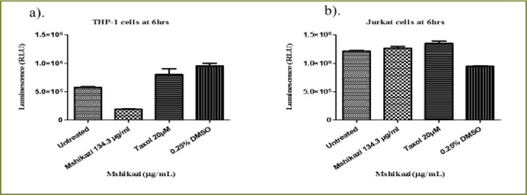
Methods: The viability of human peripheral blood mononuclear cells (PBMCs), monocytic (THP-1) and T-lymphocyte (Jurkat) cell lines exposed to varying concentrations of aqueous extracts of the three herbal medicines were assessed using adenosine triphosphate (ATP) assay.
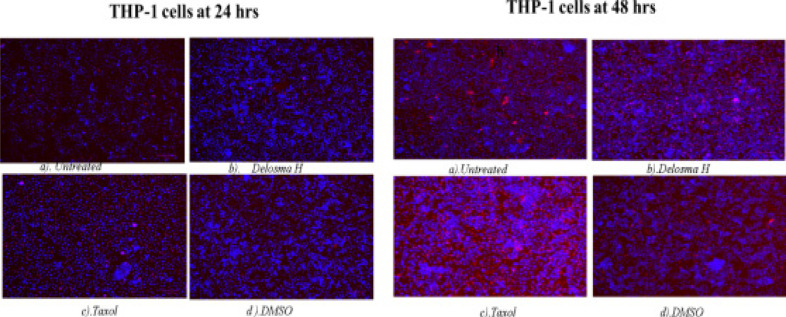
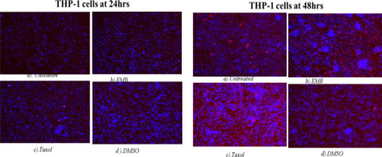
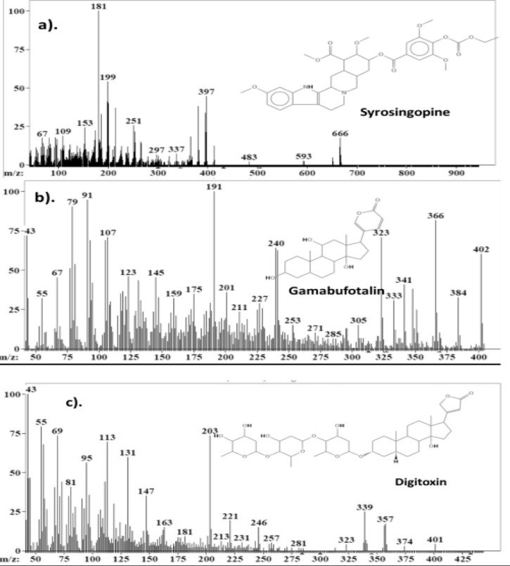
Results: All three extracts showed a dose-dependent effect on the viability of PBMCs. Cell viability decreased with increasing concentrations of extracts when compared with the untreated cells at 24 and 48 hours. The inhibitory activities (IC50) of the extract were found in the order of Mshikazi > EMB, > Delosma H. All the extracts induced apoptosis with minimal necrosis. Many bioactive compounds that have been previously reported to have anticancer effects were identified in the extracts.
Conclusion: Mshikazi extract significantly inhibited the growth of THP-1 and Jurkat cells and induced cell death through apoptosis than the other two extracts.
Keywords: Leukaemia; apoptosis; phytochemical compounds; traditional herbal medicines.
© 2023 Adeniyi JN et al.
Figures


Similar articles
-
Phytochemical profile and in vitro antioxidant activity of Emelia M (EMB), Mshikazi and Delosma H herbal medicines as demonstrated in THP-1 and Jurkat leukaemia cell lines.Afr Health Sci. 2021 Dec;21(4):1924-1937. doi: 10.4314/ahs.v21i4.51. Afr Health Sci. 2021. PMID: 35283952 Free PMC article.
-
Old plants newly discovered: Cassia sieberiana D.C. and Cassia abbreviata Oliv. Oliv. root extracts inhibit in vitro HIV-1c replication in peripheral blood mononuclear cells (PBMCs) by different modes of action.J Ethnopharmacol. 2012 May 7;141(1):48-56. doi: 10.1016/j.jep.2012.01.044. Epub 2012 Feb 2. J Ethnopharmacol. 2012. PMID: 22326358
-
Anti-tumor effect of hot aqueous extracts from Sonchus oleraceus (L.) L. and Juniperus sabina L - Two traditional medicinal plants in China.J Ethnopharmacol. 2016 Jun 5;185:289-99. doi: 10.1016/j.jep.2016.03.044. Epub 2016 Mar 19. J Ethnopharmacol. 2016. PMID: 27001625
-
A review on traditionally used South African medicinal plants, their secondary metabolites and their potential development into anticancer agents.J Ethnopharmacol. 2020 Oct 28;261:113101. doi: 10.1016/j.jep.2020.113101. Epub 2020 Jun 17. J Ethnopharmacol. 2020. PMID: 32562876 Review.
-
Application of nanotechnology to herbal antioxidants as improved phytomedicine: An expanding horizon.Biomed Pharmacother. 2022 Sep;153:113413. doi: 10.1016/j.biopha.2022.113413. Epub 2022 Aug 6. Biomed Pharmacother. 2022. PMID: 36076482 Review.
Cited by
-
Editor's choice: Covid-19 and HIV are still very much with us.Afr Health Sci. 2023 Jun;23(2):i-v. doi: 10.4314/ahs.v23i2.1. Afr Health Sci. 2023. PMID: 38223647 Free PMC article. No abstract available.
References
-
- Jemal A, Bray F, Forman D, Brien MO, Ferlay J. Cancer Burden in Africa and Opportunities for Prevention. Cancer in Africa. 2012. pp. 1–13. 10.1002/cncr.27410. - PubMed
-
- Dent J, Manner CK, Milner D, Mutebi M, Ng ang 'a A, Olopade OI, et al. Africa's Emerging Cancer Crisis: A Call to Action [Internet] 2019. Jan 10, pp. 1–8. 2017, Available from: https://bvgh.org/wp-content/uploads/2017/07/Africas-Emerging-Cancer-Cris....
-
- Herbst MC. Fact Sheet on Adult Acute Myeloid Leukaemia (AML) Cancer Association of South Africa (CANSA) 2016. pp. 1–18.
-
- Lockwood W. Leukemia: AML, CML, ALL and CLL. 2017. pp. 2–32.
MeSH terms
Substances
LinkOut - more resources
Full Text Sources
Medical