

Phenomic Imaging
- PMID: 38223684
- PMCID: PMC10781914
- DOI: 10.1007/s43657-023-00128-8
Phenomic Imaging
Abstract
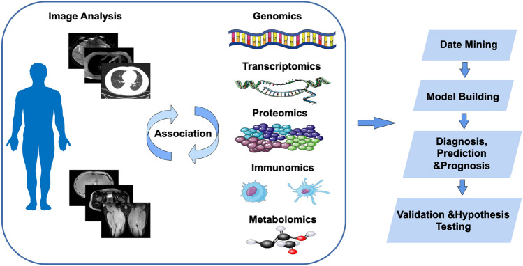
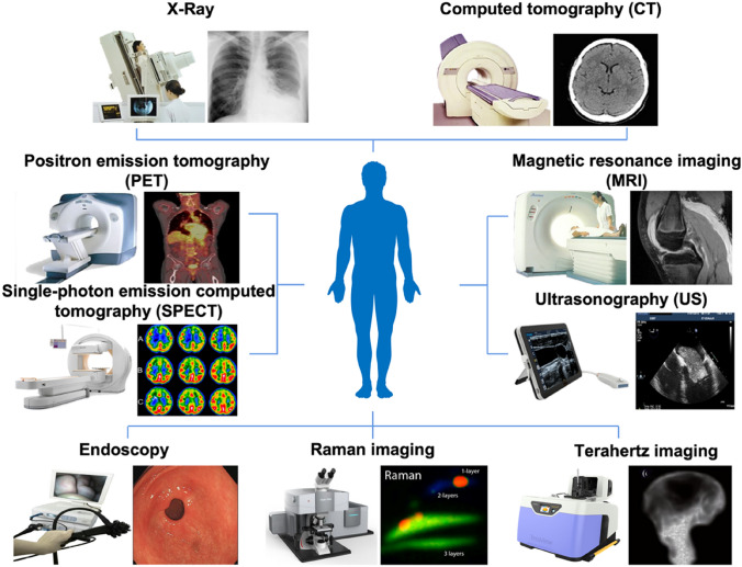
Human phenomics is defined as the comprehensive collection of observable phenotypes and characteristics influenced by a complex interplay among factors at multiple scales. These factors include genes, epigenetics at the microscopic level, organs, microbiome at the mesoscopic level, and diet and environmental exposures at the macroscopic level. "Phenomic imaging" utilizes various imaging techniques to visualize and measure anatomical structures, biological functions, metabolic processes, and biochemical activities across different scales, both in vivo and ex vivo. Unlike conventional medical imaging focused on disease diagnosis, phenomic imaging captures both normal and abnormal traits, facilitating detailed correlations between macro- and micro-phenotypes. This approach plays a crucial role in deciphering phenomes. This review provides an overview of different phenomic imaging modalities and their applications in human phenomics. Additionally, it explores the associations between phenomic imaging and other omics disciplines, including genomics, transcriptomics, proteomics, immunomics, and metabolomics. By integrating phenomic imaging with other omics data, such as genomics and metabolomics, a comprehensive understanding of biological systems can be achieved. This integration paves the way for the development of new therapeutic approaches and diagnostic tools.
Keywords: Genomics; Imaging; Immunomics; Metabolomics; Phenomics; Proteomics; Transcriptomics.
© The Author(s) 2023.
Conflict of interest statement
Conflict of InterestMei Tian is the Editorial Board Member of Phenomics, Yidan Gao is the editorial operation team member of Phenomics, and they were not involved in reviewing this paper.
Figures
References
-
- Ajoy R, Lo YC, Ho MH, Chen YY, Wang Y, Chen YH, Jing-Yuan C, Changou CA, Hsiung YC, Chen HM, Chang TH, Lee CY, Chiang YH, Chang WC, Hoffer B, Chou SY. CCL5 promotion of bioenergy metabolism is crucial for hippocampal synapse complex and memory formation. Mol Psychiatry. 2021;26(11):6451–6468. doi: 10.1038/s41380-021-01103-3. - DOI - PMC - PubMed
-
- Akbari H, Bakas S, Pisapia JM, Nasrallah MP, Rozycki M, Martinez-Lage M, Morrissette JJD, Dahmane N, O'Rourke DM, Davatzikos C. In vivo evaluation of EGFRvIII mutation in primary glioblastoma patients via complex multiparametric MRI signature. Neuro Oncol. 2018;20(8):1068–1079. doi: 10.1093/neuonc/noy033. - DOI - PMC - PubMed
-
- Aksoy O, Pencik J, Hartenbach M, Moazzami AA, Schlederer M, Balber T, Varady A, Philippe C, Baltzer PA, Mazumder B, Whitchurch JB, Roberts CJ, Haitel A, Herac M, Susani M, Mitterhauser M, Marculescu R, Stangl-Kremser J, Hassler MR, Kramer G, Shariat SF, Turner SD, Tichy B, Oppelt J, Pospisilova S, Hartenbach S, Tangermann S, Egger G, Neubauer HA, Moriggl R, Culig Z, Greiner G, Hoermann G, Hacker M, Heery DM, Merkel O, Kenner L. Thyroid and androgen receptor signaling are antagonized by μ-Crystallin in prostate cancer. Int J Cancer. 2021;148(3):731–747. doi: 10.1002/ijc.33332. - DOI - PMC - PubMed
-
- Arita H, Kinoshita M, Kawaguchi A, Takahashi M, Narita Y, Terakawa Y, Tsuyuguchi N, Okita Y, Nonaka M, Moriuchi S, Takagaki M, Fujimoto Y, Fukai J, Izumoto S, Ishibashi K, Nakajima Y, Shofuda T, Kanematsu D, Yoshioka E, Kodama Y, Mano M, Mori K, Ichimura K, Kanemura Y. Lesion location implemented magnetic resonance imaging radiomics for predicting IDH and TERT promoter mutations in grade II/III gliomas. Sci Rep. 2018;8(1):11773. doi: 10.1038/s41598-018-30273-4. - DOI - PMC - PubMed
Publication types
LinkOut - more resources
Full Text Sources