

A TA/Cu2+ Nanoparticle Enhanced Carboxymethyl Chitosan-Based Hydrogel Dressing with Antioxidant Properties and Promoting Wound Healing
- PMID: 38223881
- PMCID: PMC10788072
- DOI: 10.2147/IJN.S445844
A TA/Cu2+ Nanoparticle Enhanced Carboxymethyl Chitosan-Based Hydrogel Dressing with Antioxidant Properties and Promoting Wound Healing
Erratum in
-
Erratum: A TA/Cu2+ Nanoparticle Enhanced Carboxymethyl Chitosan-Based Hydrogel Dressing with Antioxidant Properties and Promoting Wound Healing [Corrigendum].Int J Nanomedicine. 2024 Apr 18;19:3609-3610. doi: 10.2147/IJN.S473836. eCollection 2024. Int J Nanomedicine. 2024. PMID: 38650836 Free PMC article.
Abstract
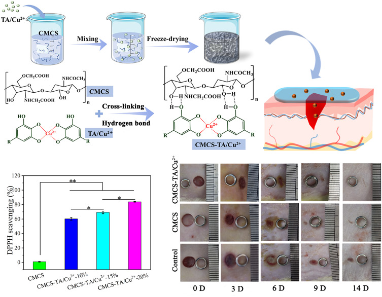
Background: As the first line of immune defense and the largest organ of body, skin is vulnerable to damage caused by surgery, burns, collisions and other factors. Wound healing in the skin is a long and complex physiological process that is influenced by a number of different factors. Proper wound care can greatly improve the speed of wound healing and reduce the generation of scars. However, traditional wound dressings (bandages, gauze, etc.) often used in clinical practice have a single function, lack of active ingredients and are limited in use. Hydrogels with three-dimensional network structure are a potential biomedical material because of their physical and chemical environment similar to extracellular matrix. In particular, hydrogel dressings with low price, good biocompatibility, degradability, antibacterial and angiogenic activity are favored by the public.
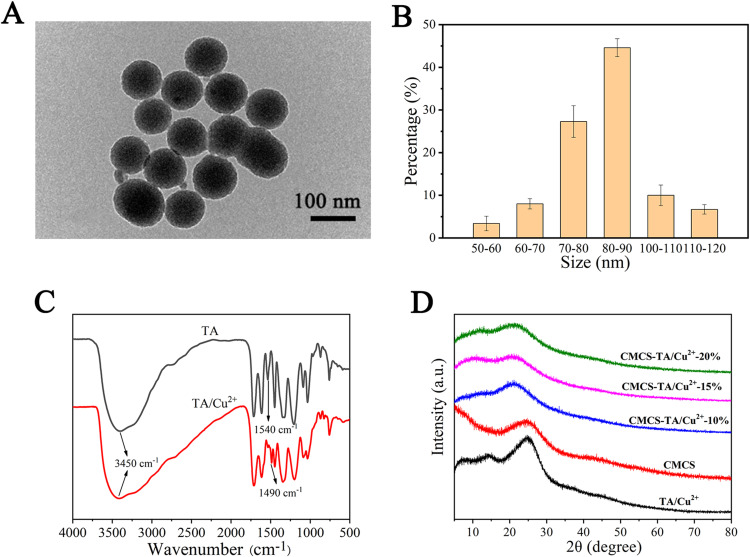
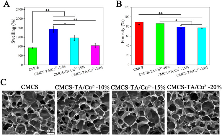
Methods: Here, a carboxymethyl chitosan-based hydrogel dressing (CMCS-TA/Cu2+) reinforced by copper ion crosslinked tannic acid (TA/Cu2+) nanoparticles was developed. This study investigated the physical and chemical characteristics, cytotoxicity, and angiogenesis of TA/Cu2+ nanoparticles and CMCS-TA/Cu2+ hydrogels. Furthermore, a full-thickness skin defect wound model was employed to assess the in vivo wound healing capacity of hydrogel dressings.
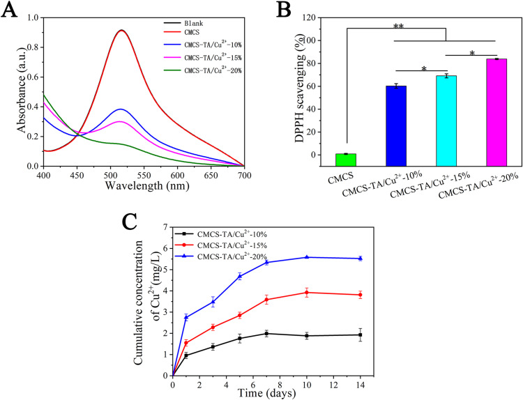
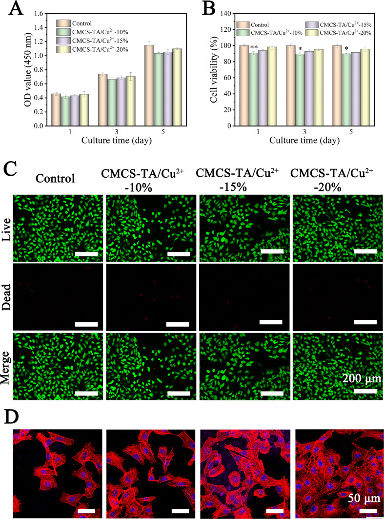
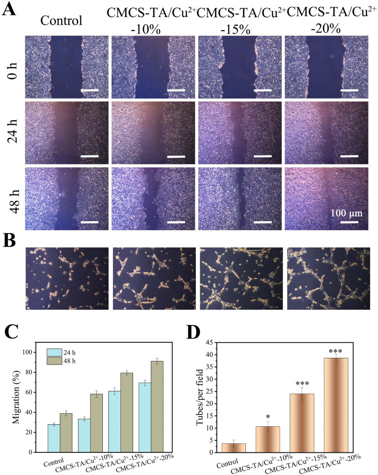
Results: The introduction of TA/Cu2+ nanoparticles not only could increase the mechanical properties of the hydrogel but also continuously releases copper ions to promote cell migration (the cell migration could reach 92% at 48 h) and tubule formation, remove free radicals and promote wound healing (repair rate could reach 90% at 9 days).
Conclusion: Experiments have proved that CMCS-TA/Cu2+ hydrogel has good cytocompatibility, antioxidant and wound healing ability, providing an advantageous solution for skin repair.
Keywords: TA/Cu2+ nanoparticles; antioxidant activity; carboxymethyl chitosan; wound dressing.
© 2024 Huang et al.
Conflict of interest statement
The authors declare that they have no potential conflicts of interest regarding the research, authorship, and/or publication of this article.
Figures

Similar articles
-
Facile preparation of polyphenol-crosslinked chitosan-based hydrogels for cutaneous wound repair.Int J Biol Macromol. 2023 Feb 15;228:99-110. doi: 10.1016/j.ijbiomac.2022.12.215. Epub 2022 Dec 22. Int J Biol Macromol. 2023. PMID: 36565830
-
An adhesive, antibacterial hydrogel wound dressing fabricated by dopamine-grafted oxidized sodium alginate and methacrylated carboxymethyl chitosan incorporated with Cu(II) complex.Biomater Adv. 2025 May;170:214217. doi: 10.1016/j.bioadv.2025.214217. Epub 2025 Feb 6. Biomater Adv. 2025. PMID: 39929017
-
Copper metal-organic framework embedded carboxymethyl chitosan-g-glutathione/polyacrylamide hydrogels for killing bacteria and promoting wound healing.Int J Biol Macromol. 2021 Sep 30;187:699-709. doi: 10.1016/j.ijbiomac.2021.07.139. Epub 2021 Jul 28. Int J Biol Macromol. 2021. PMID: 34331983
-
Chitosan-based self-healing hydrogel dressing for wound healing.Adv Colloid Interface Sci. 2024 Oct;332:103267. doi: 10.1016/j.cis.2024.103267. Epub 2024 Aug 3. Adv Colloid Interface Sci. 2024. PMID: 39121832 Review.
-
[Research advances on the role and mechanism of chitosan-based wound dressing in wound healing].Zhonghua Shao Shang Yu Chuang Mian Xiu Fu Za Zhi. 2023 Apr 20;39(4):386-390. doi: 10.3760/cma.j.cn501225-20220506-00172. Zhonghua Shao Shang Yu Chuang Mian Xiu Fu Za Zhi. 2023. PMID: 37805744 Free PMC article. Review. Chinese.
Cited by
-
Highly Biocompatible Lamellar Liquid Crystals Based on Hempseed or Flaxseed Oil with Incorporated Betamethasone Dipropionate: A Bioinspired Multi-Target Dermal Drug Delivery System for Atopic Dermatitis Treatment.Int J Nanomedicine. 2024 Dec 21;19:13687-13715. doi: 10.2147/IJN.S488684. eCollection 2024. Int J Nanomedicine. 2024. PMID: 39723176 Free PMC article.
-
Preparation and antioxidant properties of tannic acid/copper ion nanozyme hybrid nanofibrous membranes.RSC Adv. 2024 Nov 11;14(48):35743-35753. doi: 10.1039/d4ra05314a. eCollection 2024 Nov 4. RSC Adv. 2024. PMID: 39529749 Free PMC article.
-
Strong, antioxidant, and biodegradable gelatin methacryloyl composite hydrogel for oxidative stress protection in Schwann cells.Front Bioeng Biotechnol. 2025 Jun 3;13:1586380. doi: 10.3389/fbioe.2025.1586380. eCollection 2025. Front Bioeng Biotechnol. 2025. PMID: 40529174 Free PMC article.
References
-
- Chouhan D, Dey N, Bhardwaj N, et al. Emerging and innovative approaches for wound healing and skin regeneration: current status and advances. Biomaterials. 2019;216:119267. - PubMed
-
- Fiakos G, Kuang Z, Lo E. Improved skin regeneration with acellular fish skin grafts. Eng Regen. 2020;1:95–101. doi:10.1016/j.engreg.2020.09.002 - DOI
-
- Sun A, He X, Li L, et al. An injectable photopolymerized hydrogel with antimicrobial and biocompatible properties for infected skin regeneration. NPG Asia Materials. 2020;12(1): doi:10.1038/s41427-020-0206-y. - DOI
MeSH terms
Substances
LinkOut - more resources
Full Text Sources
Research Materials
