. 2024 Jul;397(7):5079-5091.
doi: 10.1007/s00210-023-02921-8. Epub 2024 Jan 15.

Cardioprotective role of diacerein in diabetic cardiomyopathy via modulation of inflammasome/caspase1/interleukin1β pathway in juvenile rats

Affiliations

Cardioprotective role of diacerein in diabetic cardiomyopathy via modulation of inflammasome/caspase1/interleukin1β pathway in juvenile rats

Marwa M M Refaie et al. Naunyn Schmiedebergs Arch Pharmacol. 2024 Jul.

Abstract

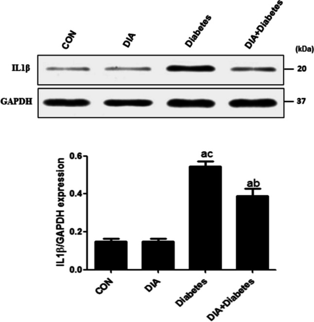
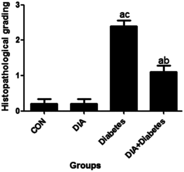
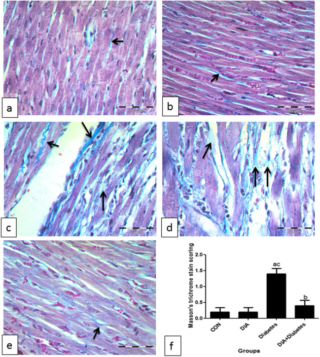
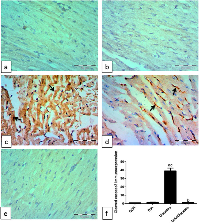
Diabetes mellitus is a common metabolic disorder affecting different body organs; one of its serious complications is diabetic cardiomyopathy (DCM). Thus, finding more cardiopreserving agents to protect the heart against such illness is a critical task. For the first time, we planned to study the suspected role of diacerein (DIA) in ameliorating DCM in juvenile rats and explore different mechanisms mediating its effect including inflammasome/caspase1/interleukin1β pathway. Four-week-aged juvenile rats were randomly divided into groups; the control group, diacerein group, diabetic group, and diabetic-treated group. Streptozotocin (45 mg/kg) single intraperitoneal (i.p.) dose was administered for induction of type 1 diabetes on the 1st day which was confirmed by detecting blood glucose level. DIA was given in a dose of 50 mg/kg/day for 6 weeks to diabetic and non-diabetic rats, then we evaluated different inflammatory, apoptotic, and oxidative stress parameters. Induction of DCM succeeded as there were significant increases in cardiac enzymes, heart weights, fasting blood glucose level (FBG), and glycosylated hemoglobin (HbA1c) associated with elevated blood pressure (BP), histopathological changes, and increased caspase 3 immunoexpression. Furthermore, there was an increase of malondialdehyde (MDA), inflammasome, caspase1, angiotensin II, nuclear factor kappa-B (NF-κB), tumor necrosis factor-α (TNFα), and interleukin 1β (IL1β). However, antioxidant parameters such as reduced glutathione (GSH) and total antioxidant capacity (TAC) significantly declined. Fortunately, DIA reversed the diabetic cardiomyopathy changes mostly due to the observed anti-inflammatory, antioxidant, and anti-apoptotic properties with regulation of blood glucose level.DIA has an ability to regulate DCM-associated biochemical and histopathological disturbances.

Keywords: Cardiomyopathy; Diabetes; Diacerein; Interleukin 1 beta.


Conflict of interest statement

The authors declare no competing interests.

Figures

Fig. 1
Fig. 1
Western blotting of IL1β expression. IL1β expression increased significantly in the diabetic group compared to the control group. However, the diabetic-treated group could significantly decrease IL1β expression if compared to the diabetic-untreated group. Results of the current study were for 10 observations represented as mean ± SD. Significance was considered if p < 0.05. aIt was given if significance was found compared to the control group. bIt was given if significance was found compared to the diabetic group. cIt was given if significance was found compared to the treated diabetic group. CON, control group; DIA, diacerein
Fig. 2
Fig. 2
Representative photomicrographs of the control group (a) and DIA group (b) of left ventricle reveal that individual cardiac myocytes were cylindrical in shape, regularly arranged, branched, and reunite forming a network. Their cytoplasm was acidophilic, crossly striated (star), and had a single central oval and vesicular nucleus (black arrows). Narrow areas of endomysium were observed between cardiac muscle cells. Flat dense nuclei of fibroblasts (yellow arrows) were seen in between of myocytes. Cardiomyocytes in diabetic group (ce) showed deformation in sizes and shapes. Areas of fiber loss (star) and disappearance (black arrow) in addition to local inflammatory cellular infiltration were seen (oval). The cytoplasm lost its striations, and some nuclei appeared smaller (red arrows). Additionally, many fibroblast nuclei apparently noticed compared with the control group (oval arrows). Wavy fibers were also noticed (dashed arrow) and extravasated red blood corpuscles (rectangle). In the diabetic-treated group (f), cardiomyocytes were apparently improved with normal appearance of the nuclei (black arrows) and scattered fibroblasts (yellow arrows). H&E ×400
Fig. 3
Fig. 3
Histopathological scoring. Histopathological grading increased in the diabetic group compared to the control group. However, it decreased in diabetic-treated group if compared to the diabetic-untreated group. Results of the current study were for 10 observations represented as mean ± SD. Significance was considered if p < 0.05. aIt was given if significance was found compared to the control group. bIt was given if significance was found compared to the diabetic group. cIt was given if significance was found compared to the treated diabetic group. CON, control group; DIA, diacerein
Fig. 4
Fig. 4
Masson trichrome staining. Representative photomicrographs of the control group (a) and DIA group (b). Masson trichrome stained sections of the heart showed little collagen fibers among cardiac muscle cells (arrows). The amount of collagen fibers obviously increased among cardiac muscle cells in the diabetic group (c, d). The amount of collagen fibers clearly decreased in the treated group (e) (Masson trichrome ×400). Scoring showed that there was significant increase of Masson trichrome stain in the diabetic group compared to the control group (f). However, the diabetic-treated group significantly decreased this scoring compared to the diabetic-untreated group. Results of the current study were for 10 observations represented as mean ± SD. Significance was considered if p < 0.05. aIt was given if significance was found compared to the control group. bIt was given if significance was found compared to the diabetic group. cIt was given if significance was found compared to the treated diabetic group. CON, control group; DIA, diacerein
Fig. 5
Fig. 5
Caspase 3 immunoexpression. Representative photomicrographs of the control group (a) and DIA group (b) immunohistochemically stained for caspase 3 show negative immunoexpression. The diabetic group shows strong positive immune expression in the cytoplasm and some nuclei (arrows) (c, d). The treated diabetic group shows noticeable decrease in expression (IHC by caspase 3 antibody ×400) (e). Scoring of caspase 3 immunoexpression shows significant increase of caspase 3 immunoexpression in the diabetic group compared to the control group (f). However, the diabetic-treated group significantly decreased its expression compared to the diabetic-untreated group. Results of the current study were for 10 observations represented as mean ± SD. Significance was considered if p < 0.05. aIt was given if significance was found compared to the control group. bIt was given if significance was found compared to the diabetic group. cIt was given if significance was found compared to the treated diabetic group. CON, control group; DIA, diacerein

Similar articles

Cited by

References

    1. Abdel-Aziz MA, Ahmed HMS, El-Nekeety AA, Sharaf HA, Abdel-Aziem SH, Abdel-Wahhab MA. Biosynthesis of gold nanoparticles for the treatment of osteoarthritis alone or in combination with diacerein((R)) in a rat model. Inflammopharmacology. 2021;29:705–719. doi: 10.1007/s10787-021-00833-8. - DOI - PubMed
    1. Agarwal V, Kaushik AS, Rehman M, Chaudhary R, Jawaid T, Kamal M, Mishra V. Interleukin-6 expression and its modulation by diacerein in a rat model of chronic stress induced cardiac dysfunction. Heliyon. 2021;7:e08522. doi: 10.1016/j.heliyon.2021.e08522. - DOI - PMC - PubMed
    1. Akhtar MS, Alavudeen SS, Raza A, Imam MT, Almalki ZS, Tabassum F, Iqbal MJ. Current understanding of structural and molecular changes in diabetic cardiomyopathy. Life Sciences. 2023;332:122087. doi: 10.1016/j.lfs.2023.122087. - DOI - PubMed
    1. Almezgagi M, Zhang Y, Hezam K, Shamsan E, Gamah M, Al-Shaebi F, Abbas AB, Shoaib M, Saif B, Han Y, Jia R, Zhang W. Diacerein: recent insight into pharmacological activities and molecular pathways. Biomed Pharmacother. 2020;131:110594. doi: 10.1016/j.biopha.2020.110594. - DOI - PubMed
    1. Alsharif KF, Elmahallawy EK, Alblihd MA, Hamad AA, Nasreldin N, Alsanie W, Aljoudi AM, Oyouni AAA, Al-Amer OM, Albarakati AJA, Lokman MS, Albrakati A, Ali FAZ. Corrigendum: melatonin ameliorates serobiochemical alterations and restores the cardio-nephro diabetic vascular and cellular alterations in streptozotocin-induced diabetic rats. Front Vet Sci. 2023;10:1240320. doi: 10.3389/fvets.2023.1240320. - DOI - PMC - PubMed

Publication types

MeSH terms