

Development and clinical application of loop-mediated isothermal amplification combined with lateral flow assay for rapid diagnosis of SARS-CoV-2
- PMID: 38225546
- PMCID: PMC10788970
- DOI: 10.1186/s12879-023-08924-3
Development and clinical application of loop-mediated isothermal amplification combined with lateral flow assay for rapid diagnosis of SARS-CoV-2
Abstract
Background: The diagnostic assay leveraging multiple reverse transcription loop-mediated isothermal amplification (RT-LAMP) could meet the requirements for rapid nucleic acid detection of severe acute respiratory syndrome coronavirus 2 (SARS-CoV-2).
Methods: The devised assay merged the lateral flow assay with the RT-LAMP technology and designed specific primers for the simultaneous detection of the target and human-derived internal reference genes within a single reaction. An inquiry into the assay's limit of detection (LOD), sensitivity, and specificity was carried out. The effectiveness of this assay was validated using 498 clinical specimens.
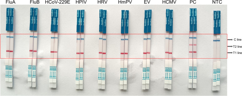
Results: This LOD of the assay was determined to be 500 copies/mL, and there was no observed cross-reaction with other respiratory pathogens. The detection results derived from clinical specimens showed substantial concordance with those from real-time reverse transcription-polymerase chain reaction (RT-qPCR) (Cohen's kappa, 0.876; 95% CI: 0.833-0.919; p<0.005). The diagnostic sensitivity and specificity were 87.1% and 100%, respectively.
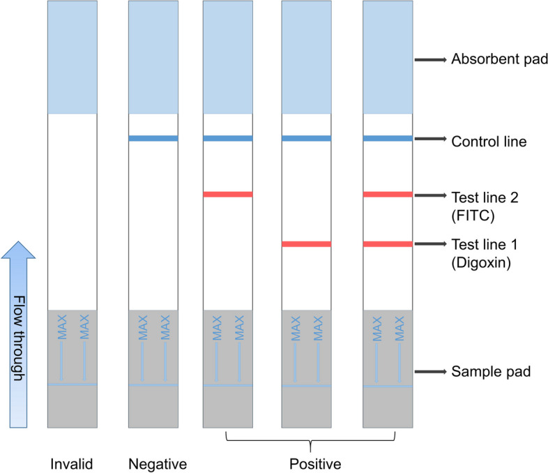
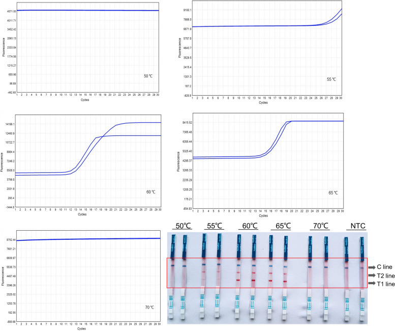
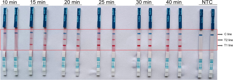
Conclusion: The RT-LAMP assay, paired with a straightforward and disposable lateral immunochromatographic strip, achieves visual detection of dual targets for SARS-CoV-2 immediatly. Moreover, the entire procedure abstains from nucleic acids extraction. The samples are lysed at room temperature and subsequently proceed directly to the RT-LAMP reaction, which can be executed within 30 minutes at a constant temperature of 60-65°C. Then, the RT-LAMP amplification products are visualized using colloidal gold test strips.
Trial registration: This study was registered at the Chinese Clinical Trial Registry (Registration number: ChiCTR2200060495, Date of registration 2022-06-03).
Keywords: COVID-19; Diagnostics; Isothermal amplification; Molecular testing; RT-LAMP; SARS-CoV-2.
© 2024. The Author(s).
Conflict of interest statement
The authors declare no competing interests.
Figures


Similar articles
-
A Multiplex and Colorimetric Reverse Transcription Loop-Mediated Isothermal Amplification Assay for Sensitive and Rapid Detection of Novel SARS-CoV-2.Front Cell Infect Microbiol. 2021 Jun 29;11:653616. doi: 10.3389/fcimb.2021.653616. eCollection 2021. Front Cell Infect Microbiol. 2021. PMID: 34268131 Free PMC article.
-
Development and Clinical Application of a Rapid and Sensitive Loop-Mediated Isothermal Amplification Test for SARS-CoV-2 Infection.mSphere. 2020 Aug 26;5(4):e00808-20. doi: 10.1128/mSphere.00808-20. mSphere. 2020. PMID: 32848011 Free PMC article.
-
Rapid and Visual Detection of SARS-CoV-2 Using Multiplex Reverse Transcription Loop-Mediated Isothermal Amplification Linked With Gold Nanoparticle-Based Lateral Flow Biosensor.Front Cell Infect Microbiol. 2021 Jul 14;11:581239. doi: 10.3389/fcimb.2021.581239. eCollection 2021. Front Cell Infect Microbiol. 2021. PMID: 34336708 Free PMC article.
-
A Recent Update on Advanced Molecular Diagnostic Techniques for COVID-19 Pandemic: An Overview.Front Immunol. 2021 Dec 14;12:732756. doi: 10.3389/fimmu.2021.732756. eCollection 2021. Front Immunol. 2021. PMID: 34970254 Free PMC article. Review.
-
Evaluation of RT-LAMP Assay for Rapid Detection of SARS-CoV-2.Lab Med. 2023 Jan 5;54(1):56-64. doi: 10.1093/labmed/lmac030. Lab Med. 2023. PMID: 35849098 Free PMC article.
Cited by
-
From Tradition to Innovation: Diverse Molecular Techniques in the Fight Against Infectious Diseases.Diagnostics (Basel). 2024 Dec 21;14(24):2876. doi: 10.3390/diagnostics14242876. Diagnostics (Basel). 2024. PMID: 39767237 Free PMC article. Review.
References
-
- Dhama K, Nainu F, Frediansyah A, Yatoo MI, Mohapatra RK, Chakraborty S, Zhou H, Islam MR, Mamada SS, Kusuma HI, Rabaan AA, Alhumaid S, Mutair AA, Iqhrammullah M, Al-Tawfiq JA, Mohaini MA, Alsalman AJ, Tuli HS, Chakraborty C, Harapan H. Global emerging Omicron variant of SARS-CoV-2: Impacts, challenges and strategies. J Infect Public Health. 2023;16(1):4–14. doi: 10.1016/j.jiph.2022.11.024. - DOI - PMC - PubMed
MeSH terms
Substances
Supplementary concepts
Grants and funding
LinkOut - more resources
Full Text Sources
Medical
Miscellaneous
