. 2023 Dec 26:37:101623.
doi: 10.1016/j.bbrep.2023.101623. eCollection 2024 Mar.

Gastrodin improves osteoblast function and adhesion to titanium surface in a high glucose environment

Affiliations

Gastrodin improves osteoblast function and adhesion to titanium surface in a high glucose environment

Yi Li et al. Biochem Biophys Rep. .

Abstract

Objective: To investigate the effects of gastrodin on the biological behavior of osteoblasts and osseointegration on the surface of the titanium plate in a high glucose environment, and to explore the possible regulatory mechanisms involved.

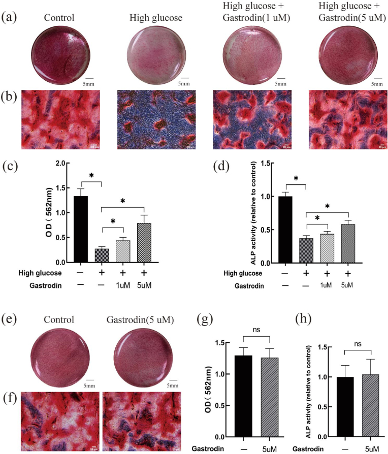
Methods: A high glucose-induced oxidative damage model of MC3T3-E1 cells was established in vitro to observe the effects of gastrodin on cellular oxidative stress, cell viability, osteogenic differentiation, mineralization, migration, and adhesion ability on the titanium surface.

Results: High glucose environment can cause oxidative stress damage to MC3T3-E1 cells, leading to a decrease in cell viability, osteogenesis, migration, adhesion and other functions. Gastrodin can upregulate the expression of antioxidant enzymes (Nrf2 and HO-1) and osteogenic differentiation related proteins (RUNX2 and BMP2) in MC3T3-E1 cells in high glucose environment, thereby inhibiting the excessive production of intracellular reactive oxygen species (ROS), reversing the decrease in cell viability, and improving the osteogenic differentiation and mineralization ability of osteoblasts. And gastrodin alleviated the decline in cell migration ability, improved the morphology of the cytoskeleton and increased the adhesion ability of osteoblasts on the surface of titanium plates in high glucose environment. However, gastrodin itself did not affect the cell viability, osteogenic differentiation and mineralization ability of osteoblasts in normal environment.

Conclusions: Gastrodin may protect MC3T3-E1 cells osteogenesis and osseointegration on the surface of the titanium plate in vitro by upregulating antioxidant enzymes expression, and attenuating high glucose-induced oxidative stress. Therefore, gastrodin may be a potential drug to address the problem of poor implant osseointegration in patients with diabetes.

Keywords: Antioxidant; Diabetes; Gastrodin; Osseointegration; Osteoporosis.


Conflict of interest statement

The authors declare that they have no known competing financial interests or personal relationships that could have appeared to influence the work reported in this paper.

Figures

Fig. 1
Fig. 1
Effect of Gastrodin on toxicity of MC3T3-E1 cells in high glucose environment. (a, b) In the 5.5 mM glucose environment, MC3T3-E1 cells were cultured with different concentrations of gastrodin alone. (c, d) MC3T3-E1 cells were pretreated gastrodin for 2 h before treatment with 25 mM glucose. Each value is reported as the mean ± standard deviation of three independent experiments. n = 9, *P < 0.05.
Fig. 2
Fig. 2
Effect of gastrodin on oxidative stress and osteogenic differentiation of MC3T3-E1 cells in high glucose environment. (a, b) Detection of ROS Levels in MC3T3 E1 cells by flow cytometry, after treatment with gastrodin and high glucose for 24 h (c, d, e, f, g) Effects of gastrodin in expression of antioxidant enzymes (NRF2, HO-1) and osteogenic differentiation-related proteins (BMP2, RNNX2) in MC3T3-E1 cells during high glucose environment. Each value is reported as the mean ± standard deviation of three independent experiments. N = 9, *P < 0.05.
Fig. 3
Fig. 3
Effect of gastrodin on osteogenic differentiation and mineralization of MC3T3-E1 cells in high glucose and normal environment. (a, b) The Alizarin Red S staining of MC3T3-E1 cells which were treated with gastrodin and high glucose for 14 days. Scale bar = 20 μm. (e, f) The Alizarin Red S staining of MC3T3-E1 cells which were treated with gastrodin alone for 14 days. Scale bar = 20 μm. (c, g) Semi-quantitative assay of the calcium nodules. (d, h) Analysis of ALP activity in MC3T3-E1 cells at day 7. Each value is reported as the mean ± standard deviation of three independent experiments. N = 9, *P < 0.05. (For interpretation of the references to color in this figure legend, the reader is referred to the Web version of this article.)
Fig. 4
Fig. 4
Effect of gastrodin on migration capacity of MC3T3-E1 cells in high glucose environment. (a, b) Cell migration and gap closure images at 0 h and 24 h. Scale bar = 100 μm. (c) The cell migration ability was quantified by gap closure rate. Each value is reported as the mean ± standard deviation of three independent experiments. N = 9, *P < 0.05.
Fig. 5
Fig. 5
Effect of gastrodin on adhesion of MC3T3-E1 cells on titanium surface in high glucose environment. (a) Observation of MC3T3-E1 cell nucleus by DAPI staining after cultured for 24 h. Scale bar = 200 μm. (b) Observation of actin cytoskeleton of MC3T3-E1 cells by TRITC phalloidin staining after cultured for 24 h. Scale bar = 50 μm. (c) Observation of MC3T3-E1 cells by HE staining after cultured for 24 h. Scale bar = 50 μm. (d) The number of MC3T3-E1 cell nucleus by DAPI staining. (e) The average number of pseudopods per cell by TRITC phalloidin staining. (f) The H&E staining area of MC3T3-E1 cells adhering on titanium surface. Each value is reported as the mean ± standard deviation of three independent experiments. N = 9, *P < 0.05.
actin.tif
actin.tif
actin_spl_marker.tif
BMP2.tif
BMP2.tif
BMP2_spl_marker.tif
BMP2_spl_marker.tif
HO-1.tif
HO-1.tif
HO-1_spl_marker.tif
HO-1_spl_marker.tif
NRF2.tif
NRF2.tif
NRF2.tif
NRF2.tif
RUNX2.tif
RUNX2.tif
RUNX2_spl_marker.tif

References

    1. Qu B., Gong K., Yang H. SIRT1 suppresses high glucose and palmitate-induced osteoclast differentiation via deacetylating p66Shc. Mol. Cell. Endocrinol. 2018;474:97–104. doi: 10.1016/j.mce.2018.02.015. - DOI - PubMed
    1. Wu Y., Ding Y., Tanaka Y. Risk factors contributing to type 2 diabetes and recent advances in the treatment and prevention. Int. J. Med. Sci. 2014;11:1185–1200. doi: 10.7150/ijms.10001. - DOI - PMC - PubMed
    1. Song Y., Zhang S. Clinical characteristics, diagnosis and treatment of dental implant repair in patients with diabetes. Chin. J. Stomatol. 2021;56:1172–1178. - PubMed
    1. Zawada A., Ratajczak A.E., Rychter A.M. Treatment of diabetes and osteoporosis—a reciprocal risk? Biomedicines. 2022;10:2191–2199. doi: 10.3390/biomedicines10092191. - DOI - PMC - PubMed
    1. Chrcanovic B.R., Albrektsson T., Wennerberg A. Diabetes and oral implant failure. J. Dent. Res. 2014;93:859–867. doi: 10.1177/0022034514538820. - DOI - PMC - PubMed

LinkOut - more resources