Review
. 2023 Dec 21:24:100920.
doi: 10.1016/j.mtbio.2023.100920. eCollection 2024 Feb.

Plant molecules reinforce bone repair: Novel insights into phenol-modified bone tissue engineering scaffolds for the treatment of bone defects

Affiliations
Review

Plant molecules reinforce bone repair: Novel insights into phenol-modified bone tissue engineering scaffolds for the treatment of bone defects

Yuhang Chen et al. Mater Today Bio. .

Abstract

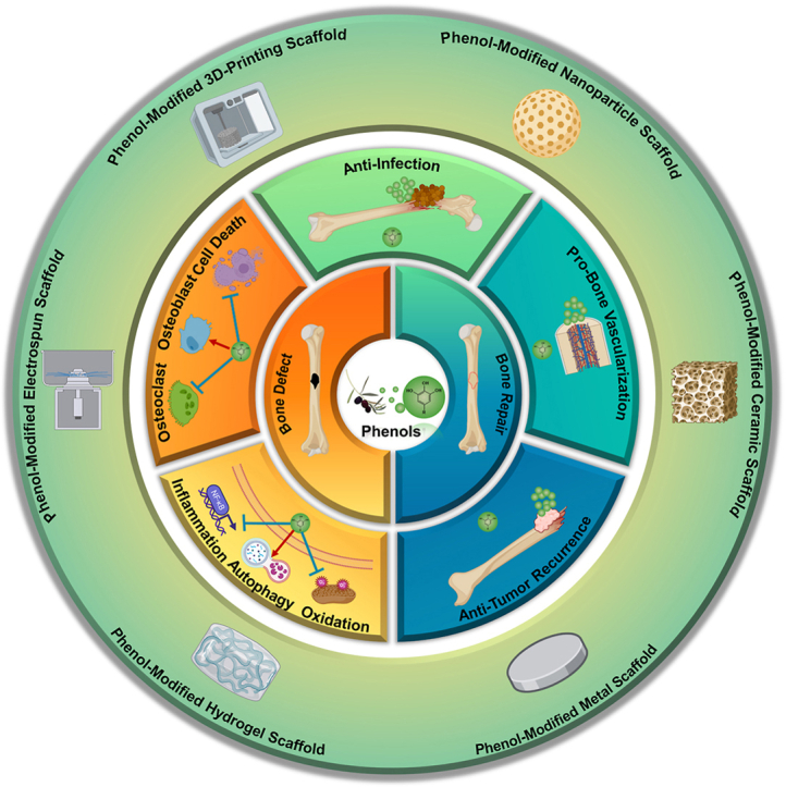
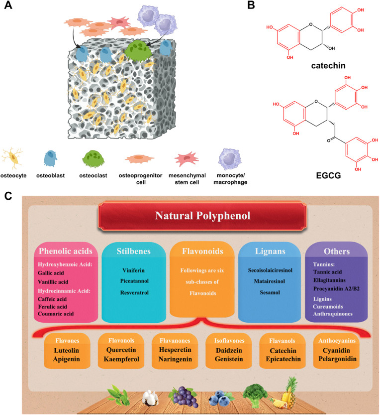
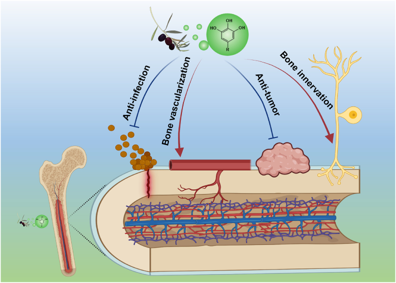
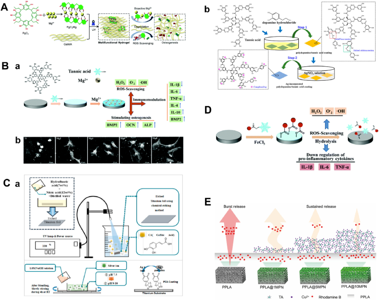
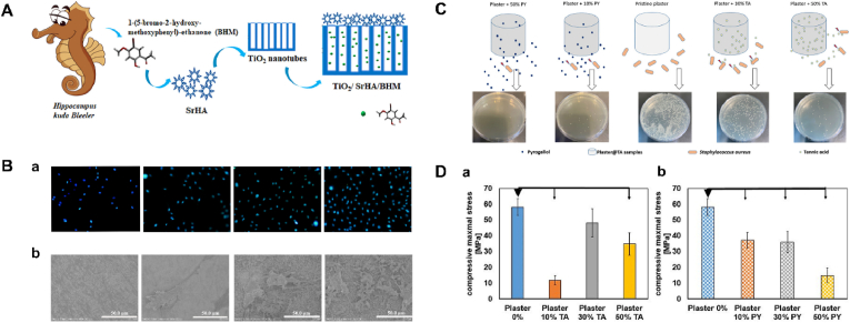
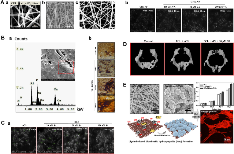
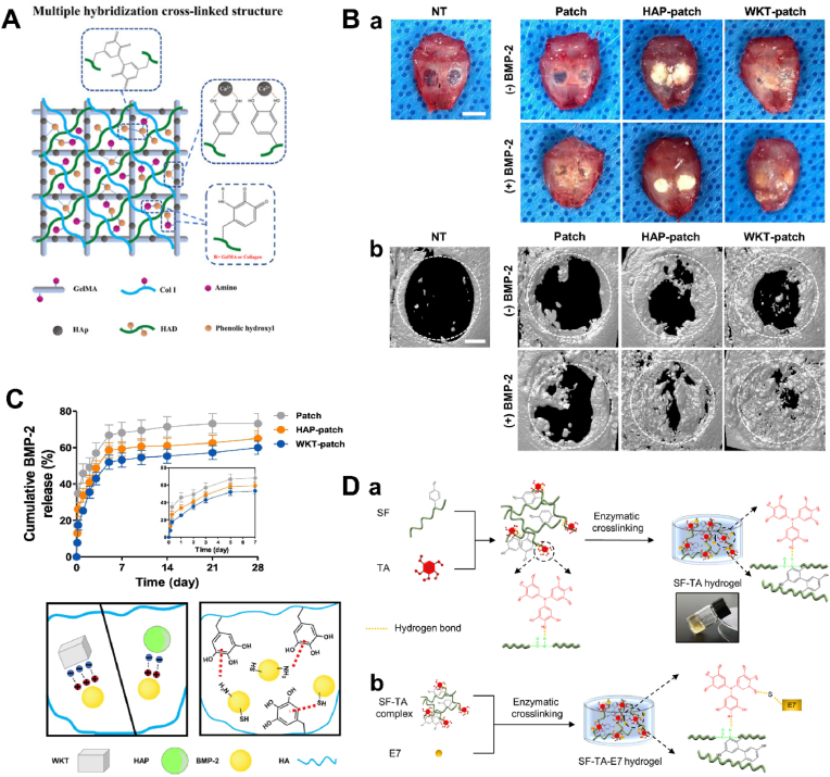
Bone defects have become a major cause of disability and death. To overcome the limitations of natural bone implants, including donor shortages and immune rejection risks, bone tissue engineering (BTE) scaffolds have emerged as a promising therapy for bone defects. Despite possessing good biocompatibility, these metal, ceramic and polymer-based scaffolds are still challenged by the harsh conditions in bone defect sites. ROS accumulation, bacterial infection, excessive inflammation, compromised blood supply deficiency and tumor recurrence negatively impact bone tissue cells (BTCs) and hinder the osteointegration of BTE scaffolds. Phenolic compounds, derived from plants and fruits, have gained growing application in treating inflammatory, infectious and aging-related diseases due to their antioxidant ability conferred by phenolic hydroxyl groups. The prevalent interactions between phenols and functional groups also facilitate their utilization in fabricating scaffolds. Consequently, phenols are increasingly incorporated into BTE scaffolds to boost therapeutic efficacy in bone defect. This review demonstrated the effects of phenols on BTCs and bone defect microenvironment, summarized the intrinsic mechanisms, presented the advances in phenol-modified BTE scaffolds and analyzed their potential risks in practical applications. Overall, phenol-modified BTE scaffolds hold great potential for repairing bone defects, offering novel patterns for BTE scaffold construction and advancing traumatological medicine.

Keywords: Bone defect; Bone tissue engineering; Osteogenesis; Phenols; Polyphenols; Scaffolds.


Conflict of interest statement

The authors declare that they have no known competing financial interests or personal relationships that could have appeared to influence the work reported in this paper.

Figures

Image 1
Graphical abstract
Fig. 1
Fig. 1
Phenols regulate BTCs. (A) The profile of bone tissue cells. Osteoprogenitor cells are a special kind of mesenchymal stem cells, which differentiate into osteoblasts and consequently mature to be osteocytes. In the meantime, osteoclasts derive from the fusion and differentiation of precursor monocytes/macrophages. Created with Biorender.com. (B) Phenolic hydroxyl groups (red) in the structure of phenols (represented by catechin and EGCG). (C) The classification of polyphenols. ((C) adapted from Ref. [22].)
Fig. 2
Fig. 2
The schematic diagram for phenols' effects on the microenvironment of bone regeneration. The regulation of phenols on bone environment involves four processes, including anti-infection, promoting the bone vascularization, antagonizing the recurrence of tumor, and potentially inducing the bone innervation. Created with Biorender.com
Fig. 3
Fig. 3
Phenol-incorporated metal BTE scaffolds. (A) Collagen hydrogels were prepared by mixing PG solution, Cell Culture Medium and collagen type I solution, then neutralizing the solution via dripping NaOH solution to form hydrogels. (B) The PG-modified hydrogel enhanced the expression of osteogenic genes COL1A1, BGLAP and RANKL and inhibited inflammatory factors IL-6, TNFA and MMP2. (C) MED-TiF facilitated the expression of the integrin family in MG63 cells and enhanced osteogenesis in vivo estimated by H&E staining. ((A) and (B) adapted from Ref. [165] (C) adapted from Ref. [166].).
Fig. 4
Fig. 4
Metal-phenolic networks for BTE. (A) PgC3 coordinated with Mg2+ to form magnesium-seamed PgC3 cages (PgC3Mg) to scavenge ROS and facilitate osteogenesis. (B) (a) Illustration of the coating process of TA/Mg2+ MPN and its potential biofunctions; (b) Macrophages' morphology after cultured on Ti plates with different coatings for 12 h; scale bar is 20 μm. (C) (a) Schematic illustration of the process of coating polycaffeic acid (PCA) on etched titanium (upper image) and synthesis of metallic silver on the Ti–PCA substrate (lower image); (b) The synthetic procedure and potential reaction mechanism of Ag-incorporated polydopamine/TA coating. (D) Illustration of the coating process of TA–IND/Fe3+ coating and its immunomodulatory mechanism. (E) The TA/Cu2+ coatings inhibited the burst release of Rhodamine B from the PPLA scaffolds in a dose-dependent manner. ((A) adapted from Ref. [2], (B) adapted from Ref. [169], (C) adapted from Refs. [126,127], (D) adapted from Ref. [171] (E) adapted from Ref. [172].).
Fig. 5
Fig. 5
Phenol-modified ceramic BTE scaffolds. (A) The compound BHM was embedded in the surface-generated SrHA on the TiO2. (B) Fluorescent staining (DAPI) (a) and SEM images (b) of MC3T3-E1 cells adhering to Ti, TiO2, TiO2/SrHA and TiO2/SrHA/BHM (from the left to the right). (C) The addition of PY or TA enhanced the antibacterial effect of plaster compared with pristine plaster in a dose-dependent manner. (D) Evolution of the maximal stress under compression for the pristine plaster, the plaster@TA (a) and the plaster@PY (b) composites (P < 0.05 Bold arrows). ((A) and (B) adapted from Ref. [178], (C) and (D) adapted from Ref. [179].).
Fig. 6
Fig. 6
Phenol-modified electrospun fiber scaffolds in BTE. (A) SEM images of PCL-based electrospun fiber scaffolds doped by CUR (a), trans-anethole (b) or pomegranate peel extract (c). (B) (a) The SEM image and EDS spectrum of the Ca and P deposited on the CUR-loaded nanofiber scaffolds; (b) The von Kossa staining of Ca on the trans-anethole-incorporated fiber scaffold. (C) SEM images of chitosan microspheres containing sinapic acid (SA) (a) and vanderatric acid (VA) (b). (D) Micro-CT images of calvarial bone defects in rats treated with the chitosan-loading PCL scaffold and the SA/chitosan-loading PCL scaffold for 4 weeks. (E) Lignin/PCL fibrous platform was prepared by electrospinning, followed by incubation in a SBF. Lignin donated abundant hydroxyl groups to bind with metal ions and facilitate the nucleation and growth of HA, leading to enhanced osteogenesis. ((A) adapted from Refs. [[181], [182], [183]], (B) adapted from Refs. [181,182], (C) adapted from Refs. [184,185], (D) adapted from Ref. [184] (E) adapted from Ref. [186].).
Fig. 7
Fig. 7
Phenol-modified hydrogel scaffolds in BTE. (A) Dopamine-modified hyaluronic acid bound to GelMA/HA scaffold via the covalent bond between phenolic hydroxyl groups and amino acids. (B) Gross images (a) and Micro-CT images (b) of mouse calvarial bone defects treated with different patches. Scale bar in (a) = 5 mm. Scale bar in (b) = 1 mm. (C) PG reduced the release of BMP2 contained in hydrogel patches through hydrogen bonds. (D) (a) TA was mixed with SF to build SF-TA hydrogel through hydrogen bonds; (b) E7 peptide was doped into SF-TA hydrogel and formed hydrogen bonds with TA. ((A) adapted from Ref. [189], (B) and (C) adapted from Ref. [190] (D) adapted from Ref. [191].).
Fig. 8
Fig. 8
Phenol-modified 3D bioprinting scaffolds in BTE. (A) Illustrations of manufacturing EGCG-coated PLLA (a) and PCL (b) scaffolds. (B) Representative SEM images showing the structure of pure PCLA, PCLA/KH-HA, PCLA/HA-EGCG and PCLA/KH-HA-EGCG scaffolds. (C) The agar plate counts (a) and SEM images (b) demonstrated that the KH-HA-EGCG coating boost the process of disinfection and mineral deposition of PLCA scaffold, respectively. (D) Schematics of the interactions between TA and collagen/proteins during gelation and degradation. ((A) adapted from Refs. [197,198], (B) and (C) adapted from Ref. [199] (D) adapted from Ref. [200].).
Fig. 9
Fig. 9
Phenol-modified nanoparticle scaffolds in BTE. (A) The illustration of mTN fabrication (a) and SEM images of mTNs (b). (B) Live/dead staining and MTT assay results of hADSCs cultured using different concentrations of TA and mTNs (scale bar = 200 μm). ∗Significantly different compared to the group with no nanoparticles (p < 0.05). (C) SEM (a) and TEM (b) images of chitosan nanoparticles (nCS), nCS + 40 μM orsellinic acid (OA), nCS + 80 μM OA, and nCS + 120 μM OA. (D) (a) The synthesis of the ROS-responsive resveratrol-loaded cyclodextrin nanomicelles (RSV-NMs); (b) TRAP staining images in top row and immunofluorescence images in bottom row identified the inhibiting effect of RSV-NMs on osteoclasts. ((A) and (B) adapted from Ref. [201], (C) adapted from Ref. [202] (D) adapted from Ref. [206].).

Similar articles

Cited by

References

    1. Laird N.Z., Acri T.M., Tingle K., Salem A.K. Gene- and RNAi-activated scaffolds for bone tissue engineering: current progress and future directions. Adv. Drug Deliv. Rev. 2021:613–627. doi: 10.1016/j.addr.2021.05.009. - DOI - PMC - PubMed
    1. Tan X., Wu J., Wang R., Wang C., Sun Y., Wang Z., Ye L. PgC(3)Mg metal-organic cages functionalized hydrogels with enhanced bioactive and ROS scavenging capabilities for accelerated bone regeneration. J. Mater. Chem. B. 2022;28:5375–5387. doi: 10.1039/d2tb00907b. - DOI - PubMed
    1. Allesina L., Mazzola M., Belluati A., Mosca S., Placella G., Salini V. Surgical treatment of critical size bone defects with Masquelet technique versus bone transport: a systematic review and meta-analysis of comparative studies. Arch. Orthop. Trauma Surg. 2023;143:7081–7096. doi: 10.1007/s00402-023-05049-9. - DOI - PubMed
    1. Pina S., Oliveira J.M., Reis R.L. Natural-based nanocomposites for bone tissue engineering and regenerative medicine: a review. Adv. Mater. 2015;7:1143–1169. doi: 10.1002/adma.201403354. - DOI - PubMed
    1. Yuan L., Ding S., Wen C. Additive manufacturing technology for porous metal implant applications and triple minimal surface structures. A review Bioact Mater. 2019;1:56–70. doi: 10.1016/j.bioactmat.2018.12.003. - DOI - PMC - PubMed

LinkOut - more resources