. 2023 Nov 25;4(1):100250.
doi: 10.1016/j.xjidi.2023.100250. eCollection 2024 Jan.

Therapeutic TNF Inhibitors Exhibit Differential Levels of Efficacy in Accelerating Cutaneous Wound Healing

Affiliations

Therapeutic TNF Inhibitors Exhibit Differential Levels of Efficacy in Accelerating Cutaneous Wound Healing

Yonghao Cao et al. JID Innov. .

Abstract

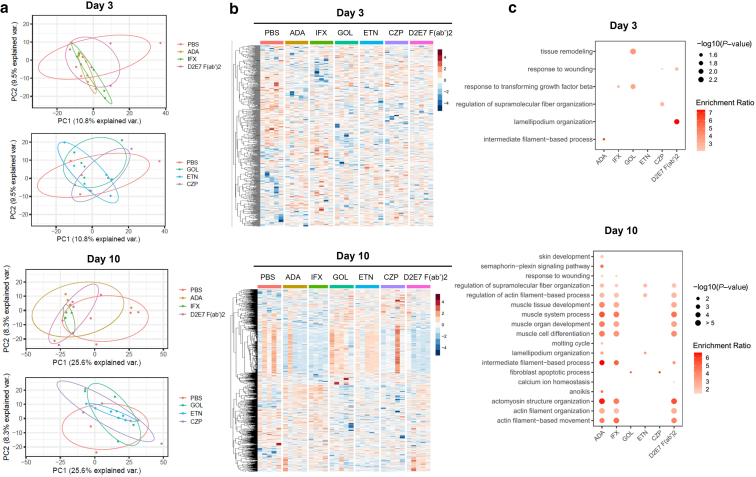
Adalimumab but neither etanercept nor certolizumab-pegol has been reported to induce a wound-healing profile in vitro by regulating macrophage differentiation and matrix metalloproteinase expression, which may underlie the differences in efficacy between various TNF-α inhibitors in impaired wound healing in patients with hidradenitis suppurativa, a chronic inflammatory skin disease. To examine and compare the efficacy of various TNF inhibitors in cutaneous wound healing in vivo, a human TNF knock-in Leprdb/db mouse model was established to model the impaired cutaneous wound healing as seen in hidradenitis suppurativa. The vehicle group exhibited severe impairments in cutaneous wound healing. In contrast, adalimumab significantly accelerated healing, confirmed by both histologic assessment and a unique healing transcriptional profile. Moreover, adalimumab and infliximab showed similar levels of efficacy, but golimumab was less effective, along with etanercept and certolizumab-pegol. In line with histologic assessments, proteomics analyses from healing wounds exposed to various TNF inhibitors revealed distinct and differential wound-healing signatures that may underlie the differential efficacy of these inhibitors in accelerating cutaneous wound healing. Taken together, these data revealed that TNF inhibitors exhibited differential levels of efficacy in accelerating cutaneous wound healing in the impaired wound-healing model in vivo.

Keywords: Hidradenitis suppurativa; TNF inhibitors; Wound healing.


Figures

Figure 1
Figure 1
Murine models of acute and impaired cutaneous wound healing. (a) Murine models were developed for studying acute and impaired cutaneous wound healing. Four 6-mm full-thickness skin punches were created on the back of the C57BL/6 or db/db mice. (b) Excisional wound sizes were evaluated by caliper measurement on C57BL/6, hTNFki, db/db, or hTNFki-db/db mice at baseline and day 7. The results represent the means of wound size from each mouse. ∗∗∗P < .001 (unpaired t-test); n = 3 mice per group from 1 of 3 representative studies. (c) Schematic representation of the murine model for studying impaired cutaneous wound healing. ns, not significant.
Figure 2
Figure 2
Immunophenotypic analysis in the periphery of hTNFki mice compared with that in C57BL/6 mice. hTNFki and C57BL/6 mice aged 6 weeks were killed to harvest the whole blood, thymus, and spleen samples. Single-cell suspensions were then prepared, and cells were counted and stained for immunophenotypic analysis with the following antibodies against CD4, CD8, B220, CD11b, CD11c, Ly6G, and Ly6C as well as with Zombie. Live single cells were gated and analyzed. (a) No significant differences (cell numbers) were observed with WBCs, RBCs, lymphocytes, neutrophils, monocytes, or eosinophils in the blood. (b) Minimal differences were observed in the thymus (total thymocyte numbers or CD4 single-positive or CD8 single-positive thymocytes). (c) Minimal differences were observed in the spleen, including the total splenocyte numbers, CD4 T cells, CD8 T cells, macrophages, monocytes, or dendritic cells. Slight reduction in B cells and neutrophils in the spleen of hTNFki mice compared with those of C57BL/6 mice but only in females was found. ∗P < .05, unpaired t-test, n = 5 mice per group. Data represent 1 of 2 independent studies. RBC, red blood cell; WBC, white blood cell.
Figure 3
Figure 3
Comparison between acute and impaired wound-healing models. (a) Evaluation of C57BL/6 mice and hTNFki mice response to R848. C57BL/6 or hTNFki mice were administered with 8C11 (an anti-mouse TNF-α antibody) or ADA, respectively, for 1 hour, followed by the administration of R848 (15 μg). Peripheral blood was taken after 2 hours of stimulation and tested for murine MCP-1, IL-6, and KC levels in the circulation of C57BL/6 or hTNFki mice in response to 8C11 or ADA, respectively. n = 8 mice per group from 1 of 2 representative studies. Four 6-mm full-thickness excisional wounds were created on the back of indicated mouse strains (b) C57BL/6, (c) hTNFki, (d) db/db, and (e) hTNFki-db/db followed by the administration of either ADA (hTNFki and hTNFki-db/db mice) or 8C11 (C57BL/6 and db/db mice) at 20 mg/kg or PBS (vehicle control), twice a week by subcutaneous injection. Wound area measurements and photographs were taken to assess wound size reduction. The results represent the means of wound size from each mouse on the indicated days. ∗P < .05 and ∗∗P < .01, unpaired t-test; n = 3 mice per group from 1 of 3 representative studies. Data represents 2 independent studies. ADA, adalimumab; KC, keratinocyte chemoattractant; MCP-1, monocyte chemoattractant protein-1; ns, not significant.
Figure 4
Figure 4
ADA accelerates excisional cutaneous wound healing in hTNFki-db/db mice. (a) Four 6-mm full-thickness excisional wounds were created on the back of hTNFki-db/db mice, followed by administration of either ADA (20 mg/kg) or PBS (vehicle control), twice a week by subcutaneous injection. Wound area measurements and photographs were taken to assess wound size reduction. Two representative mice from each group were shown. (b) Excisional wound sizes were shown on hTNFki-db/db mice administered with either ADA or PBS (n = 6 mice per group). Results represent 1 of 4 independent experiments. (c) Trichrome slides were scanned and measured with a micrometer for epidermal wound length. Epithelial closure was calculated by the percentage of original opening subtracted from 100 and then converted to the percentage of epidermal closure (percentage healed). (d) Representative images of trichrome-stained and PSR-stained histologic sections demonstrate epidermal regeneration and collagen deposition at days 7 and 14 after wounding (bar = 500 μm). The edges of the wound are designated by the arrows. The wound area with incomplete re-epithelialization is designated by the thick black line. (e) Image analysis quantitation of new (light) versus mature (dark) collagen was performed on PSR slides with VisioPharm. (f) Ki-67 staining was quantified to determine the percentage of Ki-67–positive area per section. (g) IBA-1 staining was quantified to determine the percentage of IBA-1–positive area per section. Each dot represents the mean readout of 2–4 slides from the same mice. All data are shown as mean ± SEM; n = 10 mice per group from 2 independent studies. ∗P < .05, ∗∗P < .01, ∗∗∗P < .0001, and ∗∗∗∗P < .0001; Tukey’s multiple comparisons test. ADA, adalimumab; ns, not significant; PSR, Picrosirius red.
Figure 5
Figure 5
Differentially expressed genes and pathways in wounded skin after ADA or vehicle control administration. Four 6-mm full-thickness excisional wounds were created on the back of hTNFki-db/db mice, followed by administration of either ADA (20 mg/kg) or vehicle control, twice a week by subcutaneous injection. Wounded skin tissues from each group were collected on days 0 (baseline), 3, 7, and 10 for RNA sequencing. Data show the differentially expressed genes (fold change ≥ 2; FDR < 0.05) from wounded skin samples exposed to either ADA or vehicle control compared with that at its baseline at (a) day 3 or (b) day 10. (c) Enriched pathways in wounded skin exposed to ADA or vehicle control by IPA. The differentially expressed genes were used for IPA. The heatmap shows the top 30 canonical enriched pathways (FDR < 0.05) from each treatment group on days 3, 7, and 10 compared with those at their baselines. White in the heatmap represents FDR < 0.05. For the color gradient of each heatmap, data were transformed by log2 for the logarithmic function and normalized by z score, which was defined by the mean expression of each gene across all samples divided by its SD. BL denotes baseline, and D10 denotes day 10. ADA, adalimumab; FDR, false discovery rate; IPA, Ingenuity Pathway Analysis.
Figure 6
Figure 6
TNF inhibitors exhibit differential levels of efficacy in accelerating cutaneous wound healing in vivo. (a) Four identical 6-mm skin punch wounds were created on the back of homozygous hTNFki-db/db experimental mice, followed by administration of either each TNF inhibitor (10 mg/kg) or vehicle control (PBS), twice a week by subcutaneous injection. (a) The size of excision wounds was measured and calculated. Data are shown as mean ± SEM (each error bar is constructed using 1 SEM). (b) Wound healing at day 21 for each treatment group. The results represent the means of wound size from each mouse. ∗∗∗P < .001 (unpaired t-test); n = 5 mice per group. Results represent 1 of 4 independent studies. ADA, adalimumab; ETN, etanercept; GOL, golimumab; IFX, infliximab.
Figure 7
Figure 7
Differential expression proteins and enriched pathways in healing wounds exposed to various TNF inhibitors. Principal component analysis of correlations and variations of protein profiles from healing wounds exposed to TNF inhibitors on (a) day 3 and day 10. Heatmap shows DEPs identified by proteomics from healing skin exposed to indicated TNF inhibitors or vehicle control (PBS) on (b) day 3 and day 10. DEPs were defined with P < .05 and fold change > 1.5 in the pairwise comparisons. Pathway over-representation analysis with Ontology (GO) terms of biological processes was performed with DEPs from healing skin exposed to indicated TNF inhibitors compared with that of the vehicle control on (c) day 3 and day 10. Pathways with P < .05 were shown in at least 1 comparison group. The statistical significance of pathway over-representation was indicated by the dot size, and the ratio of enrichment was indicated by the color scales. n = 5 mice per group. D2E7 F(ab’)2 denotes F(ab’)2 fragments of ADA; D2E7-hCa1 denotes IgA1 Fc-type of ADA. ADA, adalimumab; CZP, certolizumab-pegol; DEP, differential expression protein; ETN, etanercept; GO, Gene Ontology; GOL, golimumab; IFX, infliximab; PC1, principal component 1; PC2, principal component 2.

References

    1. Abreu-Blanco M.T., Watts J.J., Verboon J.M., Parkhurst S.M. Cytoskeleton responses in wound repair. Cell Mol Life Sci. 2012;69:2469–2483. - PMC - PubMed
    1. Adams D.R., Yankura J.A., Fogelberg A.C., Anderson B.E. Treatment of hidradenitis suppurativa with Etanercept injection. Arch Dermatol. 2010;146:501–504. - PubMed
    1. Ashcroft G.S., Jeong M.J., Ashworth J.J., Hardman M., Jin W., Moutsopoulos N., et al. Tumor necrosis factor-alpha (TNF-α) is a therapeutic target for impaired cutaneous wound healing. Wound Repair Regen. 2012;20:38–49. - PMC - PubMed
    1. Bechara F.G., Podda M., Prens E.P., Horváth B., Giamarellos-Bourboulis E.J., Alavi A., et al. Efficacy and safety of adalimumab in conjunction with surgery in moderate to severe hidradenitis suppurativa: the SHARPS randomized clinical trial. JAMA Surg. 2021;156:1001–1009. - PMC - PubMed
    1. Butler D.M., Malfait A.M., Mason L.J., Warden P.J., Kollias G., Maini R.N., et al. DBA/1 mice expressing the human TNF-alpha transgene develop a severe, erosive arthritis: characterization of the cytokine cascade and cellular composition. J Immunol. 1997;159:2867–2876. - PubMed

LinkOut - more resources