. 2024 Apr;29(3):339-351.
doi: 10.1007/s00775-023-02040-y. Epub 2024 Jan 16.

Flexible active-site loops fine-tune substrate specificity of hyperthermophilic metallo-oxidases

Affiliations

Flexible active-site loops fine-tune substrate specificity of hyperthermophilic metallo-oxidases

Vânia Brissos et al. J Biol Inorg Chem. 2024 Apr.

Abstract

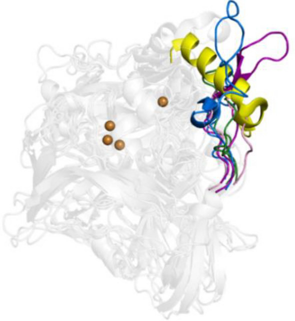
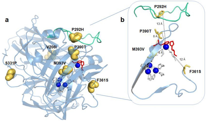
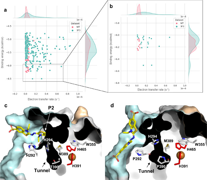
Hyperthermophilic ('superheat-loving') archaea found in high-temperature environments such as Pyrobaculum aerophilum contain multicopper oxidases (MCOs) with remarkable efficiency for oxidizing cuprous and ferrous ions. In this work, directed evolution was used to expand the substrate specificity of P. aerophilum McoP for organic substrates. Six rounds of error-prone PCR and DNA shuffling followed by high-throughput screening lead to the identification of a hit variant with a 220-fold increased efficiency (kcat/Km) than the wild-type for 2,2'-azino-bis(3-ethylbenzothiazoline-6-sulphonic acid) (ABTS) without compromising its intrinsic activity for metal ions. The analysis of the X-ray crystal structure reveals four proximal mutations close to the T1Cu active site. One of these mutations is within the 23-residues loop that occludes this site, a distinctive feature of prokaryotic MCOs. The increased flexibility of this loop results in an enlarged tunnel and one additional pocket that facilitates bulky substrate-enzyme interactions. These findings underscore the synergy between mutations that modulate the dynamics of the active-site loop enabling enhanced catalytic function. This study highlights the potential of targeting loops close to the T1Cu for engineering improvements suitable for biotechnological applications.

Keywords: Directed evolution; Enzyme engineering; Enzyme specificity; Laccases; Multicopper oxidases.


Conflict of interest statement

The authors declare no competing financial interests.

Figures

Scheme 1
Scheme 1
The general reaction of substrates oxidation and oxygen reduction by Mcos
Fig. 1
Fig. 1
Superimposition of cartoons highlighting the major secondary structure elements occluding the access to the T1Cu center in prokaryotic MCOs, Escherichia coli CueO (yellow, PDB 1KV7), Campylobacter jejuni McoC (light pink, PDB 3ZX1), Pyrobaculum aerophilum McoP (green, PDB 3AW5), Thermus thermophilus Tth (purple, 2XU9), and Aquifex aeolicus McoA (blue, PDB 6SYY). Copper ions are represented as dark orange spheres
Fig. 2
Fig. 2
Lineage of McoP variants after six rounds of directed evolution. In the 1st round, ~ 20,000 clones were screened using “activity-on-plate” with 20 mM ABTS as substrate. A209 was chosen as a parent for the second round of evolution, and ~ 7000 clones were screened using 5 mM ABTS as substrate. Variant G9 was selected for the third round, where ~ 12,000 variants were screened using 2.5 mM ABTS. Variant 1B4 was selected for the fourth round, where ~ 13,000 clones were screened using 2.5 mM ABTS as substrate, and 810 variants showing the highest activity were rescreened in liquid assays in 96-well plates. Variant 4B9 was chosen for the fifth round of evolution, where ~ 10,000 variants were screened using 2 mM of ABTS as substrate, and 436 variants were rescreened in 96-well plates. Variant 1B5 was selected, and after DNA-shuffling with wild-type gene, ~ 4000 variants were screened using 2 mM of ABTS as substrate, 378 variants were rescreened in 96-well plates, and variant 3F3 was selected. We have observed the introduction of at least one mutation close to the copper centers in each round of evolution: E446G, M393V, F361S, P292H, and P390T (in bold; see Fig. 3)
Fig. 3
Fig. 3
Mapping mutations in the 3F3 variant. a Transparent cartoon representation of the 3F3 crystal structure colored in blue. The mutations V206I, P292H, S331P, F361S, P390T, and M393V are thick golden sticks. b Zoomed view of the closest mutations to the T1Cu, displayed as thin golden sticks. The loop 288–310 is colored in green cyan. The copper atoms are represented in Klein blue spheres. The T1Cu ligands (H391 and H465) and TNC are in red and grey sticks, respectively. The mutations P390T and M393V, closer to T1Cu, show the lowest accessible surface areas (ASA), 8% and 1%, respectively, whereas the remaining mutations display ASA values between 29 and 39%
Fig. 4
Fig. 4
Molecular access to the T1Cu center. Cutaway view of the solvent-accessible surface in wild-type (PDB 3AW5) (a) and variant 3F3 (b) showing the tunnel and cavity (with pockets P1 and P2). The T1Cu histidine ligands (H391 and H465) are red, while the other TNC ligands are grey. The W355 residue is shown as purple sticks. The mutated residues P292H, F361S, P390T, and M393V are labeled as yellow. The diameter of tunnels, in wild-type and 3F3 variant, and the distance H294-H292 are shown as light grey dashed lines. The solvent-accessible surface of McoP (c) and variant 3F3 (d) shows the loop 288–310 (blue cyan) surrounding the tunnel. The cavity (beige) shows solvent-exposed T1Cu ligands H465 (red) and W355 (purple). The pocket P1 is labeled in wild-type (c), and both P1 (exposing H465 and W355) and P2 (exposing W355) are visible in 3F3 (d). Surface mutations P292H and F361S are shown in yellow. A rotation of 90 degrees in the y-axis (right panel in (d)) allows a more precise view of the P2 main entrance in the 3F3 variant, while in wild-type, it is occluded (right panel in (c))
Fig. 5
Fig. 5
Docking ABTS to wild-type and 3F3 variant. Binding energy (kcal/mol) vs. electron transfer rate is shown for wild-type and 3F3 variant (a). The critical substrate positions for the wild-type are colored pink, while those for the 3F3 variant are colored blue. Only docking positions closer than 3.0 Å to suitable solvent-exposed residues near T1Cu (H292, H294, W355, or H465) were considered. A zoomed view of the most efficient ABTS binding positions for ET is shown (b). Cutaway view of the solvent-accessible surface of representative enzymes with lower binding energy and higher ET rates: variant 3F3 (c) and wild-type (PDB 3AW5) (d), showing the possible residues involved in ET. The T1Cu histidine ligands (H391 and H465) are colored in red, while the carbon atoms of the other residues are shown in grey. The oxygen and nitrogen atoms are colored in red and blue, respectively. The copper atom is shown as an orange sphere

References

    1. Solomon EI, Sundaram UM, Machonkin TE. Multicopper oxidases and oxygenases. Chem Rev. 1996;96:2563–2605. doi: 10.1021/cr950046o. - DOI - PubMed
    1. Kosman DJ. Multicopper oxidases: a workshop on copper coordination chemistry, electron transfer, and metallophysiology. J Biol Inorg Chem. 2010;15:15–28. doi: 10.1007/s00775-009-0590-9. - DOI - PubMed
    1. Lindley PF. Multi-copper oxidases. In: Bertini I, Sigel A, Sigel H, editors. Handbook on metalloproteins. New York: Marcel Dekker; 2001. pp. 763–811.
    1. Hakulinen N, Rouvinen J. Three-dimensional structures of laccases. Cell Mol Life Sci. 2015;72:857–868. doi: 10.1007/s00018-014-1827-5. - DOI - PMC - PubMed
    1. Jones SM, Solomon EI. Electron transfer and reaction mechanism of laccases. Cell Mol Life Sci. 2015;72:869–883. doi: 10.1007/s00018-014-1826-6. - DOI - PMC - PubMed

Publication types

Substances

LinkOut - more resources