. 2024 Sep 1;19(9):2050-2056.
doi: 10.4103/1673-5374.390953. Epub 2023 Dec 15.

Biochanin A attenuates spinal cord injury in rats during early stages by inhibiting oxidative stress and inflammasome activation

Affiliations

Biochanin A attenuates spinal cord injury in rats during early stages by inhibiting oxidative stress and inflammasome activation

Xigong Li et al. Neural Regen Res. .

Abstract

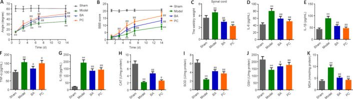
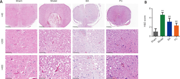
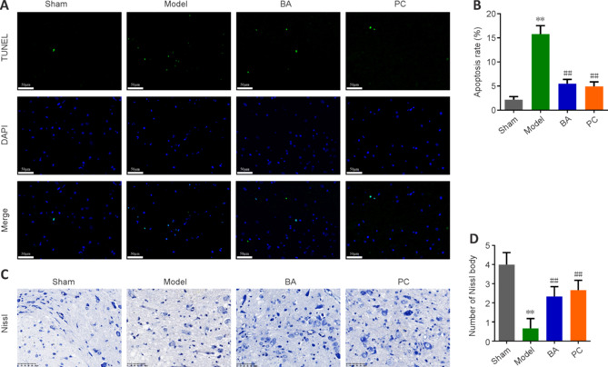
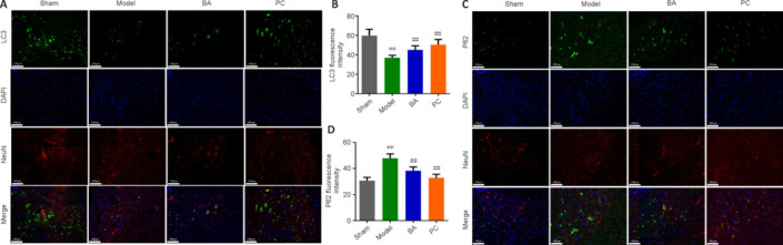
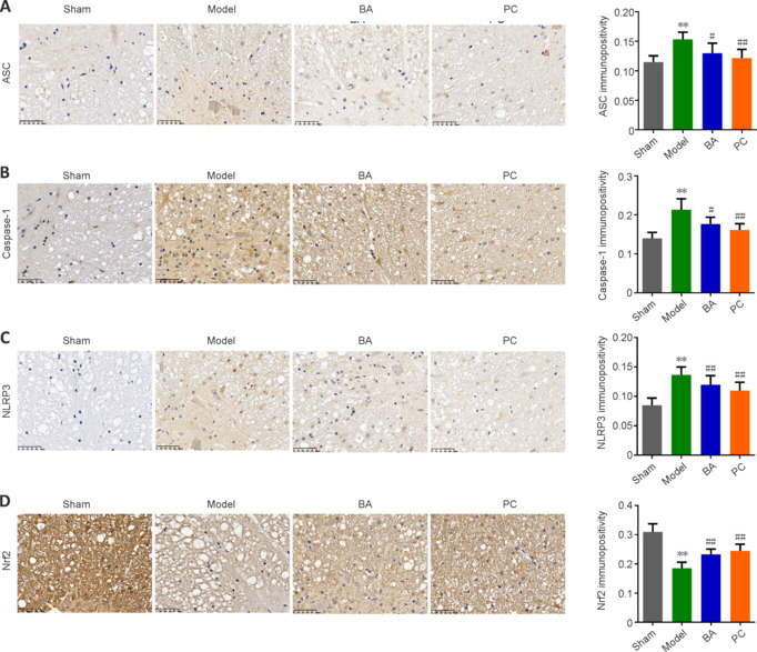
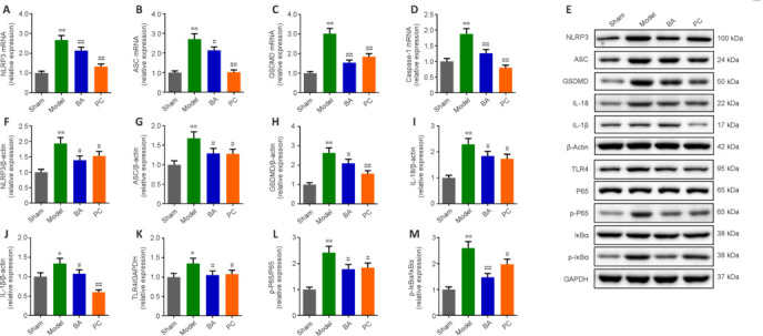
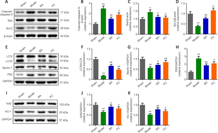
JOURNAL/nrgr/04.03/01300535-202409000-00038/figure1/v/2024-01-16T170235Z/r/image-tiff Previous studies have shown that Biochanin A, a flavonoid compound with estrogenic effects, can serve as a neuroprotective agent in the context of cerebral ischemia/reperfusion injury; however, its effect on spinal cord injury is still unclear. In this study, a rat model of spinal cord injury was established using the heavy object impact method, and the rats were then treated with Biochanin A (40 mg/kg) via intraperitoneal injection for 14 consecutive days. The results showed that Biochanin A effectively alleviated spinal cord neuronal injury and spinal cord tissue injury, reduced inflammation and oxidative stress in spinal cord neurons, and reduced apoptosis and pyroptosis. In addition, Biochanin A inhibited the expression of inflammasome-related proteins (ASC, NLRP3, and GSDMD) and the Toll-like receptor 4/nuclear factor-κB pathway, activated the Nrf2/heme oxygenase 1 signaling pathway, and increased the expression of the autophagy markers LC3 II, Beclin-1, and P62. Moreover, the therapeutic effects of Biochanin A on early post-spinal cord injury were similar to those of methylprednisolone. These findings suggest that Biochanin A protected neurons in the injured spinal cord through the Toll-like receptor 4/nuclear factor κB and Nrf2/heme oxygenase 1 signaling pathways. These findings suggest that Biochanin A can alleviate post-spinal cord injury at an early stage.


Conflict of interest statement

Conflicts of interest: All authors declare no conflicts of interest.

Figures

Figure 1
Figure 1
The effect of BA on hindlimb recovery, edema, inflammatory factors, and oxidative stress in SCI rats. (A, B) On the 1st, 3rd, 5th, 7th, and 14th days after operation, BA-mediated promotion of hindlimb strength and motor function recovery in SCI rats was verified by inclined plane test (A) and BBB score (B), respectively. (C) On the 15th day, the wet/dry weight ratio of the spinal cord was used to evaluate the effect of BA on spinal cord edema. (D–K) Expression levels of the inflammatory factors IL-6 (D), IL-1β (E), TNF-α (F), and IL-18 (G) and the oxidative stress markers CAT (H), SOD (I), GSH (J), and MDA (K) in the spinal cord were determined by ELISA on the 15th day. Data are expressed as mean ± SD (n = 6). **P < 0.01, vs. sham group; #P < 0.05, ##P < 0.01, vs. model group (one-way analysis of variance followed by Tukey's post hoc test). BA: Biochanin A; BBB: Basso Beattie Bresnahan; CAT: catalase; ELISA: enzyme-linked immunosorbent assay; GSH: glutathione; IL-18: interleukin-18; IL-1β: interleukin-1β; IL-6: interleukin-6; MDA: malondialdehyde; PC: positive control; SCI: spinal cord injury; SOD: superoxide dismutase; TNF-α: tumor necrosis factor-α.
Figure 2
Figure 2
BA ameliorates injury at the histopathological levels in the spinal cord of SCI rats. H&E staining was performed on days 15–20 after SCI. (A, B) Representative photographs (original magnification 40×, 200×, 400×, scale bars: 400 μm, 100 μm, 50 μm, A) and semi-quantitative scores (B) for H&E staining of the spinal cord. BA and methylprednisolone treatment improved damage at the histopathological level, including neuronal necrosis and tissue cavity formation, disruption of gray and white matter structure, disruption of neuronal structure, and inflammatory cell infiltration, in the spinal cord of SCI rats. The semi-quantitative scores were proportional to the degree of histopathological damage. Data are expressed as mean ± SD (n = 6). **P < 0.01, vs. sham group; ##P < 0.01, vs. model group (one-way analysis of variance followed by Tukey's post hoc test). BA: Biochanin A; H&E: hematoxylin-eosin; SCI: spinal cord injury; PC: positive control.
Figure 3
Figure 3
On day 21 after injury, the effects of BA on neuronal apoptosis and neuron damage in SCI rats were observed. (A, B) The degree of apoptosis in the spinal cord of SCI rats was measured by TUNEL staining (original magnification 400×). The green fluorescence signal was strongest in the model group. BA and methylprednisolone treatment decreased the green fluorescence intensity. Scale bars: 50 μm. (C, D) The number of Nissl bodies in the anterior horn of the spinal cord in SCI rats was determined by Nissl staining (original magnification 400×). The model group exhibited fewer Nissl bodies than the sham group. BA and methylprednisolone treatment increased the number of Nissl bodies. Scale bars: 50 μm. Data are expressed as mean ± SD (n = 6). **P < 0.01, vs. sham group; ##P < 0.01, vs. model group (one-way analysis of variance followed by Tukey's post hoc test). BA: Biochanin A; PC: positive control; SCI: spinal cord injury; TUNEL: terminal deoxynucleotidyl transferase dUTP nick end labeling.
Figure 4
Figure 4
On day 21 after injury, the promoting effect of BA on neuronal autophagy in the spinal cord tissue of SCI rats was observed. (A, B) Representative immunofluorescence images (A, original magnification 400×) and statistical analysis (B) of LC3 fluorescence intensity. LC3 fluorescence intensity (green Alexa Fluor® 488) was decreased in neurons (marked by NeuN, red, Alexa Fluor® 647) in the model group compared with that in the sham group; BA and methylprednisolone treatment enhanced LC3 fluorescence intensity (green, Alexa Fluor® 488). Scale bars: 100 μm. (C, D) Representative immunofluorescence images (C, original magnification 400×) and statistical analysis of P62 fluorescence intensity (D). Scale bars: 100 μm. P62 fluorescence intensity (green, Alexa Fluor® 488) in neurons (marked by NeuN, red, Alexa Fluor® 647) was stronger in the model group than in the sham group; BA and methylprednisolone treatment decreased P62 fluorescence intensity (green Alexa Fluor® 488). Data are expressed as mean ± SD (n = 6). **P < 0.01, vs. sham group; ##P < 0.01, vs. model group (one-way analysis of variance followed by Tukey's post hoc test). BA: Biochanin A; DAPI: 4,6-diamino-2-phenylindole; LC3: microtube-associated protein 1 light-chain 3; P62: sequestosome-1; PC: positive control; SCI: spinal cord injury.
Figure 5
Figure 5
BA suppresses pyroptosis and promotes anti-oxidation in the spinal cord of SCI rats. On day 22 after injury, immunohistochemistry was performed. (A–D) ASC (A), caspase-1 (B), NLRP3 (C), and Nrf2 (D) expression in the spinal cord was detected by immunohistochemistry (original magnification 400×). ASC, Caspase-1, and NLRP3 immunopositivity was higher in the model group than in the sham group, and BA and methylprednisolone treatment decreased the immunopositivity of all three proteins; while Nrf2 immunopositivity was decreased in model group and increased in rats treated with BA and methylprednisolone treatment. Scale bars: 50 μm. Data are expressed as mean ± SD (n = 6). **P < 0.01, vs. sham group; #P < 0.05, ##P < 0.01, vs. model group (one-way analysis of variance followed by Tukey's post hoc test). AOD: Average optical density; ASC: apoptosis-associated speck-like protein containing CARD; BA: Biochanin A; NLRP3: NOD-like receptor thermal protein domain associated protein 3; Nrf2: nuclear factor erythroid2-related factor 2; PC: positive control; SCI: spinal cord injury.
Figure 6
Figure 6
BA alleviates pyroptosis and inflammation in the spinal cord tissue of SCI rats. On day 18 after SCI, Western blot assay was performed. (A–D) NLRP3 (A), ASC (B), GSDMD (C), and Caspase-1 (D) mRNA expression levels were measured by quantitative reverse transcription-polymerase chain reaction. (E) Western blot analysis of inflammation- and pyroptosis-associated protein expression levels. β-Actin was used as an internal reference for NLRP3, ASC, GADMD, IL-18, and IL-1β. GAPDH was used as an internal reference for TLR4, P65, p-P65, IκBα, and p-IκBα. (F–M) NLRP3 (F), ASC (G), GSDMD (H), IL-18 (I), IL-1β (J), TLR4 (K), p-P65/P65 (L), and p-IκBα/IκBα (M) protein expression levels were measured by Western blot assay. All western blot data were normalized to the sham group. Data are expressed as mean ± SD (n = 3). *P < 0.05, **P < 0.01, vs. sham group; #P < 0.05, ##P < 0.01, vs. model group (one-way analysis of variance followed by Tukey's post hoc test). ASC: Apoptosis-associated speck-like protein containing CARD; BA: Biochanin A; GAPDH: glyceraldehyde-3-phosphate dehydrogenase; GSDMD: gasdermin D; IL-18: interleukin-18; IL-1β: interleukin-1β; NLRP3: NOD-like receptor thermal protein domain associated protein 3; PC: positive control; p-P65: phospho-NF-κB P65; p-IκBα: phospho-NF-kappa-B inhibitor alpha; SCI: spinal cord injury; TLR4: Toll-like receptor 4.
Figure 7
Figure 7
BA inhibits apoptosis and oxidative stress and promotes autophagy in the spinal cord tissue of SCI rats. (A) Western blot analysis of apoptosis-related protein expression levels. (B–D) Quantification of cleaved-caspase-3 (B), Bax (C), and Bcl-2 (D) protein levels from the western blot data. (E) Western blot analysis of autophagy-related protein expression levels. (F–H) Quantification of LC3II/LC3I (F), Beclin-1 (G), and P62 (H) protein levels from the western blot data. (I) Western blot analysis of oxidative stress-related protein expression levels. β-Actin was used as an internal reference for cleaved-caspase-3, Bax, and Bcl-2. GAPDH was used as an internal reference for LC3I, LC3II, Beclin-1, P62, Nrf2, and HO-1. (J, K) Western blot analysis of Nrf2 (J) and HO-1 (K) protein expression levels. All western blot data were normalized to the sham group. Data are expressed as mean ± SD (n = 3). **P < 0.01, vs. sham group; #P < 0.05, ##P < 0.01, vs. model group (one-way analysis of variance followed by Tukey's post hoc test). BA: Biochanin A; HO-1: heme oxygenase-1; LC3: microtube-associated protein 1 light-chain 3; Nrf2: nuclear factor erythroid2-related factor 2; P62: sequestosome-1; PC: positive control; SCI: spinal cord injury.

Similar articles

Cited by

References

    1. Abdel-Wahab BA, Alkahtani SA, Elagab EAM. Tadalafil alleviates cisplatin-induced reproductive toxicity through the activation of the Nrf2/HO-1 pathway and the inhibition of oxidative stress and apoptosis in male rats. Reprod Toxicol. 2020;96:165–174. - PubMed
    1. Anjum A, Yazid MD, Fauzi Daud M, Idris J, Ng AMH, Selvi Naicker A, Ismail OHR, Athi Kumar RK, Lokanathan Y. Spinal cord injury: pathophysiology, multimolecular interactions, and underlying recovery mechanisms. Int J Mol Sci. 2020;21:7533. - PMC - PubMed
    1. Bai Y, Li Z, Liu W, Gao D, Liu M, Zhang P. Biochanin A attenuates myocardial ischemia/reperfusion injury through the TLR4/NF-κB/NLRP3 signaling pathway. Acta Cir Bras. 2019;34:e201901104. - PMC - PubMed
    1. Basso DM, Beattie MS, Bresnahan JC. A sensitive and reliable locomotor rating scale for open field testing in rats. J Neurotrauma. 1995;12:1–21. - PubMed
    1. Biasizzo M, Kopitar-Jerala N. Interplay between NLRP3 inflammasome and autophagy. Front Immunol. 2020;11:591803. - PMC - PubMed