. 2024 Jan 23;121(4):e2317283121.
doi: 10.1073/pnas.2317283121. Epub 2024 Jan 16.

Tumor resistance to anti-mesothelin CAR-T cells caused by binding to shed mesothelin is overcome by targeting a juxtamembrane epitope

Affiliations

Tumor resistance to anti-mesothelin CAR-T cells caused by binding to shed mesothelin is overcome by targeting a juxtamembrane epitope

X F Liu et al. Proc Natl Acad Sci U S A. .

Abstract

Despite many clinical trials, CAR-T cells are not yet approved for human solid tumor therapy. One popular target is mesothelin (MSLN) which is highly expressed on the surface of about 30% of cancers including mesothelioma and cancers of the ovary, pancreas, and lung. MSLN is shed by proteases that cleave near the C terminus, leaving a short peptide attached to the cell. Most anti-MSLN antibodies bind to shed MSLN, which can prevent their binding to target cells. To overcome this limitation, we developed an antibody (15B6) that binds next to the membrane at the protease-sensitive region, does not bind to shed MSLN, and makes CAR-T cells that have much higher anti-tumor activity than a CAR-T that binds to shed MSLN. We have now humanized the Fv (h15B6), so the CAR-T can be used to treat patients and show that h15B6 CAR-T produces complete regressions in a hard-to-treat pancreatic cancer patient derived xenograft model, whereas CAR-T targeting a shed epitope (SS1) have no anti-tumor activity. In these pancreatic cancers, the h15B6 CAR-T replicates and replaces the cancer cells, whereas there are no CAR-T cells in the tumors receiving SS1 CAR-T. To determine the mechanism accounting for high activity, we used an OVCAR-8 intraperitoneal model to show that poorly active SS1-CAR-T cells are bound to shed MSLN, whereas highly active h15B6 CAR-T do not contain bound MSLN enabling them to bind to and kill cancer cells.

Keywords: antibody; cancer; immunotherapy; ovarian cancer; pancreatic cancer.


Conflict of interest statement

Competing interests statement:X.F.L., M.O., M.H., and I.P. are inventors on patents on 15B6 antibody and have assigned all rights to NIH. One of the reviewers (M.G.R.) of this paper is working with a company that has licensed an anti-MSLN antibody (designated SD1) from NIH that was discovered by NIH employee M.H., a co-author of this paper.

Figures

Fig. 1.
Fig. 1.
Illustration of MSLN and CAR-Ts organization. (A and B) Structural model of MSLN connected to GPI anchor bound by the two humanized scFvs: 15B6 and SS1, used in CAR-T studies. The MSLN (PDB ID 7UED_M) is in olive green; the C-terminal MSLN residues N584 to S598 (PDB ID 7U8C_BA2) in orange are the epitopes where 15B6 binds. The light chain, linker, and heavy chain of the humanized 15B6 and SS1 scFv, modeled by AlphaFold2, are shown in ribbon and surface drawing in light blue, dark gray, blue, pink, dark gray, and red, respectively. The complex was made by superposing scFv15B6 and scFvSS1 models to 7U8C, 4F3F, and 7UED. (C) Comparison of mouse and humanized 15B6 structures. The MSLN C-terminal peptide N584 – S598 is in orange, complexed with mouse 15B6 Fv in green (PDB ID 7U8C). Humanized scFv 15B6 modeled by AlphaFold2 is in blue, superposed with mouse 15B6 Fv. CDR regions are in darker colors and labeled. The RMSD between the two 15B6s calculated by TM-align is 0.85 Å with TM-score 0.98 and all 223 Fv residues aligned, except the 15 residues linker colored in gray. (D) Schematic showing expression vectors used to make CAR-T cells. SS, signal sequence; hEGFRt contains domains III and IV and transmembrane domain.
Fig. 2.
Fig. 2.
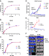
Humanized 15B6 CAR-T cells have the same activity as CAR-T with mouse Fv. (AC) Cell killing assays comparing h15B6 with either mouse 15B6 or SS1 CAR-T using OVCAR 8 (A), KLM1 (B) or RH29 cells (C). n = 3 (AC). h15B6 CAR-T cause complete regressions of KLM1 tumors in NSG mice. SS1 CAR-T and control T cells have no activity, n = 5. (D) h15B6 CAR-T give complete responses in KLM1 subcutaneous tumors expressing MSLN but not in a MSLN knock out line (KLM1 E10), n = 5. (E) h15B6 CAR-T give complete responses in an intraperitoneal OVCAR 8 model. KLM1 injected 5 million, KLM1E10 injected 0.1 million. Tumor size was measured twice a week. OVCAR8-luc cells were implanted IP. Five millions of h15B6 CAR-T were injected on day 7. Tumors were imaged every week as described in the Methods section.
Fig. 3.
Fig. 3.
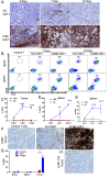
h15B6 CAR-T cells, but not SS1 CAR-T cells, accumulate in tumors. (A) Mice received 5 million OVCAR-8 cells I.P., and on day 21, the mice were imaged and treated with 10 million of either SS1 or h15B6 CAR-T cells. After 4, 7, or 10 d of treatment, tumors were removed for IHC analysis with anti-CD3. (B) On days 7 and 10, spleen and blood cells were analyzed by flow cytometry using anti-CD3 and anti-CD45. All cells were first gated with DAPI for live cells; then, CD3 and CD45 double-positive cells were identified as indicated by circles. (C) In the Left panel, CD3-positive cells from A were analyzed using HALO imaging, and % of CD3-positive cells is shown in the graph. (D and E) show % CAR-T in blood and spleen (N = 3). (F) Mice with KLM1 subcutaneous tumors were injected with either control T cells, SS1 CAR-T, or h15B6 CAR-T 7 d (when tumor reached 100 mm3) after tumor implantation. Seven days later, tumors were removed and analyzed by IHC staining with anti-CD3, (G) Percentage of CD3-positive cells in blood, spleen, or tumor (n = 3). (H) KLM1 E10 MSLN KO cells were implanted as in A, and after 7 d of 15B6 CAR-T treatment, tumors were removed and stained with anti-CD3, showing that there is no h15B6 CAR-T present in the tumor. **P < 0.01; ***P < 0.001.
Fig. 4.
Fig. 4.
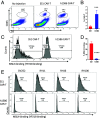
Binding of shed MSLN to CAR-T cells: h15B6 CAR-T cells in mouse ascites do not contain shed-MSLN and do not bind to shed MSLN in ascites of patients. (A and B) Mice with OVCAR 8 I.P. tumors were treated 21 d after tumor implantation with either h15B6 or SS1 CAR-T. Seven days later, the cells in ascites were stained for anti-CD3 and anti-CD45. For flow analysis, live cells were first gated with DAPI; then, CD3 and CD45 double-positives cells were identified and are circled. (B) Percentage of CD3 and CD45 double-positive cells (n = 6). (C) CD3 and CD45 double-positive cells from A were analyzed for binding to YP218-Alexa 647. (D) Summary of MSLN containing cells identified by Mab YP218 from 2 different experiments (n = 6). (E) Ascites or pleural fluid from ovarian patient (OV252) or mesothelioma patients (RH11, RH16, and RH100) were incubated with either 15B6 or SS1 CAR-T cells. The presence of shed MSLN on the CAR-T cells was detected by YP218-alexa 647. Refer SI Appendix, Fig. S5 for CAR-T positive cell identification. **P < 0.01; ***P < 0.001.
Fig. 5.
Fig. 5.
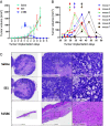
Pancreatic Cancer PDX studies in mice. Human PDX pancreatic cancers were implanted subcutaneously, and the mice were treated with CAR-T cells when tumors reached 50 mm3. (A) Average tumor volume: arrow indicates injection periods; 7 mice /group. (B) Individual mouse response to h15B6 CAR-T treatment. Arrows indicate the injection days for individual mice. (C) H.E. staining of xenografts of saline- and SS1 CAR-T-treated mice showing characteristic central necrosis at the end point. A collagenous scar is all that remains in the 15B6-treated mice.
Fig. 6.
Fig. 6.
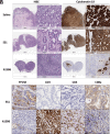
Histological Analysis of PDX tumors. (A) H and E analysis on day 7 shows glandular structures that are Cytokeratin 19 positive in saline and SS1 CAR-T cells groups as well as dense matrix. h15B6 CAR-T-treated tumors show infiltration with inflammatory cells and very few remaining Cytokeratin-positive tumor cells. (B) Antibody staining of tumors shows that SS1 CAR-T tumors are MSLN positive (YP158) and CD3, CD4, and CD8 negative. H15B6 CAR-T-treated tumors are MSLN negative and very positive for CD3, CD4, and CD8.
Fig. 7.
Fig. 7.
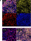
Proliferation of h15B6 CAR-T cells in PDX pancreatic cancers on 7-d treatment time point. (A) Immunofluorescence study showing colocalization of Ki-67 (red) and CD3 (yellow) and DAPI (red) merged image (pink) in PDX cancers. (B) Fig. 6B shows that the SS1 CAR-T-treated tumors are mostly composed of replicating tumor cells that are DAPI and Ki67 positive. Very few tumor cells are present in the h15B6 CAR-T-treated group, and CD3-positive, Ki67-positive cells are present. Ki-67 positivity (pink) in neoplastic ductal epithelium in the SS1 CAR-T-treated mice and Ki-67 (pink) positivity in CAR-T cells (yellow) in the h15B6 CAR-T-treated mice.
Fig. 8.
Fig. 8.
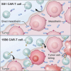
Cartoon demonstrating why CAR-T cells targeting a distal MSLN epitope have very low anti-tumor activity and CAR-T targeting a non-shed epitope have high activity. MSLN (blue and red) on tumor cells is cut by proteases leaving a short stalk (red) attached to the cell and releasing the shed portion (blue) into the extra-cellular fluid of the tumor and then into the blood. CAR-T cells made with SS1 Fv recognize a distal epitope of MSLN present on shed MSLN, bind to shed MSLN as they enter the tumor or pass in capillaries through the tumor and do not accumulate in the tumor or reach tumor cells. CAR-T cells, made with 15B6 Fv, do not bind to shed MSLN. Therefore, after tumor entry, they can bind to and kill cancer cells. They are also activated and they expand as well.

Similar articles

Cited by

References

    1. Zou Y., Xu W., Li J., Chimeric antigen receptor-modified T cell therapy in chronic lymphocytic leukemia. J. Hematol. Oncol. 11, 130 (2018). - PMC - PubMed
    1. Kai R., Jain M. D., Smith E. L., Mechanisms of resistance and treatment of relapse after CAR-T-cell therapy for large B-cell lymphoma and multiple myeloma. Transplant. Cell. Ther. 29, 418–428 (2023), 10.1016/j.jtct.2023.04.007. - DOI - PMC - PubMed
    1. Neelapu S. S., et al. , Axicabtagene ciloleucel CAR-T-cell therapy in refractory large B-cell lymphoma. N. Engl. J. Med. 377, 2531–2544 (2017). - PMC - PubMed
    1. Jogalekar M. P., et al. , CAR-T-Cell-Based gene therapy for cancers: New perspectives, challenges, and clinical developments. Front. Immunol. 13, 925985 (2022). - PMC - PubMed
    1. Ma S., et al. , Current progress in CAR-T cell therapy for solid tumors. Int. J. Biol. Sci. 15, 2548–2560 (2019), 10.7150/ijbs.34213. - DOI - PMC - PubMed

LinkOut - more resources