Review
. 2024 Jan 16;149(3):251-266.
doi: 10.1161/CIRCULATIONAHA.123.065657. Epub 2024 Jan 16.

Coronary Artery Calcification: Current Concepts and Clinical Implications

Affiliations
Review

Coronary Artery Calcification: Current Concepts and Clinical Implications

Carlotta Onnis et al. Circulation. .

Abstract

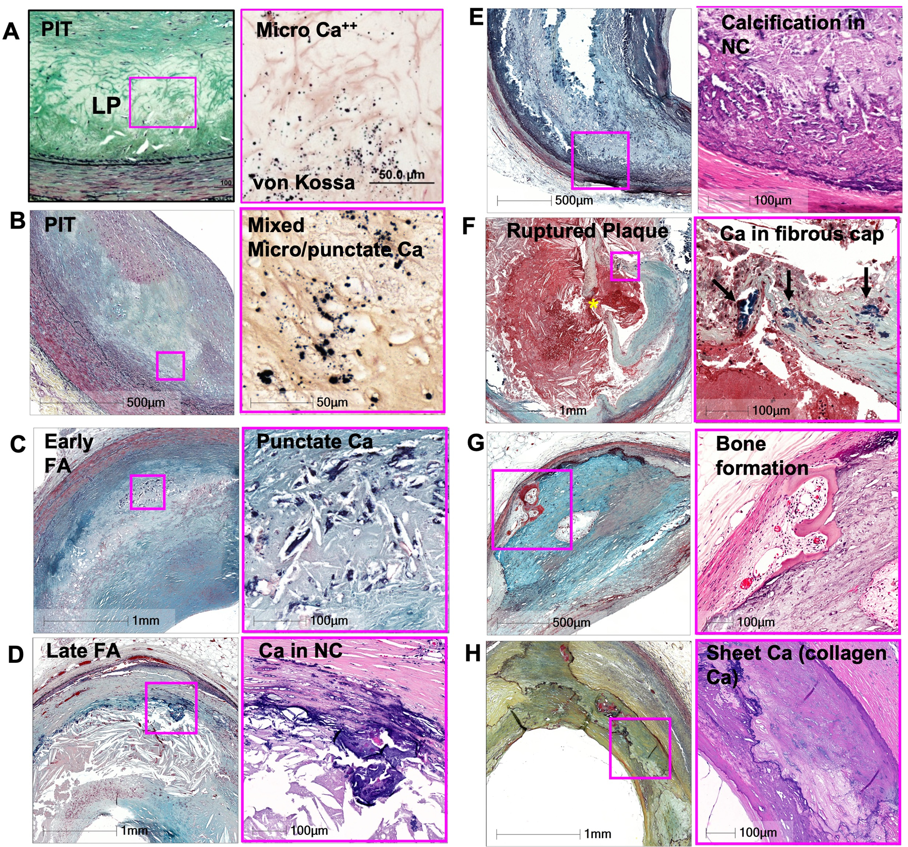
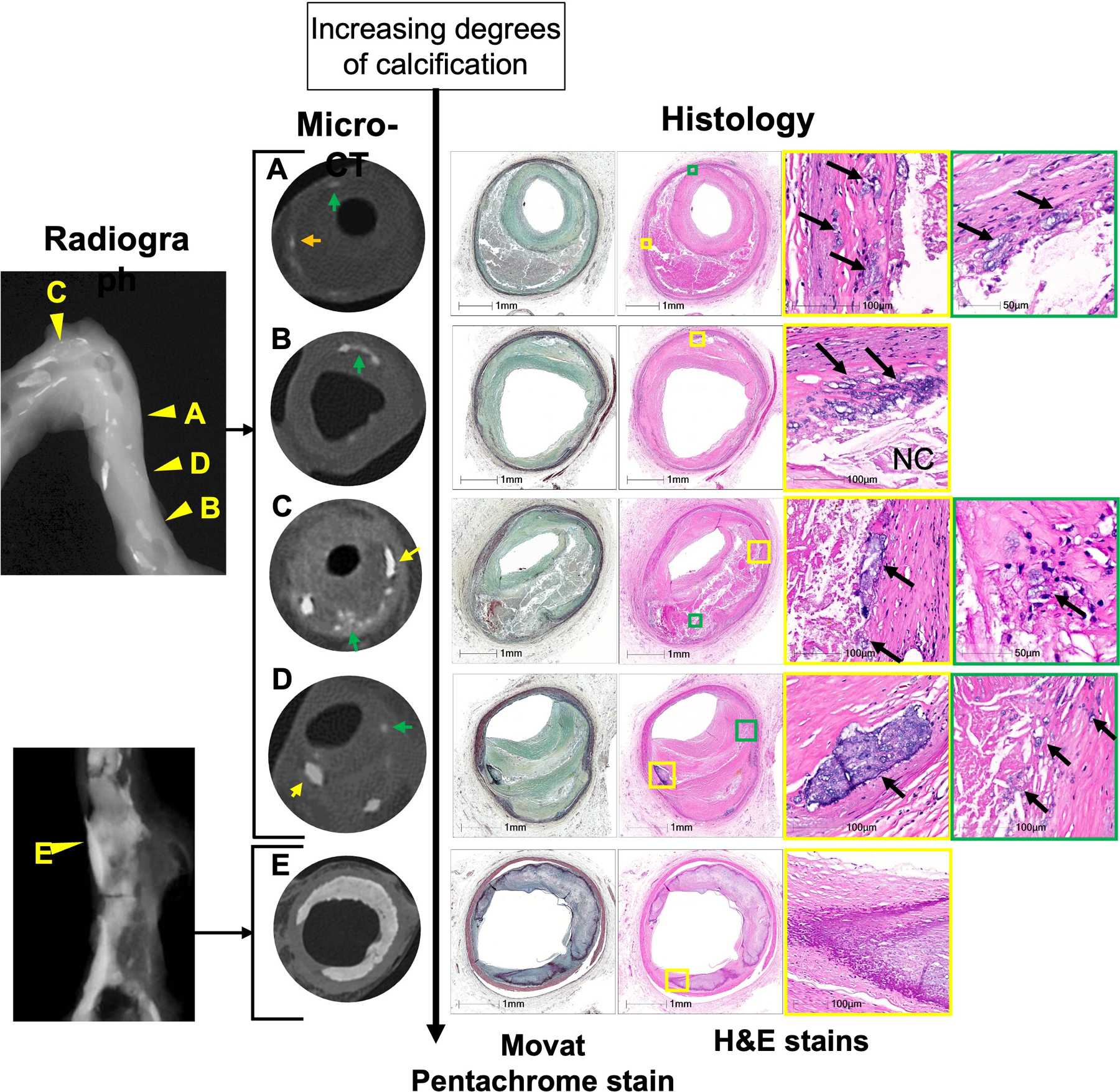
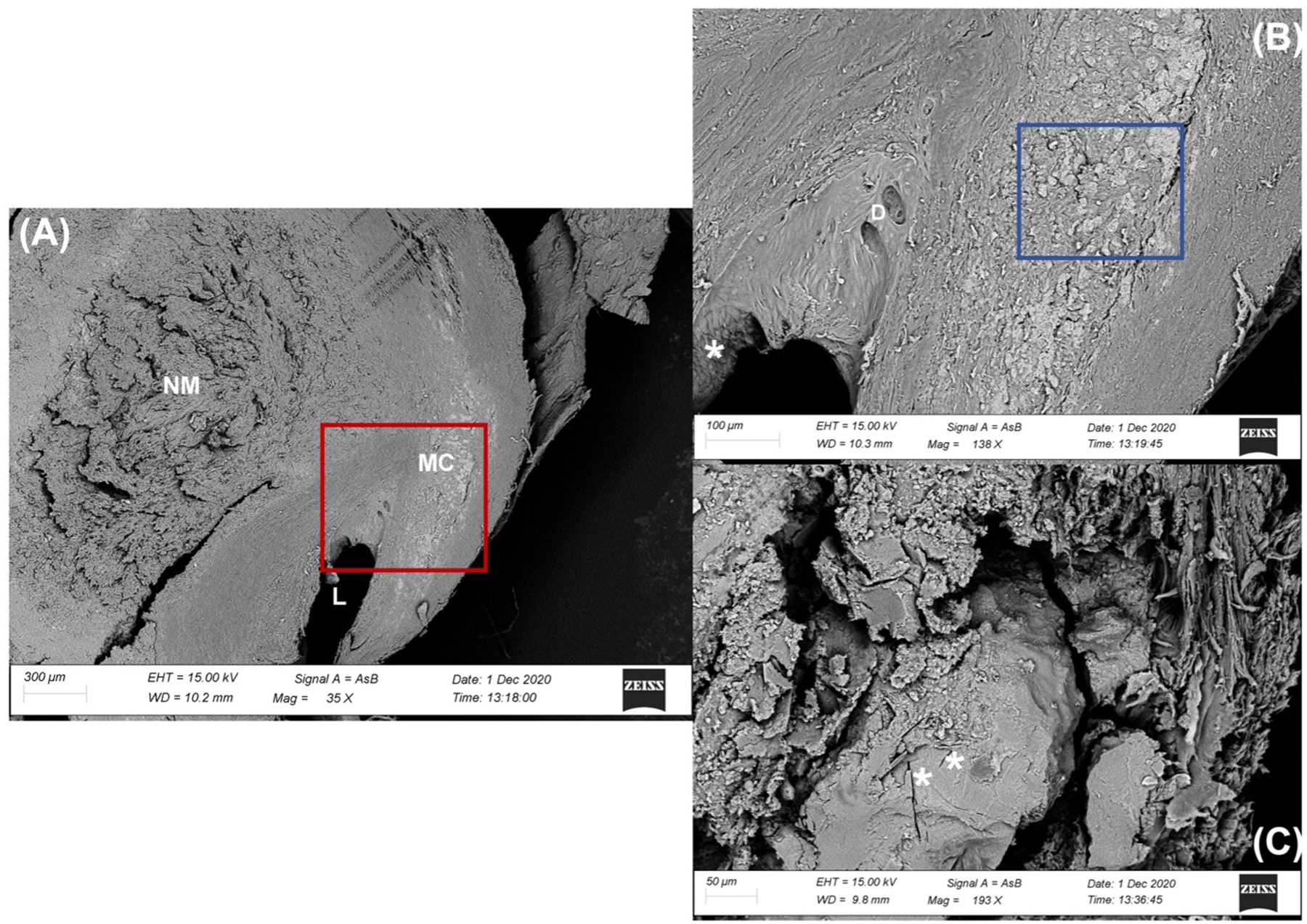
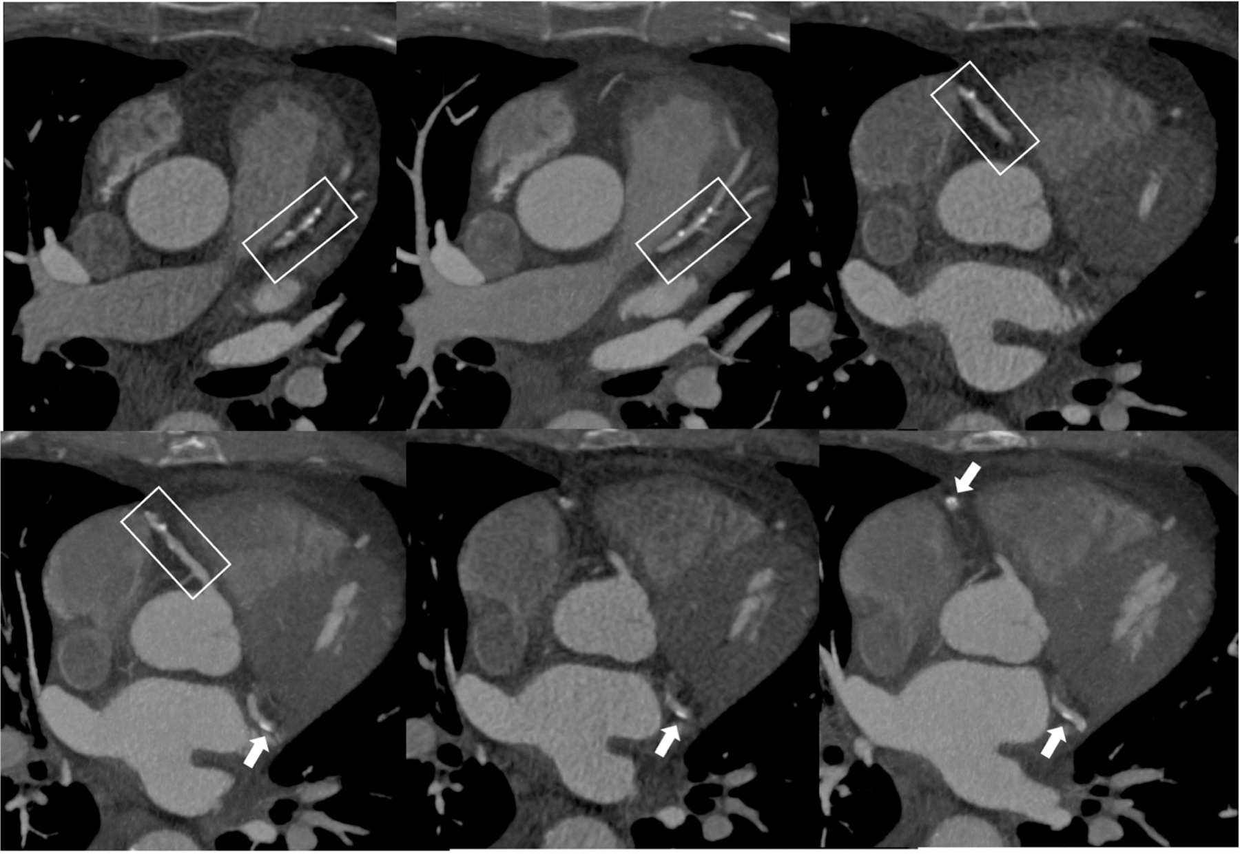
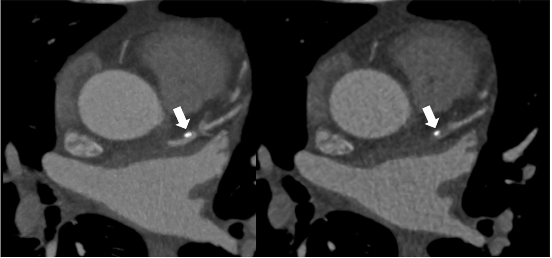
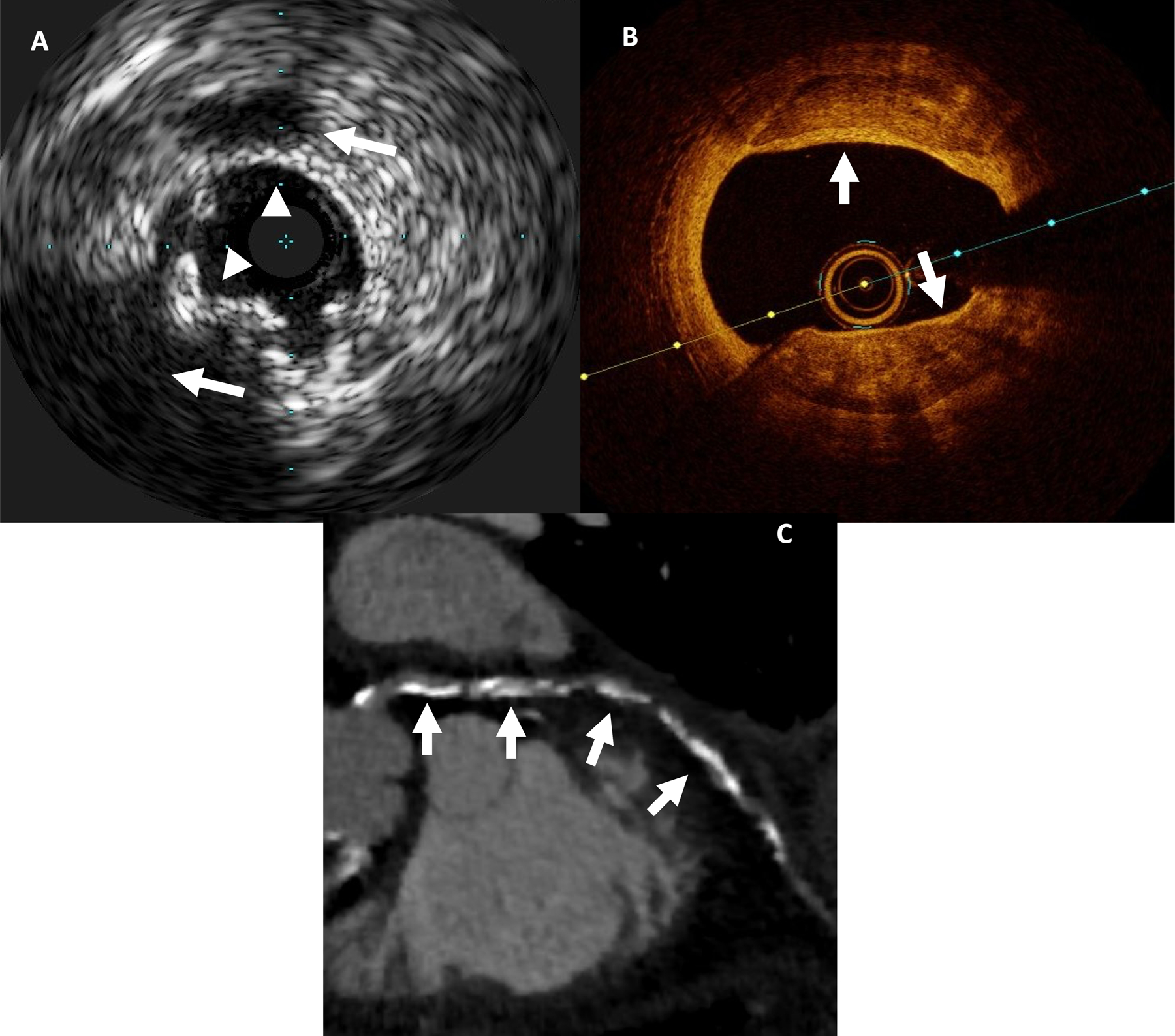
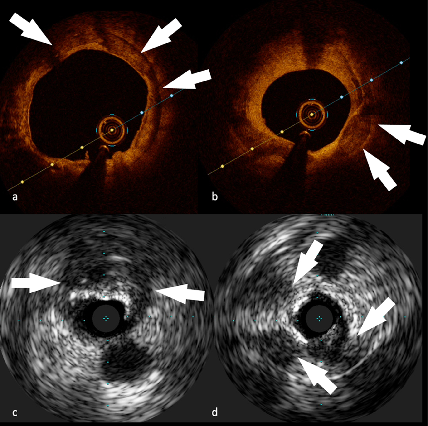
Coronary artery calcification (CAC) accompanies the development of advanced atherosclerosis. Its role in atherosclerosis holds great interest because the presence and burden of coronary calcification provide direct evidence of the presence and extent of coronary artery disease; furthermore, CAC predicts future events independently of concomitant conventional cardiovascular risk factors and to a greater extent than any other noninvasive biomarker of this disease. Nevertheless, the relationship between CAC and the susceptibility of a plaque to provoke a thrombotic event remains incompletely understood. This review summarizes the current understanding and literature on CAC. It outlines the pathophysiology of CAC and reviews laboratory, histopathological, and genetic studies, as well as imaging findings, to characterize different types of calcification and to elucidate their implications. Some patterns of calcification such as microcalcification portend increased risk of rupture and cardiovascular events and may improve prognosis assessment noninvasively. However, contemporary computed tomography cannot assess early microcalcification. Limited spatial resolution and blooming artifacts may hinder estimation of degree of coronary artery stenosis. Technical advances such as photon counting detectors and combination with nuclear approaches (eg, NaF imaging) promise to improve the performance of cardiac computed tomography. These innovations may speed achieving the ultimate goal of providing noninvasively specific and clinically actionable information.

Keywords: atherosclerosis; calcinosis; cardiac imaging techniques; coronary angiography; coronary vessels; plaque, atherosclerotic; vascular calcification.


Conflict of interest statement

Disclosures Dr Libby is an unpaid consultant to, or involved in clinical trials for Amgen, AstraZeneca, Baim Institute, Beren Therapeutics, Esperion Therapeutics, Genentech, Kancera, Kowa Pharmaceuticals, Medimmune, Merck, Moderna, Novo Nordisk, Novartis, Pfizer, and Sanofi-Regeneron. Dr Libby is a member of the scientific advisory board for Amgen, Caristo Diagnostics, Cartesian Therapeutics, CSL Behring, DalCor Pharmaceuticals, Dewpoint Therapeutics, Eulicid Bioimaging, Kancera, Kowa Pharmaceuticals, Olatec Therapeutics, Medimmune, Novartis, PlaqueTec, TenSixteen Bio, Soley Thereapeutics, and XBiotech, Inc. Dr Libby’s laboratory has received research funding in the last 2 years from Novartis, Novo Nordisk, and Genentech. Dr Libby is on the Board of Directors of XBiotech, Inc. Dr Libby has a financial interest in Xbiotech, a company developing therapeutic human antibodies, in TenSixteen Bio, a company targeting somatic mosaicism and clonal hematopoiesis of indeterminate potential (CHIP) to discover and develop novel therapeutics to treat age-related diseases, and in Soley Therapeutics, a biotechnology company that is combining artificial intelligence with molecular and cellular response detection for discovering and developing new drugs, currently focusing on cancer therapeutics. Dr Libby’s interests were reviewed and are managed by Brigham and Women’s Hospital and Mass General Brigham in accordance with their conflict-of-interest policies. Dr Libby reports the following patents: Il-1beta binding antibodies for use in treating cancer (US20230220063A1 - Pending); and Treatment of brain ischemia-reperfusion injury (US20220041710A1 - Pending).

Figures

Figure 1
Figure 1
Calcification in various types of plaque. (A) Microcalcification (varies from 0.5 to15um) in PIT. (B) Mixed punctate and microcalcification may be observed in PIT. (C) Punctate Calcification in early FA. (D) Calcified in late FA, occurs as fragmented calcification and is seen near the media involving collagen and necrotic core. (E) NC calcification. (F) Plaque rupture showing calcification in the fibrous cap. Asterisk indicate the site of ruptured fibrous cap. (G) Bone formation near the border of medial layer. (H) Sheet collagen calcification (collagen Ca). All low power images is shown with Movat pentachrome stain. High power images are shown with Von Kossa stain in A and B, Movat pentachrome stain in C and E, and H&E in D, F, G, and H. Abbreviations: PIT = pathologic intimal thickening, FA = fibro atheroma, NC = necrotic core. (Modified from Otsuka F, et al. Arterioscler Thromb Vasc Biol 2014;34:724-736)
Figure 2
Figure 2
Detection of calcification by various modalities (radiograph, micro-CT, and histology). Image A to E shows severity of calcification from punctate calcification to sheet calcification by micro-CT; on the right are shown the corresponding histologic images and on the left is shown the radiograph of the artery; yellow arrows show the area of calcification in micro-CT and arrowheads in radiograph show the matching areas. (A) Micro-CT image can detect punctate calcification (on radiograph speckled) in the border area of a large NC with adjoining fibrous tissue in late FA. (B) Micro-CT image shows speckled calcification composed of an aggregate of punctate calcification in the early FA. (C, D) Micro-CT show varying sizes of fragmented calcification (yellow arrows) and speckled calcification (green arrows) in the late FA (C) and healed plaque rupture (D). (E) Micro-CT showing sheet calcification. Abbreviations: FA = fibroatheroma, NC = necrotic core.
Figure 3
Figure 3
Electron microscopy images. Low (A), medium (B [red boxed area in (A)]) and high (C [blue boxed area in (B)]) magnification scans from a transversal section of carotid artery observed by Back Scattered Electron probe (BSE). (A) The lumen (L) of the vessel is remarkably reduced and eccentrically constricted by the large necrotic mass (NM) in the thickness of the Tunica Media. The Tunica Intima appears strongly altered even at low magnification. The BSE mode, utilized for analyzing differences in tissue density, highlights an area with numerous microcalcifications (MC) within the Tunica Media with a light gray-white tone. (B) A greater enlargement of the vessel lumen shows the presence of small diverticula (D) and important differences in the shape of the endothelial cells, some of them in fact appear taller and less distended (white asterisk *) according to the blood flow. In the thickness of the tunica media, the micro-calcifications of the plaque are distinguished by definition and size. (C) Partially sectioned microcalcification useful to show the internal structure. The calcified mass in calcium phosphate appears fairly homogeneous centrally, with some traces of the presence of cholesterol crystals (white asterisk *). In the periphery, small centers of crystalline aggregation are visible, which suggest a progressive growth of the calcified mass.
Figure 4
Figure 4
Atherosclerotic plaque as seen in CCTA. Male patient, 67 years old, history of diabetes. CCTA images show diffuse mixed-atherosclerotic plaques, with a three-vessel distribution (box and arrows).
Figure 5
Figure 5
Atherosclerotic plaque as seen in CCTA. Intermediate-risk 71-year-old patient presenting with acute chest pain and no known history of CAD. CCTA image shows highly dense calcified plaque (arrows).
Figure 6
Figure 6
Invasive and non-invasive imaging of atherosclerotic plaque. 72-year-old male patient with CAC score of 357: (A) IVUS: multiple calcifications (white arrowheads) with shadowing (white arrows) (B) OCT: multiple superficial calcifications, seen as signal-poor heterogeneous regions with well delineated borders (white arrows) (C) CCTA: diffuse mixed plaque, predominantly calcified (white arrows)
Figure 7
Figure 7
Coronary atherosclerotic plaque images obtained with invasive imaging techniques. OCT (A-B) and IVUS (C-D), showing different types of calcifications: (A) Concentric calcification (white arrows) on RCA in a 74-year-old male patient (B) Superficial calcification (white arrows) on LAD in a 67-year-old male patient (C) Big superficial calcification with posterior cone of shadow (white arrows) on LCx in a 72-year-old male patient (D) Multiple clusters of calcifications (white arrows) on RCA in a 62-year-old male patient

Similar articles

Cited by

References

    1. WHO- World Health Organization, “Cardiovascular diseases (CVDs)”, 11th June 2021, who.int, https://www.who.int/news-room/fact-sheets/detail/cardiovascular-diseases... Accessed 12th December 2022
    1. Nicoll R, Wiklund U, Zhao Y, Diederichsen A, Mickley H, Ovrehus K, Zamorano P, Gueret P, Schmermund A, Maffei E, et al. The coronary calcium score is a more accurate predictor of significant coronary stenosis than conventional risk factors in symptomatic patients: Euro-CCAD study. Int J Cardiol 2016;207:13–19. - PubMed
    1. Osawa K, Nakanishi R, Budoff M. Coronary Artery Calcification. Glob Heart 2016;11(3):287–293. - PMC - PubMed
    1. Detrano R, Guerci AD, Carr JJ, Bild DE, Burke G, Folsom AR, Liu K, Shea S, Szklo M, Bluemke DA, et al. Coronary calcium as a predictor of coronary events in four racial or ethnic groups. N Engl J Med 2008;358(13):1336–1345. - PubMed
    1. Madhavan MV, Tarigopula M, Mintz GS, Maehara A, Stone GW, Généreux P. Coronary artery calcification: pathogenesis and prognostic implications. J Am Coll Cardiol 2014;63(17):1703–1714. - PubMed

Publication types

MeSH terms