. 2024 Feb 14;32(2):181-190.e9.
doi: 10.1016/j.chom.2023.12.014. Epub 2024 Jan 15.

A bacterial sialidase mediates early-life colonization by a pioneering gut commensal

Affiliations

A bacterial sialidase mediates early-life colonization by a pioneering gut commensal

Ekaterina Buzun et al. Cell Host Microbe. .

Abstract

The early microbial colonization of the gastrointestinal tract can have long-term impacts on development and health. Keystone species, including Bacteroides spp., are prominent in early life and play crucial roles in maintaining the structure of the intestinal ecosystem. However, the process by which a resilient community is curated during early life remains inadequately understood. Here, we show that a single sialidase, NanH, in Bacteroides fragilis mediates stable occupancy of the intestinal mucosa in early life and regulates a commensal colonization program. This program is triggered by sialylated glycans, including those found in human milk oligosaccharides and intestinal mucus. NanH is required for vertical transmission from dams to pups and promotes B. fragilis dominance during early life. Furthermore, NanH facilitates commensal resilience and recovery after antibiotic treatment in a defined microbial community. Collectively, our study reveals a co-evolutionary mechanism between the host and microbiota mediated through host-derived glycans to promote stable colonization.

Keywords: commensal colonization; early-life microbiome; host-microbe interactions; human milk oligosaccharides.


Conflict of interest statement

Declaration of interests The authors declare no competing interests.

Figures
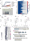
Figure 1.
Figure 1.. Defining HMO utilization system in B. fragilis
(A) Growth of seven Bacteroides species: B. fragilis NCTC 9343 (Bf), B. thetaiotaomicron VPI-8482 (Bt), P. vulgatus ATCC 8482 (Pv), B. ovatus ATCC 8483 (Bo), B. salyersiae DSM 18765 (Bs), B. uniformis ATCC 8492 (Bu), and B. acidifaciens JCM 10556 (Ba) in defined medium with 1.5 % pooled HMOs (pHMOs). Solid line represents mean ± SD. (B) TLC of cell-free supernatant collected from stationary phase cultures. Controls: sialic acid/Neu5Ac (SA); lactose (Lac); galactose (Gal), glucose (Glc); fucose (Fuc). (C) HPLC analysis of the supernatant collected during B. fragilis growth on pHMOs over time. (D) Proteomic analysis of pHMO-grown whole B. fragilis cells, pHMOs vs glucose fold change at early-, mid-, and late-log phases. Significance: log2-fold change ≥2, P-value < 0.05. (E) Relative abundance of proteins with a max log10-fold change ≥ 2 and a Benjamini Hochenburg corrected P-value < 0.05 across growth phases. Hierarchical clustering was performed using Euclidean distance. Scale bar: protein abundance from high (dark purple) to low (white). (F) Organization of significantly abundant polysaccharide utilization loci (PULs) in B. fragilis during growth on pHMO, gene labeling corresponds to locus tags BFxxxx: N-acetyl neuraminic acid utilization (Nan) operon (pink diamond); HMO PUL1 (maroon diamond); Domain of N-glycan (Don) utilization operon (teal circle); Commensal colonization factor (CCF) operon (black circle). See also Figure S1, Figure S2, and Tables S1-S3.
Figure 2.
Figure 2.. NanH is required for growth on HMOs and promotes intestinal colonization.
(A – G) Growth curves and area under the curve (AUC) of Bf WT (blue) and Bf ΔnanH (brown) on (A) BHI-S; (B) Pooled HMOs (pHMOs); (C) Diagrammatic structures of sialylated (top) and non-sialylated (bottom) HMOs. Sugar and linkage keys are shown; (D) Pooled sialylated HMOs; (E) Pooled neutral HMOs; (F) 6’-sialylactose (6’SL); (G) 3’-sialylactose (3’SL). Data is shown as mean ± SD of 3 biological replicates. Solid line represents mean ± SD. (H – I) Germ-free mice were orally gavaged with either B. fragilis WT or ΔnanH (n=10). Abundance of each strain was quantified in CFU/mg of (G) feces and (I) colonic mucus. Limit of detection (LOD) = 5 CFU/mg. (J– K) Germ-free mice (n=5) were orally gavaged with 1:1 mixture of B. fragilis WT and ΔnanH strains. (J) Quantification of Bf WT and Bf ΔnanH in feces of co-colonized mice. (K) The competitive index (log10) as indicated by input/output ratios. Differentiation between the Bf WT and Bf ΔnanH was performed as described in Methods. Data are shown as geometric mean ± SD, representative of two independent experiments. *P < 0.05; **P < 0.01; ***P < 0.001; ns – not significant; (Kruskal-Wallis and Mann-Whitney tests); ND – not detected. See also Figure S3, Figure S4.
Figure 3.
Figure 3.. NanH mediates stable niche occupancy and early life colonization.
(A) Germ-free mice were mono-associated with Bf WT (initial) and challenged with Bf ΔnanH on day 8; n=5. (B) Germ-free mice were mono-associated with Bf ΔnanH (initial) and challenged with Bf WT on day 8; n=5.LOD = 10 CFU/mg of feces (C-D) CFU/mg of the initial and the challenge strains in (C) fecal pellets and (D) colonic mucus 27 days post-challenge; n=5. Data are shown as geometric mean ± SD, representative of two independent experiments. LOD = 5 CFU/mg of feces or mucus; *P < 0.05; ns – not significant; Mann-Whitney tests. (E) Vertical transmission schematic (left) and the abundance of Bf WT and Bf ΔnanH (right) in the feces of adult breeders (dark gray) and newborn suckling pups (light gray). n=16 pups. (F) Images of stomachs collected from suckling pups (top). TLC analysis of the homogenized stomachs (bottom). Controls: 6’SL, 3’SL, lactose (Lac), glucose (Glc), galactose (Gal), and fucose (Fuc). (G) HPLC analysis of the stomach contents. Peaks in the shaded area indicate the peaks of 3’SL and 6’SL.
Figure 4.
Figure 4.. B. fragilis NanH defines resilience and competitive fitness.
(A-B) Abundance of (A) Bf WT and B. thetaiotaomicron (n=12 pups) and (B) Bf ΔnanH and B. thetaiotaomicron (n=20 pups) in the feces of adult breeders (♀♂) and newborn suckling pups at designated ages (days old). (C – D) Germ free mice were inoculated with (C) Bf WT and B. thetaiotaomicron WT (Bt) or (D) Bf ΔnanH and B. thetaiotaomicron WT (Bt) at 1:1 ratio; n=5 per group. (E – G) Mice harboring the Simplified Human Intestinal Microbiota (SIHUMIx) were challenged with WT and Bf ΔnanH (1:1) on day 0. (G) The abundance of Bf WT and Bf ΔnanH was determined by qPCR, relative to total microbial 16S before (day 10), after (day 15), and during the recovery period (days 20 and 27) from the ciprofloxacin treatment; n=4. (F) Fecal load (CFU/mg) of Bf WT and Bf ΔnanH before and after the ciprofloxacin treatment. Differentiation between strains is described in Methods. (G) Abundance of Bf WT and Bf ΔnanH in colonic mucus, determined by qPCR; n= 4. Data show mean ± SD, LOD = 5 CFU/mg. *P < 0.05; ns – not significant; Mann-Whitney test. Data represents at least two independent experiments. (H) Growth of B. fragilis strains isolated from human infants on 15 mg/ml pHMOs. Solid line represents the mean ± SD. See also Figure S4.

Update of

References

    1. Gensollen T, Iyer SS, Kasper DL, and Blumberg RS (2016). How colonization by microbiota in early life shapes the immune system. Science 352, 539–544. 10.1126/science.aad9378. - DOI - PMC - PubMed
    1. Lou YC, Olm MR, Diamond S, Crits-Christoph A, Firek BA, Baker R, Morowitz MJ, and Banfield JF (2021). Infant gut strain persistence is associated with maternal origin, phylogeny, and traits including surface adhesion and iron acquisition. Cell Reports Medicine 2, 100393. 10.1016/j.xcrm.2021.100393. - DOI - PMC - PubMed
    1. Mitchell CM, Mazzoni C, Hogstrom L, Bryant A, Bergerat A, Cher A, Pochan S, Herman P, Carrigan M, Sharp K, et al. (2020). Delivery Mode Affects Stability of Early Infant Gut Microbiota. Cell Reports Medicine 1, 100156. 10.1016/j.xcrm.2020.100156. - DOI - PMC - PubMed
    1. Stewart CJ, Ajami NJ, O’Brien JL, Hutchinson DS, Smith DP, Wong MC, Ross MC, Lloyd RE, Doddapaneni H, Metcalf GA, et al. (2018). Temporal development of the gut microbiome in early childhood from the TEDDY study. Nature 562, 583–588. 10.1038/s41586-018-0617-x. - DOI - PMC - PubMed
    1. Bäckhed F, Roswall J, Peng Y, Feng Q, Jia H, Kovatcheva-Datchary P, Li Y, Xia Y, Xie H, Zhong H, et al. (2015). Dynamics and Stabilization of the Human Gut Microbiome during the First Year of Life. Cell Host & Microbe 17, 690–703. 10.1016/j.chom.2015.04.004. - DOI - PubMed