Review
. 2024 Mar;260(Pt 1):129495.
doi: 10.1016/j.ijbiomac.2024.129495. Epub 2024 Jan 14.

Improving DNA nanostructure stability: A review of the biomedical applications and approaches

Affiliations
Review

Improving DNA nanostructure stability: A review of the biomedical applications and approaches

Mahboobeh Nasiri et al. Int J Biol Macromol. 2024 Mar.

Abstract

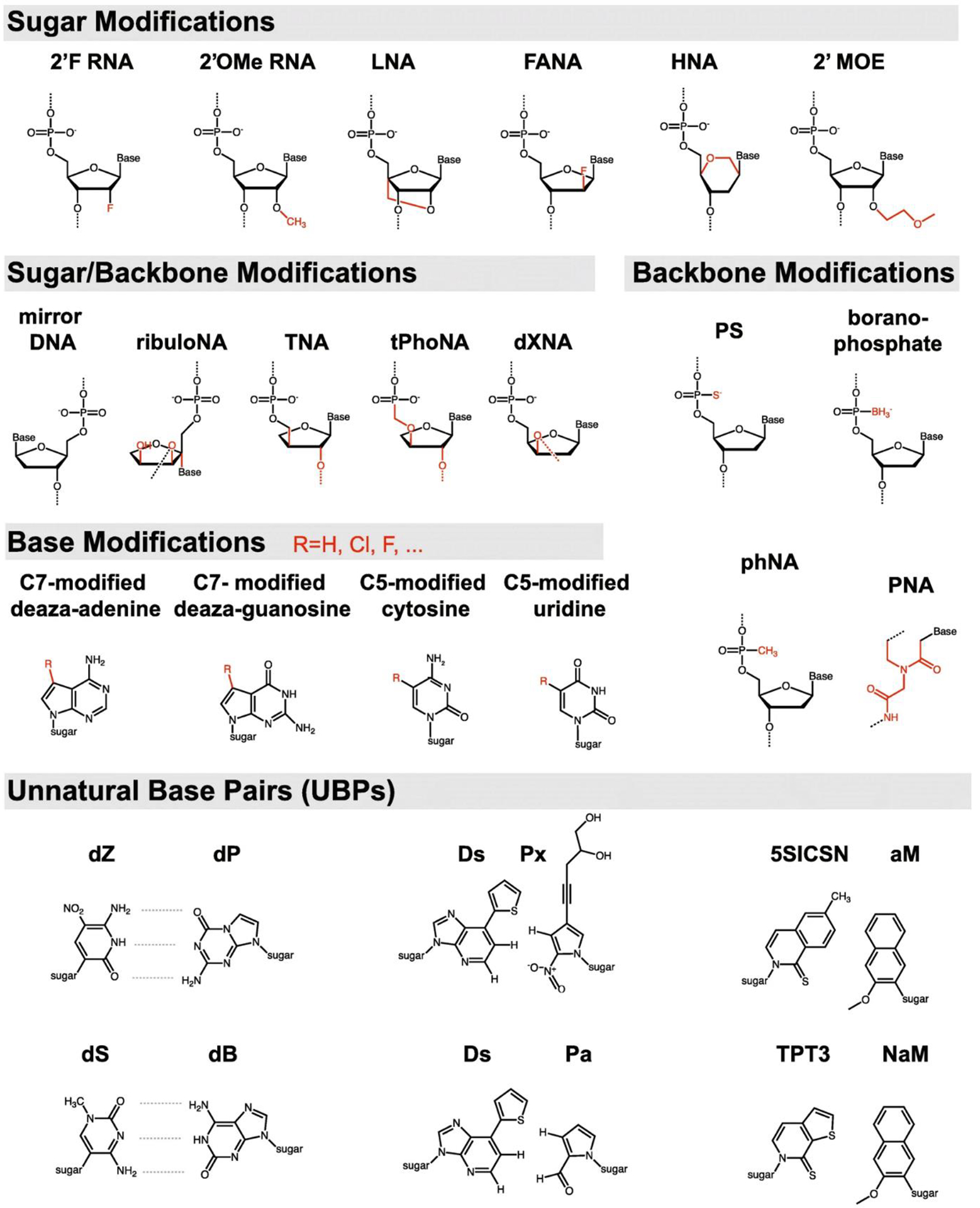
DNA's programmable, predictable, and precise self-assembly properties enable structural DNA nanotechnology. DNA nanostructures have a wide range of applications in drug delivery, bioimaging, biosensing, and theranostics. However, physiological conditions, including low cationic ions and the presence of nucleases in biological systems, can limit the efficacy of DNA nanostructures. Several strategies for stabilizing DNA nanostructures have been developed, including i) coating them with biomolecules or polymers, ii) chemical cross-linking of the DNA strands, and iii) modifications of the nucleotides and nucleic acids backbone. These methods significantly enhance the structural stability of DNA nanostructures and thus enable in vivo and in vitro applications. This study reviews the present perspective on the distinctive properties of the DNA molecule and explains various DNA nanostructures, their advantages, and their disadvantages. We provide a brief overview of the biomedical applications of DNA nanostructures and comprehensively discuss possible approaches to improve their biostability. Finally, the shortcomings and challenges of the current biostability approaches are examined.

Keywords: Biomedical applications; Biostability; DNA nanostructures; DNA nucleases.


Conflict of interest statement

Declaration of competing interest The authors declare that they have no known competing financial interests or personal relationships that could have appeared to influence the work reported in this paper.

Figures

Fig. 1.
Fig. 1.
Classification of DNA nanostructures. a) 2D lattice-like DNA nanostructure forms through sticky-end cohesion of Four-way junction DNA with sticky ends. Reprinted from [6], Copyright (2023), with permission from Elsevier. b) DNA origami [8]. c) DNA tetrahedron (left). Modified and reproduced from ref [58] with permission, DNA cage (right). Modified and reproduced from ref [16] with permission. d) different helix bundle nanotubes built from the alignment of DNA duplexes to form channel-like structures. Modified and reproduced from ref [33] with permission. Copyright (2019) Wiley-VCH Verlag GmbH & Co. KGaA, Weinheim. e) Spherical nucleic acid-nanoparticle conjugate [59]. f) Tripodna, tetrapodna, hexapodna, and octapodna DNA polypods. Reprinted (adapted) with permission from ref [47]. Copyright (2012), American Chemical Society. g) pH and temperature-responsive hydrogel for drug delivery. Modified and reproduced from ref [30] with permission. Copyright (2020), American Chemical Society.
Fig. 2.
Fig. 2.
Examples of drug delivery. a) Co-delivery of the anticancer doxorubicin and CpG oligodeoxynucleotides to cancer cells. Reprinted (adapted) with permission from ref [65]. Copyright (2022) American Chemical Society. b) targeted delivery of siRNA to glioma cells [64].
Fig. 3.
Fig. 3.
a) General mechanism of actions of DNA-based biosensors. Modified and reproduced from ref [73]. Copyright (2021) Wiley-VCH GmbH. b) Fluorescently labeled aptamer loaded on DNA octahedron to sense two tumor biomarkers (TK1 mRNA and GalNAc-T mRNA) in tumor cells. The complementary base-pairing of the mRNA with aptamer increases the distance between fluorescent dyes and quenchers and the detecting signal emitted. Adapted with permission from ref [71]. Copyright (2018) American Chemical Society. c) Detection of AMP and lysozyme through conformational changes in the DNAzyme-aptamer hybrid structure. Reprinted (adapted) with permission from ref [66]. Copyright (2009) American Chemical Society.
Fig. 4.
Fig. 4.
Bioimaging using functionalized DNA nanostructures. a) Schematic illustration of DNA micelles encapsulating a target-sensitive molecular beacon aptamer. Reprinted (adapted) with permission from ref [80]. Copyright (2013) American Chemical Society. b) Functionalized DNA tetrahedron for detection of surface nucleolin. Adapted with permission from ref [77]. Copyright (2022) Wiley-VCH GmbH.
Fig. 5.
Fig. 5.
Protective coating of DNA nanostructures. a) Schematic representation of the fate of naked and polyethylene glycol-oligolysine-functionalized DNA nanostructure in physiological conditions (low salt, 10% fetal bovine serum). Oligolysine protects DNA nanostructure dissociation, and polyethylene glycol-oligolysine completely protects DNA nanostructure in physiological conditions [114]. Copyright (2017). b) Schematic represents the sensitivity of DNA to Mg2+-depleted media and the presence of nucleases. Cationic polymers stabilize DNA nanostructures from dissociation and degradation in physiological mimic conditions. Reproduced from Ref [118] with permission from the Royal Society of Chemistry. c) Virus envelope mimic (lipid-DNA conjugate) protects DNA nanostructures from harsh physiological conditions to deliver their cargo to the target sites. Reprinted with permission from ref [112]. All rights reserved. d) Bovine serum albumin-dendron (BSA-G2) protects the 60-helix bundle from degradation in DNase I-rich media. These conjugates are efficiently uptake by cells and diminish immune responses. Reprinted (adapted) with permission from ref [107]. Copyright (2014) American Chemical Society.
Fig. 6.
Fig. 6.
Schematic of different types of DNA crosslinkers. a) Psoralen links thymines of the complementary strands to form DNA adducts. Adapted with permission from ref [147]. Copyright (2018) Elsevier Ltd. All rights reserved. B) The linkage between azide and alkyne at the termini of the DNA strand forms DNA catenane. Modified and reproduced from ref [141]. Copyright (2015) WILEY-VCH Verlag GmbH & Co. KGaA, Weinheim. c) SNS binds to cytosines and covalently connects DNA complementary strands. Reproduced from Ref [146] with permission from the Royal Society of Chemistry.
Fig 7.
Fig 7.
Expanded nucleic acid chemistry. 2’-F RNA: 2’-Fluoro RNA; 2’OMe RNA: 2’-O-methylation RNA; LNA: Locked nucleic acid; FANA: 2’-Fluoro-arabinonucleic acid; HNA: Hexitol nucleic acid; 2’MOE: 2-Methoxyethyl; PS: Phosphorothioate; PNA: Peptide nucleic acid [160]. Copyright 2020.

Similar articles

Cited by

References

    1. Kuang H, Xu C, DNA-based chiral nanostructures, in: Chiral Nanomater. Prep. Prop. Appl, 2017: pp. 179–221. 10.1002/9783527682782.ch7. - DOI
    1. Liu W, Halverson J, Tian Y, V Tkachenko A, Gang O, Self-organized architectures from assorted DNA-framed nanoparticles, Nat. Chem 8 (2016) 867–873. 10.1038/nchem.2540. - DOI - PubMed
    1. Lee DS, Qian H, Tay CY, Leong D, Cellular processing and destinies of artificial DNA nanostructures., Chem Soc Rev. 45 (2016) 4199–4225. 10.1039/C5CS00700C. - DOI - PubMed
    1. Hong F, Zhang F, Liu Y, Yan H, DNA Origami: Scaffolds for Creating Higher Order Structures, 2017. 10.1021/acs.chemrev.6b00825. - DOI - PubMed
    1. Sharma A, Vaghasiya K, Verma RK, Yadav AB, DNA nanostructures: Chemistry, self-assembly, and applications, Elsevier Inc., 2018. 10.1016/B978-0-323-51254-1.00003-8. - DOI

LinkOut - more resources