. 2024 Feb 21;44(8):e1560232024.
doi: 10.1523/JNEUROSCI.1560-23.2024.

Amnesia after Repeated Head Impact Is Caused by Impaired Synaptic Plasticity in the Memory Engram

Affiliations

Amnesia after Repeated Head Impact Is Caused by Impaired Synaptic Plasticity in the Memory Engram

Daniel P Chapman et al. J Neurosci. .

Abstract

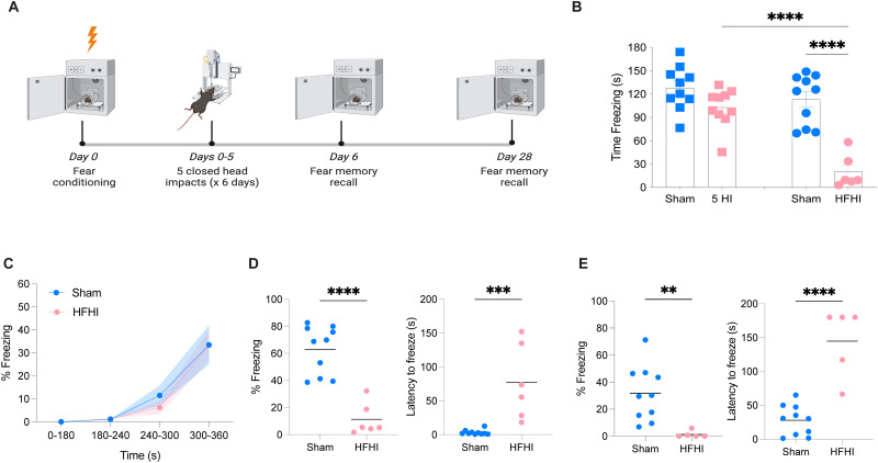
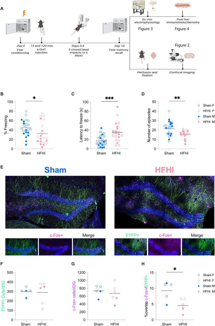
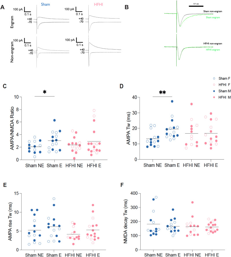
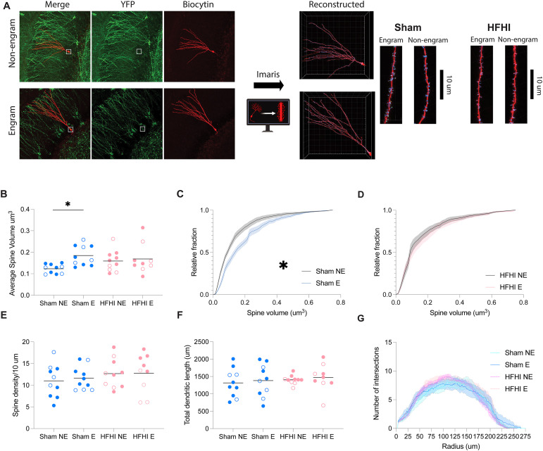
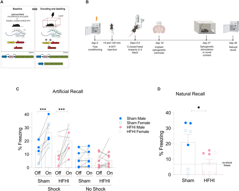
Subconcussive head impacts are associated with the development of acute and chronic cognitive deficits. We recently reported that high-frequency head impact (HFHI) causes chronic cognitive deficits in mice through synaptic changes. To better understand the mechanisms underlying HFHI-induced memory decline, we used TRAP2/Ai32 transgenic mice to enable visualization and manipulation of memory engrams. We labeled the fear memory engram in male and female mice exposed to an aversive experience and subjected them to sham or HFHI. Upon subsequent exposure to natural memory recall cues, sham, but not HFHI, mice successfully retrieved fearful memories. In sham mice the hippocampal engram neurons exhibited synaptic plasticity, evident in amplified AMPA:NMDA ratio, enhanced AMPA-weighted tau, and increased dendritic spine volume compared with nonengram neurons. In contrast, although HFHI mice retained a comparable number of hippocampal engram neurons, these neurons did not undergo synaptic plasticity. This lack of plasticity coincided with impaired activation of the engram network, leading to retrograde amnesia in HFHI mice. We validated that the memory deficits induced by HFHI stem from synaptic plasticity impairments by artificially activating the engram using optogenetics and found that stimulated memory recall was identical in both sham and HFHI mice. Our work shows that chronic cognitive impairment after HFHI is a result of deficiencies in synaptic plasticity instead of a loss in neuronal infrastructure, and we can reinstate a forgotten memory in the amnestic brain by stimulating the memory engram. Targeting synaptic plasticity may have therapeutic potential for treating memory impairments caused by repeated head impacts.

Keywords: amnesia; brain trauma; concussion; engram; head impact optogenetics.


Conflict of interest statement

The authors declare no competing financial interests.

Figures

Figure 1.
Figure 1.
HFHI mice display chronic retrograde amnesia following contextual fear conditioning. A, Schematic of the experimental design. C57Bl/6 male mice were fear conditioned, exposed to sham or HFHI protocols, and then tested for natural recall on day 6 (24 h after the final impact) and day 28. B, Multiple days of head impact are required to induce retrograde amnesia. A single day of five head impact (5HI) does not cause memory impairments; however, HFHI exposure over 6 d causes a significant decrease in memory recall (ANOVA with Tukey's post hoc test; ****p < 0.001). C, Sham and HFHI mice display similar learning curves, demonstrating that both groups of mice successfully acquired the fear memory prior to the HFHI protocols. D, At day 6, HFHI showed significantly decreased time freezing and increased latency to freeze compared with sham mice. E, At day 28, HFHI showed significantly decreased time freezing and increased latency to freeze. Unpaired t test, **p < 0.01, ***p < 0.001, ****p < 0.0001.
Figure 2.
Figure 2.
HFHI prevents reactivation of the engram without affecting engram size. A, Schematic of the experimental design for TRAP2/Ai32 mice used for the electrophysiology and immunohistochemistry experiments. TRAP2/Ai32 mice were fear conditioned, injected with 4-OHT, and randomly selected for HFHI or sham protocols. At day 10 a natural recall test was performed, and mice were prepared for ex vivo electrophysiology or perfused and fixed for IHC. B–D, HFHI mice display a retrograde amnesia phenotype compared with sham mice, as measured by percent freezing, latency to freeze, and the number of freezing episodes. Unpaired t test, *p < 0.05, **p < 0.01, ***p < 0.001. E, Confocal images of the dentate gyrus engram from sham (left) and HFHI (right) mice. green, ChR2-EYFP + neurons that were active at memory acquisition; magenta, c-Fos + neurons that were active at memory recall; blue, DAPI. F, The number of ChR2-EYFP + neurons in the dDG is similar between sham and HFHI mice. G, The number of c-Fos + neurons in the dDG is similar between sham and HFHI mice. H, The percent of overlapping c-Fos/ChR2-EYFP neurons is significantly reduced in HFHI mice compared with sham controls. Unpaired t test, *p < 0.05. Closed circles represent male animals and open circles represent female animals.
Figure 3.
Figure 3.
Engram-specific excitatory synaptic changes are abolished after HFHI. A, Example traces from sham (left) and HFHI (right) engram (top) and nonengram (bottom) cell AMPA/NMDA ratios. B, Normalized trace of AMPA current shows slowed decay in sham engram neurons compared with sham nonengram neurons. Sham (top) and HFHI (bottom). C, Quantification of the AMPA/NMDA ratio show that sham engram neurons have an increased ratio compared with nonengram neurons. There is no change in the AMPA/NMDA ratio in HFHI engram neurons. D, Quantification of AMPA Tw. Sham engram neurons display an increase in AMPA Tw compared with sham nonengram neurons. HFHI mice display no engram specific changes to AMPA Tw. E, No NMDA decay time changes occurred in neurons in either sham or HFHI mice. F, No AMPA rise time changes occurred in engram neurons in either sham or HFHI mice. Unpaired t test, *p < 0.05, **p < 0.01. Closed circles represent male animals and open circles represent female animals. Black circles represent mean ± SEM.
Figure 4.
Figure 4.
HFHI abolishes engram-specific increases in spine volume. A, After electrophysiological recordings, nonengram (top) and engram (bottom) neurons were filled with biocytin for post hoc immunohistochemistry. B, Dendritic spines from sham engram neurons had significantly higher spine volume compared with nonengram neurons, but this differentiation did not occur in HFHI mouse brains. Two-way ANOVA with Šídák's multiple-comparisons test, *p < 0.05. C, D, Cumulative distribution plots of dendritic volume reveal the differentiation of engram and nonengram dendritic spines in sham, but not HFHI mice. Mean ± SEM, two-way ANOVA, *p < 0.05. There were no differences in (E) spine density, (F) total dendritic length, or (G) dendritic complexity between engram and nonengram neurons in either sham or HFHI. Closed circles represent male animals and open circles represent female animals.
Figure 5.
Figure 5.
Amnesia after HFHI is recovered by activating the DG engram. A, Diagram for transgenic engram labeling strategy using TRAP2/Ai32 mice. B, Schematic of the experimental design. TRAP2/Ai32 mice were fear conditioned and engram neurons labeled with 4-OHT. Following sham or HFHI procedures, bilateral hippocampal cannulae were surgically implanted and mice were allowed to recover for 2 weeks before being tested for optogenetic stimulation in a novel context and natural recall. C, For optogenetic stimulation of the memory engram, mice were placed in novel Context B and the engram stimulated with nested theta/gamma stimulation of the dentate gyrus. Percent freeze was quantified for a 3 min light off epoch and a 3 min light on epoch. Optogenetic stimulation elicited a similar increase in the freezing in both sham and HFHI mice. This response is absent in no-shock (NS) controls. Three-way repeated measures ANOVA with Šídák's multiple-comparisons test, ***p < 0.001. Closed circles represent male animals and open circles represent female animals. D, Mice were tested for natural recall at day 30. Quantification of the percent time freezing demonstrate that HFHI animals have retrograde amnesia compared with sham mice. Dotted line represents average no-shock freezing time in Context A. Unpaired t test, *p < 0.05.

Similar articles

Cited by

References

    1. Allen WE, DeNardo LA, Chen MZ, Liu CD, Loh KM, Fenno LE, Ramakrishnan C, Deisseroth K, Luo L (2017) Thirst-associated preoptic neurons encode an aversive motivational drive. Science 357:1149–1155. 10.1126/science.aan6747 - DOI - PMC - PubMed
    1. Bernier BE, Lacagnina AF, Ayoub A, Shue F, Zemelman BV, Krasne FB, Drew MR (2017) Dentate gyrus contributes to retrieval as well as encoding: evidence from context fear conditioning, recall, and extinction. J Neurosci 37:6359–6371. 10.1523/JNEUROSCI.3029-16.2017 - DOI - PMC - PubMed
    1. Bolsius YG, Heckman PRA, Paraciani C, Wilhelm S, Raven F, Meijer EL, Kas MJH, Ramirez S, Meerlo P, Havekes R (2023) Recovering object–location memories after sleep deprivation-induced amnesia. Curr Biol 33:298–308.e5. 10.1016/j.cub.2022.12.006 - DOI - PubMed
    1. Chater TE, Goda Y (2014) The role of AMPA receptors in postsynaptic mechanisms of synaptic plasticity. Front Cell Neurosci 8:401. 10.3389/fncel.2014.00401 - DOI - PMC - PubMed
    1. Chen X, Zhang KL, Yang SY, Dong JF, Zhang JN (2009) Glucocorticoids aggravate retrograde memory deficiency associated with traumatic brain injury in rats. J Neurotrauma 26:253–260. 10.1089/neu.2007.0504 - DOI - PubMed

Publication types

Substances

LinkOut - more resources