Review
. 2024 Jun;77(3):289-305.
doi: 10.4097/kja.23843. Epub 2024 Jan 17.

Intraoperative pediatric electroencephalography monitoring: an updated review

Affiliations
Review

Intraoperative pediatric electroencephalography monitoring: an updated review

Ian Yuan et al. Korean J Anesthesiol. 2024 Jun.

Abstract

Intraoperative electroencephalography (EEG) monitoring under pediatric anesthesia has begun to attract increasing interest, driven by the availability of pediatric-specific EEG monitors and the realization that traditional dosing methods based on patient movement or changes in hemodynamic response often lead to imprecise dosing, especially in younger infants who may experience adverse events (e.g., hypotension) due to excess anesthesia. EEG directly measures the effects of anesthetics on the brain, which is the target end-organ responsible for inducing loss of consciousness. Over the past ten years, research on anesthesia and computational neuroscience has improved our understanding of intraoperative pediatric EEG monitoring and expanded the utility of EEG in clinical practice. We now have better insights into neurodevelopmental changes in the developing pediatric brain, functional connectivity, the use of non-proprietary EEG parameters to guide anesthetic dosing, epileptiform EEG changes during induction, EEG changes from spinal/regional anesthesia, EEG discontinuity, and the use of EEG to improve clinical outcomes. This review article summarizes the recent literature on EEG monitoring in perioperative pediatric anesthesia, highlighting several of the topics mentioned above.

Keywords: Anesthesia; EEG; Electroencephalogram; Electroencephalography; Pediatric anesthesia; Pediatric..


Conflict of interest statement

Conflicts of Interest

No potential conflict of interest relevant to this article was reported.

Figures

Fig. 1.
Fig. 1.
EEG parameters on a commercial EEG monitor. (A) EEG waveforms shows real-time 4-channel recordings from the left and right sides of the forehead, (B) percentage of EMG interference as number and graphic recordings, (C) PSI, a numeric proprietary index used by Masimo Inc., (D) percentage of BSR, (E) percentage of artifact, (F) SEF95 from left and right sides of the forehead, and (G) DSA showing left (top) and right (bottom) of the forehead (white line indicates the spectral edge frequency SEF95). This figure was adopted with permission [7]). EEG: electroencephalography, EMG: electromyography, PSI: patient state index, BSR: burst suppression ratio, SEF: spectral edge frequency, DSA: density spectral array.
Fig. 2.
Fig. 2.
Time domain trace (raw EEG waveform) on Sedline monitor showing burst suppression. Burst suppression is recognized on EEG as cycles of short periods of flat (isoelectric) activity (A) and short periods of high-amplitude activity (bursts) (B), switching back and forth every few seconds. Burst suppression reflects a profound state of brain inactivation during deep anesthesia and indicates a state of over-sedation. EEG: electroencephalography.
Fig. 3.
Fig. 3.
Screenshot of Sedline monitor indicating oversedation. (A) Insertion of the laryngeal mask airway after induction, (B) progressive increase in SEF95 from 8–12 Hz, indicating a decrease in hypnotic levels in response to decreasing sevoflurane concentrations, (C) a bolus of propofol was administered to treat laryngospasm caused by inadequate hypnotic levels in response to a change in stimulation, which led to burst suppression (indicated by the striated black lines between 08:59 and 09:01), and (D) patient is out of bust suppression and SEF95 gradually increased to approximately 10 Hz, indicating a return to a decreased hypnotic level. SEF: spectral edge frequency.
Fig. 4.
Fig. 4.
Combination of three serial screenshots of Sedline monitor indicating undersedation. A combination of three serial screenshots of a 7-year-old girl with cyanotic heart disease undergoing diagnostic cardiac catheterization. (A) The patient is hemodynamically stable with a sevoflurane concentration of 2.0%, (B) cannulation of the femoral vessels: SEF95 increased from 14–18 Hz with a decrease in slow/delta power, indicating a decrease in the hypnotic level, (C) sevoflurane increased to 2.2%, SEF95 decreased to 15 Hz with an increase in slow/delta power, indicating an increase in hypnotic depth, (D) sevoflurane concentration was reduced to 1.8% in response to decreasing blood pressure, (E) sudden severe hypotension and bradycardia, leading to EEG burst suppression, (F) EEG activity returned: SEF95 is 24 Hz with low alpha power and minimal slow/delta power, indicating arousal, and (G) blood pressure stabilized, allowing for the sevoflurane concentration to increase to 1.8%, SEF decreased to 19 Hz with increased slow/delta power, indicating an increase in hypnotic depth. EEG: electroencephalography, SEF: spectral edge frequency.
Fig. 5.
Fig. 5.
Combination of three serial Sedline screenshots of in a 4-month-old infant. Combination of three serial screenshots of a 4-month-old receiving sevoflurane anesthesia. (A) eSevo 2.4%, EEG waveform shows predominantly slow/delta oscillations, DSA shows high slow/delta power, SEF95 is 7–9 Hz, (B) surgical incision, eSevo 2.7%, minimal change in EEG, heart rate, and blood pressure, (C) pneumoperitoneum, eSevo 2.05%, EEG waveform showing higher frequency oscillations, SEF95 increased to 16 Hz and slow/delta power decreased on the DSA, indicating arousal, (D) eSevo 3%, SEF95 decreased to 10 Hz, DSA shows increased delta power, indicating an increased hypnotic level, (E) sevoflurane stopped, progressive increase in SEF95 and loss of delta power, and (F) patient extubated awake. EEG: electroencephalography, SEF: spectral edge frequency, DSA: density spectral array.
Fig. 6.
Fig. 6.
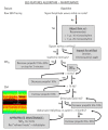
EEG-guided propofol TIVA dosing algorithm for anesthetic maintenance. This decision-making flowchart was provided to learners as a visual representation of the teaching points to help guide clinicians through EEG interpretation. The algorithm focuses on using raw EEG, SEF, and DSA to assess propofol Ce and titrate the propofol dose to the desired Ce to ensure an appropriate depth of hypnosis during the maintenance of general anesthesia (Adopted with permission [44]). EEG: electroencephalography, TIVA: total intravenous anesthesia, SEF: spectral edge frequency, DSA: density spectral array, propofol Ce: propofol concentration.

References

    1. Lee U, Mashour GA. Role of network science in the study of anesthetic state transitions. Anesthesiology. 2018;129:1029–44. - PMC - PubMed
    1. Purdon PL, Sampson A, Pavone KJ, Brown EN. Clinical electroencephalography for anesthesiologists: Part I: background and basic signatures. Anesthesiology. 2015;123:937–60. - PMC - PubMed
    1. Chan MT, Hedrick TL, Egan TD, García PS, Koch S, Purdon PL, et al. American Society for Enhanced Recovery and Perioperative Quality Initiative Joint Consensus Statement on the role of neuromonitoring in perioperative outcomes: electroencephalography. Anesth Analg. 2020;130:1278–91. - PubMed
    1. Nimmo AF, Absalom AR, Bagshaw O, Biswas A, Cook TM, Costello A, et al. Guidelines for the safe practice of total intravenous anaesthesia (TIVA): Joint Guidelines from the Association of Anaesthetists and the Society for Intravenous Anaesthesia. Anaesthesia. 2019;74:211–24. - PubMed
    1. Disma N, Veyckemans F, Virag K, Hansen TG, Becke K, Harlet P, et al. Morbidity and mortality after anaesthesia in early life: results of the European prospective multicentre observational study, neonate and children audit of anaesthesia practice in Europe (NECTARINE) Br J Anaesth. 2021;126:1157–72. - PubMed

Publication types

LinkOut - more resources