. 2024 Jan 16;14(1):1448.
doi: 10.1038/s41598-024-51945-4.

Unveiling breast cancer metastasis through an advanced X-ray imaging approach

Affiliations

Unveiling breast cancer metastasis through an advanced X-ray imaging approach

Andre L C Conceição et al. Sci Rep. .

Abstract

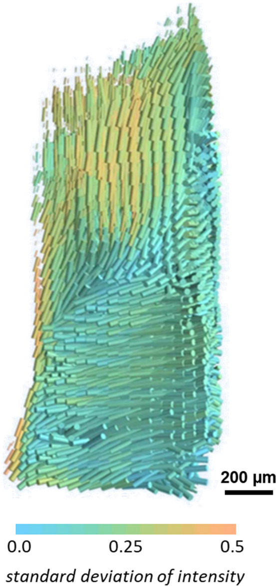
Breast cancer is a significant global health burden, causing a substantial number of deaths. Systemic metastatic tumour cell dissemination is a major cause of poor outcomes. Understanding the mechanisms underlying metastasis is crucial for effective interventions. Changes in the extracellular matrix play a pivotal role in breast cancer metastasis. In this work, we present an advanced multimodal X-ray computed tomography, by combining Small-angle X-ray Scattering Tensor Tomography (SAXS-TT) and X-ray Fluorescence Computed Tomography (XRF-CT). This approach likely brings out valuable information about the breast cancer metastasis cascade. Initial results from its application on a breast cancer specimen reveal the collective influence of key molecules in the metastatic mechanism, identifying a strong correlation between zinc accumulation (associated with matrix metalloproteinases MMPs) and highly oriented collagen. MMPs trigger collagen alignment, facilitating breast cancer cell intravasation, while iron accumulation, linked to angiogenesis and vascular endothelial growth factor VEGF, supports cell proliferation and metastasis. Therefore, these findings highlight the potential of the advanced multimodal X-ray computed tomography approach and pave the way for in-depth investigation of breast cancer metastasis, which may guide the development of novel therapeutic approaches and enable personalised treatment strategies, ultimately improving patient outcomes in breast cancer management.


Conflict of interest statement

The authors declare no competing interests.

Figures

Figure 1
Figure 1
SAXS-TT Reconstructed orientation map of the collagen fibrils. 3D representation of the spatially resolved direction of the collagen anisotropy based on the SAXS evaluation at q = 0.48 nm−1. The direction of the cylinders shows the direction of collagen orientation and the color scale the degree of orientation. Lower standard deviation means a weaker degree of orientation while higher standard deviation reflects stronger alignment of the collagen fibers in the voxel. Sample dimension: ~ 1.2 mm diameter and ~ 5.0 mm height.
Figure 2
Figure 2
Multimodal X-ray tomography. (a) Three-dimensional map of the average scattering of the collagen fibrils, reconstructed at the q = 0.48 nm−1. The dashed rectangle indicates the higher scattering intensity region. The green area inside the dashed rectangle was masked out for further maps due to none or very low collagen content; (b) the distribution of the calculated collagen fibril diameter within the region of interest (ROI); (c) the orientation map retrieved from the SAXSTT reconstruction in Fig. 1. The orientation is colour-coded according to the colour wheel inset. Highly oriented collagen fibrils show up with bright colours, whereas dense fibrils without preferential orientation are darker; (d) and (e) normalized distribution of Fe and Zn, respectively. The brown ellipse dashed area in (c), (d) and (e) highlights a potential metastatic site.
Figure 3
Figure 3
X-ray multimodal computed tomography. (a) sketch of the multimodal setup showing the motion system with all the degrees of freedom required for the SAXS Tensor Tomography technique and the coordinate system. The energy dispersive detector for XRF is positioned as close as possible to the sample perpendicularly to the incoming X-ray beam. Finally, the SAXS detector is mounted downstream to the beam at a 5.08 m distance from the sample. A 6.0 mm diameter active beamstop is used to protect the detector of the primary beam as well as to record the transmitted intensity. reconstructed transmission image; (b) Photo of the top view of the interior of the chamber filled up with He, presenting the dedicated sample environment for scanning-based tomography at the SAXSMAT beamline at the Deutsches Elektronen-Synchrotron DESY, Hamburg, Germany.

References

    1. Sung H, et al. Global cancer statistics 2020: GLOBOCAN estimates of incidence and mortality worldwide for 36 cancers in 185 countries. CA. Cancer J. Clin. 2021;71:209–249. doi: 10.3322/caac.21660. - DOI - PubMed
    1. Chaffer CL, Weinberg RA. A perspective on cancer cell metastasis. Science. 2011;331:1559–1564. doi: 10.1126/science.1203543. - DOI - PubMed
    1. Wang, L., Zhang, S. & Wang, X. The metabolic mechanisms of breast cancer metastasis. 10.3389/fonc.2020.602416. - PMC - PubMed
    1. Fidler IJ. The pathogenesis of cancer metastasis: The ‘seed and soil’ hypothesis revisited. Nat. Rev. Cancer. 2003;3:453–458. doi: 10.1038/nrc1098. - DOI - PubMed
    1. Pupa SM, Ménard S, Forti S, Tagliabue E. New insights into the role of extracellular matrix during tumor onset and progression. J. Cell. Physiol. 2002;192:259–267. doi: 10.1002/jcp.10142. - DOI - PubMed

Substances