Review
. 2024 Oct;39(5):270-290.
doi: 10.1111/omi.12450. Epub 2024 Jan 16.

Tobacco-enhanced biofilm formation by Porphyromonas gingivalis and other oral microbes

Affiliations
Review

Tobacco-enhanced biofilm formation by Porphyromonas gingivalis and other oral microbes

Jinlian Tan et al. Mol Oral Microbiol. 2024 Oct.

Abstract

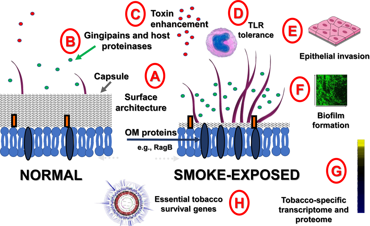
Microbial biofilms promote pathogenesis by disguising antigens, facilitating immune evasion, providing protection against antibiotics and other antimicrobials and, generally, fostering survival and persistence. Environmental fluxes are known to influence biofilm formation and composition, with recent data suggesting that tobacco and tobacco-derived stimuli are particularly important mediators of biofilm initiation and development in vitro and determinants of polymicrobial communities in vivo. The evidence for tobacco-augmented biofilm formation by oral bacteria, tobacco-induced oral dysbiosis, tobacco-resistance strategies, and bacterial physiology is summarized herein. A general overview is provided alongside specific insights gained through studies of the model and archetypal, anaerobic, Gram-negative oral pathobiont, Porphyromonas gingivalis.

Keywords: biofilms; dysbiosis; periodontitis; tobacco.


Figures

Figure 1:
Figure 1:. The influence of cigarette smoke on P. gingivalis physiology and resultant host interactions.
(A) The surface architecture of P. gingivalis is altered upon smoke-exposure. Expression of several outer membrane proteins, including RagA and RagB is upregulated under smoke-induced stress (Bagaitkaret al 2009b; Cogoet al 2012). Concomitantly, the capsule production is down-regulated, further increasing the bioavailability of surface exposed molecules (Bagaitkaret al 2010; Bagaitkaret al 2009b). (B) CSE also induces gingipain release while gingipains can degrade the primary endogenous MMP suppressor, TIMP (Bondy-Careyet al 2013). (C) Early studies suggested that nicotine exposure enhanced P. gingivalis virulence, by unknown mechanisms, in a chick embryo lethality set-up (Sayerset al 1997; Sayerset al 1999). (D) Nicotinic engagement of the cholinergic anti-inflammatory pathway; reduced pro-inflammatory potential of smoke-exposed P. gingivalis cells; and FimA-induced TLR tolerance each contribute to a tobacco-associated reduction in the innate immune response (Bagaitkaret al 2011b; Bagaitkaret al 2010; Bagaitkaret al 2009b). (E) At the same time, tobacco, or smoke components, assist in epithelial cell association with and/or invasion of epithelial cells by P. gingivalis (Cogoet al 2009; Imamuraet al 2015). (F) CSE augments biofilm formation by P. gingivalis alone and in consort with S. gordonii (Bagaitkaret al 2011b; Bagaitkaret al 2010; Bagaitkaret al 2009b). (G) A distinct tobacco-related transcriptome and proteome has been noted for P. gingivalis (Bagaitkaret al 2009b; Cogoet al 2012) while, (H) recent studies are providiing insight into the specific genes that are conditionally essential for P. gingivalis survival in a tobacco-rich niche (Hutchersonet al 2015b; Hutchersonet al 2020;Miller and Scott 2021). Further details of (A) through (H) are provided in the main text. Figure 1 is updated and adapted from a figure originally published in (Hutchersonet al 2015c) under a creative commons license (https://creativecommons.org/licenses/by/4.0/).
Figure 2:
Figure 2:. Cigarette smoke components augment P. gingivalis epithelial invasion and biofilm formation but suppress pro-inflammatory efficacy CSC exposure augments epithelial invasion
(A, B): Invasion of (A) untreated (solvent control D only) or (B) CSC‐treated (1 μg/mL) Ca9‐22 epithelial cells by P. gingivalis, as visualized by confocal scanning laser microscopy. P. gingivalis 33277 invading Ca9‐22 cells were stained red, while extracellular bacteria were detected as green–yellow. The host cell cytoskeletons stained with phalloidin appear blue. An increase in the number of intracellular bacteria was observed in CSC‐treated cells Bar: 20 μm. Reproduced from (Imamuraet al 2015), with permission. CSE exposure augments P. gingivalis - S. gordonii biofilm formation (C, D): The total number (C) and height of microcolonies (D) formed between S. gordonii and P. gingivalis under CSE-exposed (black bars) or unexposed (white bars) conditions, as examined by confocal microscopy and quantified from 6 randomly chosen microscopic fields per biofilm, as described in (Bagaitkaret al 2011a). An 80 amino acid sequence (SspB Adherence Region, BAR) on the SspB protein of S. gordonii is critical for interactions between this streptococci and the minor fimbriae of P. gingivalis (Demuth et al 2001). Pretreatment with a synthetic BAR peptide, but not the inverse construct, abrogated S. gordonii and P. gingivalis associations. Figure 2C,D is reproduced from (Bagaitkaret al 2011a) under a creative commons license (https://creativecommons.org/licenses/by/4.0/). CSE suppresses pro-inflammatory efficacy of P. gingivalis (E): Reduced TNF release from human monocytes stimulated with CSE-treated P. gingivalis is apparent. Primary human monocytes (0.5 × 10⁶) were stimulated with 106-109 cells of control P. gingivalis (black bars) or P. gingivalis cells grown in medium conditioned with cigarette smoke extract (white bars). 20 h Cell-free supernatants were harvested by centrifugation and levels of TNF determined by ELISA. Error bars represent the mean (SD) of three experiments. Reproduced from (Bagaitkaret al 2009b), with permission. ** p < 0.01.

References

    1. (2014) In: The Health Consequences of Smoking-50 Years of Progress: A Report of the Surgeon General(ed.)^(eds). Atlanta (GA).
    1. Abdelmalek SMA, Alhadad S, Abu-Omar O, Afaneh M, Abu-Qatouseh L, and Collier PJ (2022) The effect of cigarette smoke condensate (CSC) on Pseudomonas aeruginosa virulence and antibiotic sensitivity. J Appl Microbiol 132: 3951–3958. - PubMed
    1. Abreu MGL, Kawamoto D, Mayer MPA, Pascoal VDB, Caiaffa KS, Zuza EP, Duque C, and Camargo G (2019) Frequency of Porphyromonas gingivalis fimA in smokers and nonsmokers after periodontal therapy. J Appl Oral Sci 27: e20180205. - PMC - PubMed
    1. Adamowicz K, Wang H, Jotwani R, Zeller I, Potempa J, and Scott D (2012) Inhibition of GSK3 abolishes bacterial-induced periodontal bone loss in mice. Mol Med: 1190–1196. - PMC - PubMed
    1. Alawadhi NB, Lippert F, and Gregory RL (2020) Effects of casein phosphopeptide-amorphous calcium phosphate creme on nicotine-induced Streptococcus mutans biofilm in vitro. Clin Oral Investig 24: 3513–3518. - PubMed

LinkOut - more resources