. 2024 Jan 16;12(1):10.
doi: 10.1186/s40478-023-01701-y.

MANF protein expression is upregulated in immune cells in the ischemic human brain and systemic recombinant MANF delivery in rat ischemic stroke model demonstrates anti-inflammatory effects

Affiliations

MANF protein expression is upregulated in immune cells in the ischemic human brain and systemic recombinant MANF delivery in rat ischemic stroke model demonstrates anti-inflammatory effects

Jenni E Anttila et al. Acta Neuropathol Commun. .

Abstract

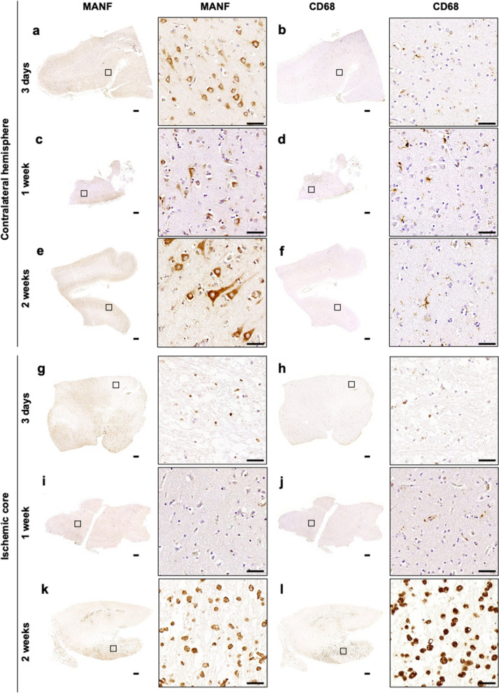
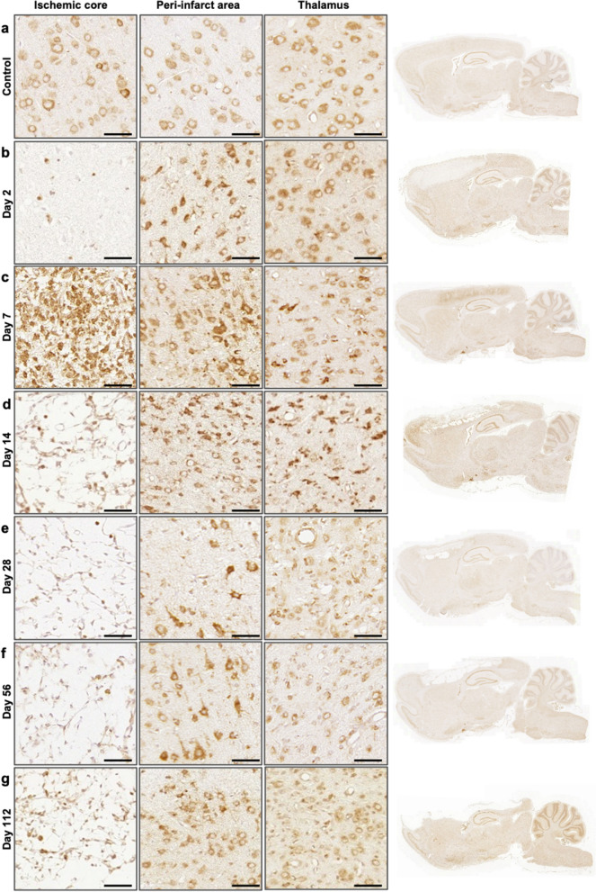
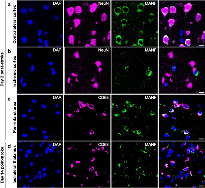
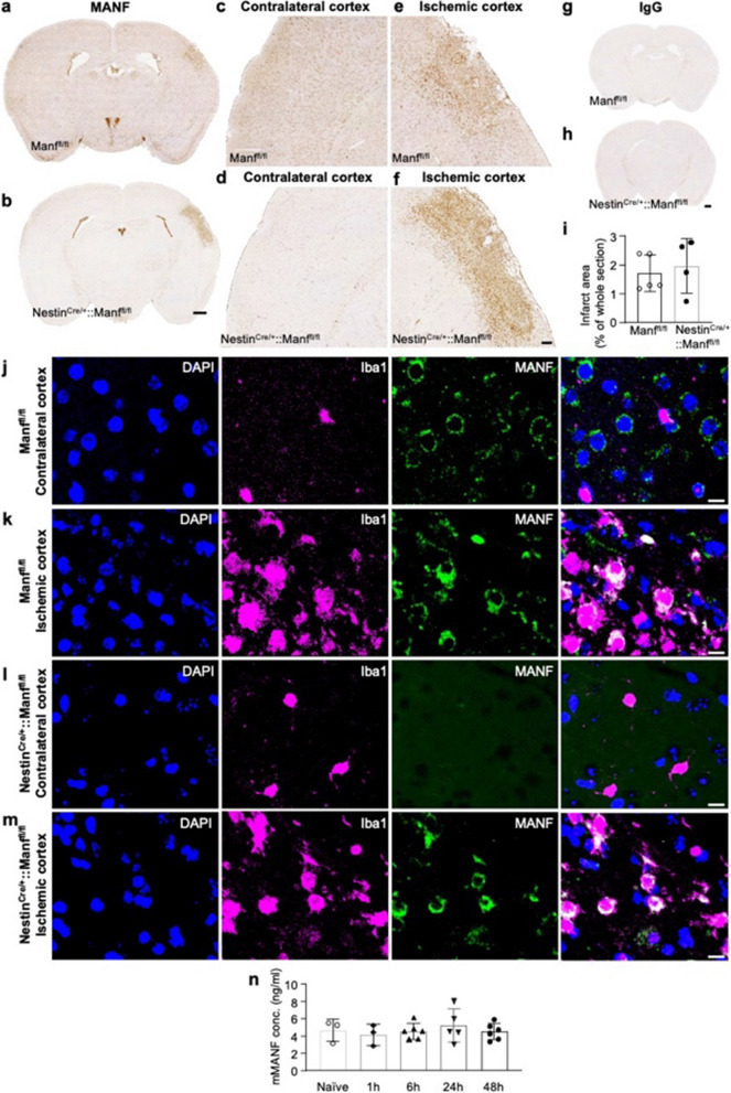
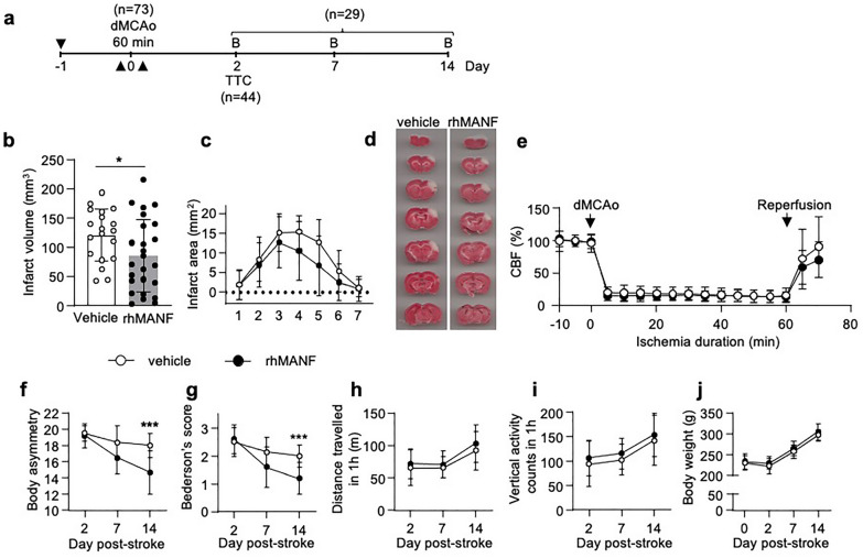
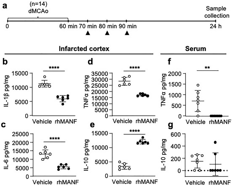
Mesencephalic astrocyte-derived neurotrophic factor (MANF) has cytoprotective effects on various injuries, including cerebral ischemia, and it can promote recovery even when delivered intracranially several days after ischemic stroke. In the uninjured rodent brain, MANF protein is expressed almost exclusively in neurons, but post-ischemic MANF expression has not been characterized. We aimed to investigate how endogenous cerebral MANF protein expression evolves in infarcted human brains and rodent ischemic stroke models. During infarct progression, the cerebral MANF expression pattern both in human and rat brains shifted drastically from neurons to expression in inflammatory cells. Intense MANF immunoreactivity took place in phagocytic microglia/macrophages in the ischemic territory, peaking at two weeks post-stroke in human and one-week post-stroke in rat ischemic cortex. Using double immunofluorescence and mice lacking MANF gene and protein from neuronal stem cells, neurons, astrocytes, and oligodendrocytes, we verified that MANF expression was induced in microglia/macrophage cells in the ischemic hemisphere. Embarking on the drastic expression transition towards inflammatory cells and the impact of blood-borne inflammation in stroke, we hypothesized that exogenously delivered MANF protein can modulate tissue recovery processes. In an attempt to enhance recovery, we designed a set of proof-of-concept studies using systemic delivery of recombinant MANF in a rat model of cortical ischemic stroke. Intranasal recombinant MANF treatment decreased infarct volume and reduced the severity of neurological deficits. Intravenous recombinant MANF treatment decreased the levels of pro-inflammatory cytokines and increased the levels of anti-inflammatory cytokine IL-10 in the infarcted cortex one-day post-stroke. In conclusion, MANF protein expression is induced in activated microglia/macrophage cells in infarcted human and rodent brains, and this could implicate MANF's involvement in the regulation of post-stroke inflammation in patients and experimental animals. Moreover, systemic delivery of recombinant MANF shows promising immunomodulatory effects and therapeutic potential in experimental ischemic stroke.

Keywords: Distal middle cerebral artery occlusion; Inflammation; Ischemia; Mesencephalic astrocyte-derived neurotrophic factor; Neuroprotection.


Conflict of interest statement

The authors declare there are no conflicts of interest.

Figures

Fig. 1
Fig. 1
MANF protein expression correlates with CD68 expression at 2 weeks after ischemic stroke in the human brain. Anti-MANF and anti-CD68 immunostaining of human cerebral tissue from ischemic stroke patients at 3d, 1wk and 2wk post-stroke. The patient and sample details are shown in Table 2. a-f Contralateral hemisphere of the ischemic brain (a, b Patient 1, frontal region; c, d Patient 4, frontal region; e, f Patient 7, parietal region) g-l The ischemic core (g, h Patient 1, pons; i, j Patient 4, frontal region; k, l Patient 7, parietal region. Scale bar is 1000 µm and 50 µm. Location of high magnification images indicated in the whole section images
Fig. 2
Fig. 2
Temporal and spatial MANF protein expression in the rat brain post-stroke. Representative images of anti-MANF immunostaining from ischemic core in the cortex, peri-infarct area, thalamus, and the whole brain sagittal sections at 2 (b), 7 (c), 14 (d), 28 (e), 56 (f), and 112 (g) days after 90-min dMCAO in rat. Control images (a) are from the contralateral hemisphere of the stroke brain. Scale bar is 50 µm
Fig. 3
Fig. 3
MANF is expressed in CD68 + cells in the ischemic cortex and ipsilateral thalamus. Double immunofluorescence staining of rat brain paraffin sections 2 (a, b, c) and 14 (d) days after 90-min dMCAo. NeuN and MANF colocalize in the contralateral (a) but not in the ischemic cortex (b). CD68 and MANF colocalize in the peri-infarct area (c) and in the ipsilateral thalamus (d). Scale bar is 10 µm
Fig. 4
Fig. 4
MANF protein expression is induced in Iba1 + cells of the NestinCre/+::Manffl/fl knockout mouse brain after ischemic stroke. Anti-MANF immunostaining of Manffl/fl control (a, c, e) and NestinCre/+::Manffl/fl knockout (b, d, f) mouse brain at 14d after permanent dMCAo. g-h Anti-rabbit IgG immunostaining of the same brain as in a, b to show the specificity of the rabbit anti-MANF antibody. i Average infarct area at 14d after permanent dMCAo in Manffl/fl wild type and NestinCre/+::Manffl/fl knockout mouse. a, b, g, h: scale bar is 500 µm. c-f: scale bar is 100 µm. j-m Anti-MANF and anti-Iba1 double immunofluorescence staining of the peri-infarct cortex of Manffl/fl control (j, k) and NestinCre/+::Manffl/fl knockout (l, m) mice at 14d after permanent dMCAo. Scale bar is 10 µm. n Endogenous MANF serum levels are not altered after permanent cerebral ischemia. Endogenous MANF levels were measured from wild type mouse serum with mouse MANF ELISA at different time points after permanent dMCAo, n = 3–6 per group. The values are expressed as mean ± SD. dMCAo = distal middle cerebral artery occlusion
Fig. 5
Fig. 5
Pretreatment with intranasally delivered recombinant human MANF decreases infarct volume and promotes behavioral recovery after stroke. a Timeline of the experiment. The black arrows indicate the intranasal administration of rhMANF or vehicle. RhMANF or PBS was administered to the rats 3 times: 12 h before a 60-min dMCAo, immediately before dMCAo and immediately after reperfusion. The total MANF dose was 20 µg or 60 µg. dMCAo = distal middle cerebral artery occlusion, B = behavioral assay, TTC = 2,3,5-triphenyltetrazolium chloride staining. b Infarction volume was determined 2 days after dMCAo by TTC staining, n = 19–25 per group, (*p < 0.05), Student’s t-test. c Distribution of infarction along the rostrocaudal axis, n = 19–25 per group. d Representative images of TTC stained brain sections from the vehicle and rhMANF groups. e RhMANF treatment had no effect on cortical cerebral blood flow (CBF). CBF was measured with laser Doppler flowmetry before ischemia, during ischemia (dMCAo) and after reperfusion. Rats received intranasal rhMANF (n = 10) or vehicle (n = 9) immediately before CBF measurement. f Body asymmetry test, g Bederson’s score, h horizontal activity, i vertical activity, and j body weight at different time points after 60-min dMCAo in vehicle (n = 14) and rhMANF (n = 15) treated rats. The total MANF dose was 20 µg. In fg (***p < 0.001) indicate comparison between vehicle and rhMANF groups with Mann–Whitney U test, corrected for multiple comparisons. The values are expressed as mean ± SD
Fig. 6
Fig. 6
Post-reperfusion treatment with intravenous recombinant human MANF decreases infarct volume. a Timeline of the experiment. The black arrow indicates intravenous administration of rhMANF or vehicle. RhMANF (1.5 µg or saline was administered to the rats 15 min after reperfusion. dMCAo = distal middle cerebral artery occlusion, TTC = 2,3,5-triphenyltetrazolium chloride staining. b Infarction volume was determined 2 days after dMCAo by TTC staining, n = 16 per group, (**p < 0.01), Student’s t-test. c Distribution of infarction along the rostrocaudal axis.d Representative images of TTC stained brain sections from vehicle and rhMANF groups. The values are expressed as mean ± SD
Fig. 7
Fig. 7
Intravenous administration of recombinant human MANF alters cytokine levels in the brain and blood after ischemic stroke. a Timeline of the experiment. Vehicle (saline) or rhMANF (1.5 µg × 3) was administered intravenously (i.v.) to rats that had undergone a 60-min distal middle cerebral artery occlusion. Arrowheads point the time of i.v. administration. After 24 h, the infarcted cortical tissue and serum were collected. be Concentration of IL-1β, IL-6, IL-10, and TNFα in the infarcted cortex 24 h post-dMCAo, normalized to the sample protein concentration (n = 6). (****p < 0.0001), Student’s t-test. f-g Concentration of TNFα and IL-10 in serum 24 h post-dMCAo, normalized to the serum protein concentration (n = 7). (**p < 0.01), Mann–Whitney U test. The values are expressed as mean ± SD

References

    1. Abo-Ramadan U, Durukan A, Pitkonen M, Marinkovic I, Tatlisumak E, Pedrono E, Soinne L, Strbian D, Tatlisumak T. Post-ischemic leakiness of the blood-brain barrier: a quantitative and systematic assessment by Patlak plots. Exp Neurol. 2009;219:328–333. doi: 10.1016/j.expneurol.2009.06.002. - DOI - PubMed
    1. Airavaara M, Chiocco MJ, Howard DB, Zuchowski KL, Peranen J, Liu C, Fang S, Hoffer BJ, Wang Y, Harvey BK. Widespread cortical expression of MANF by AAV serotype 7: localization and protection against ischemic brain injury. Exp Neurol. 2010;225:104–113. doi: 10.1016/j.expneurol.2010.05.020. - DOI - PMC - PubMed
    1. Airavaara M, Shen H, Kuo CC, Peranen J, Saarma M, Hoffer B, Wang Y. Mesencephalic astrocyte-derived neurotrophic factor reduces ischemic brain injury and promotes behavioral recovery in rats. J Comp Neurol. 2009;515:116–124. doi: 10.1002/cne.22039. - DOI - PMC - PubMed
    1. Andrews RJ. Transhemispheric diaschisis. A review and comment. Stroke. 1991;22:943–949. doi: 10.1161/01.str.22.7.943. - DOI - PubMed
    1. Anttila JE, Albert K, Wires ES, Matlik K, Loram LC, Watkins LR, Rice KC, Wang Y, Harvey BK, Airavaara M. Post-stroke intranasal (+)-naloxone delivery reduces microglial activation and improves behavioral recovery from ischemic injury. eNeuro. 2018 doi: 10.1523/ENEURO.0395-17.2018. - DOI - PMC - PubMed

Publication types