. 2023 Dec;299(12):105408.
doi: 10.1016/j.jbc.2023.105408. Epub 2023 Oct 28.

Spleen Tyrosine Kinase phosphorylates VE-cadherin to cause endothelial barrier disruption in acute lung injury

Affiliations

Spleen Tyrosine Kinase phosphorylates VE-cadherin to cause endothelial barrier disruption in acute lung injury

Mohammad Shadab et al. J Biol Chem. 2023 Dec.

Abstract

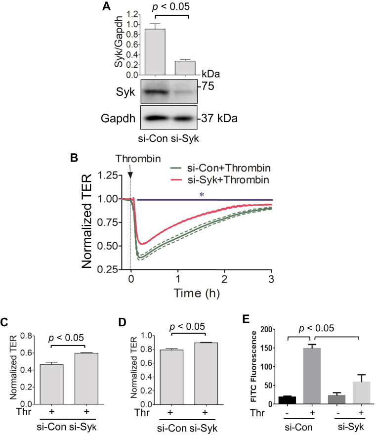
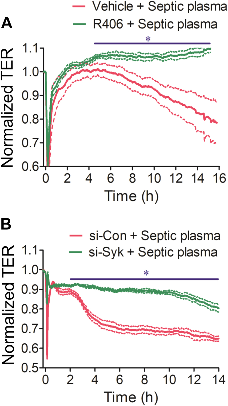
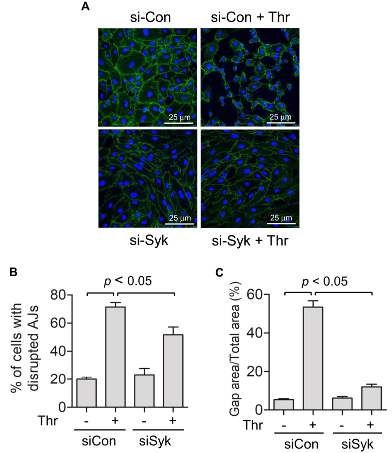
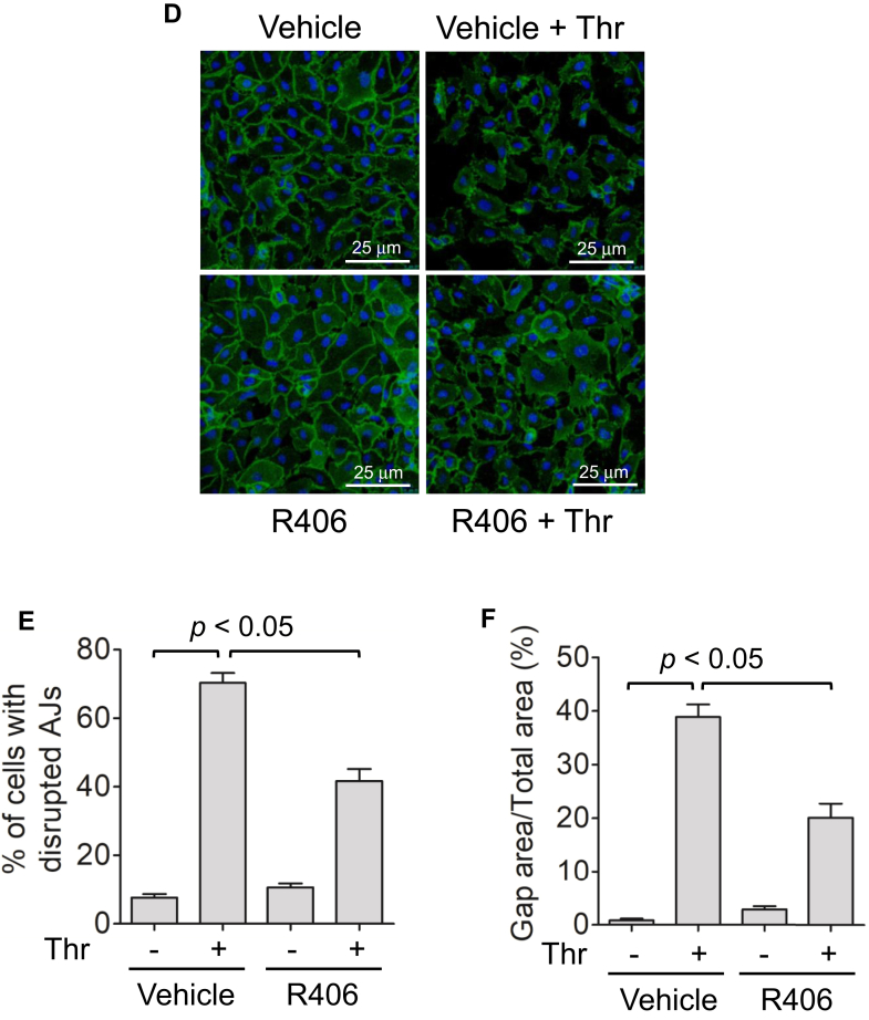
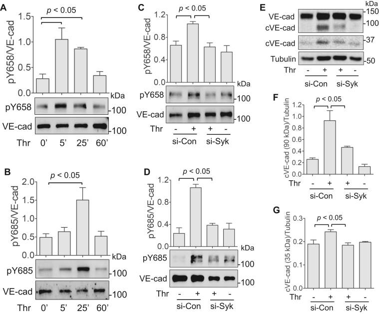
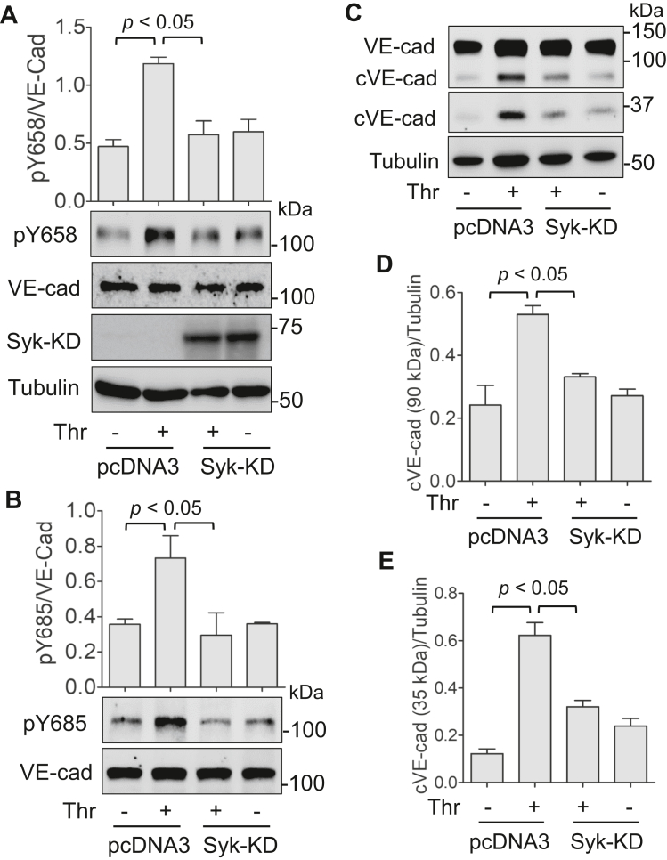
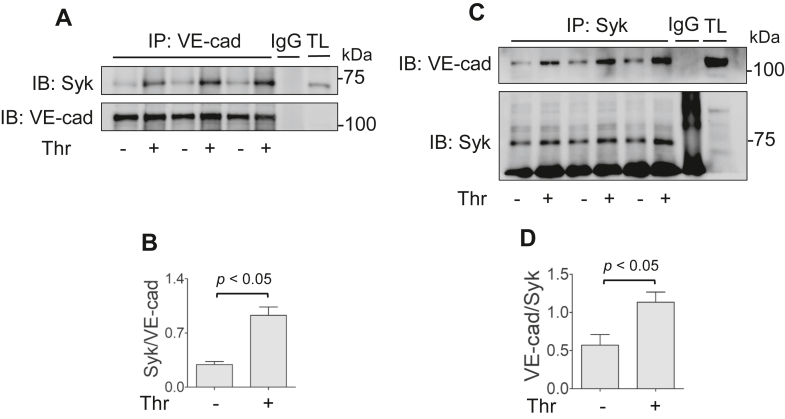
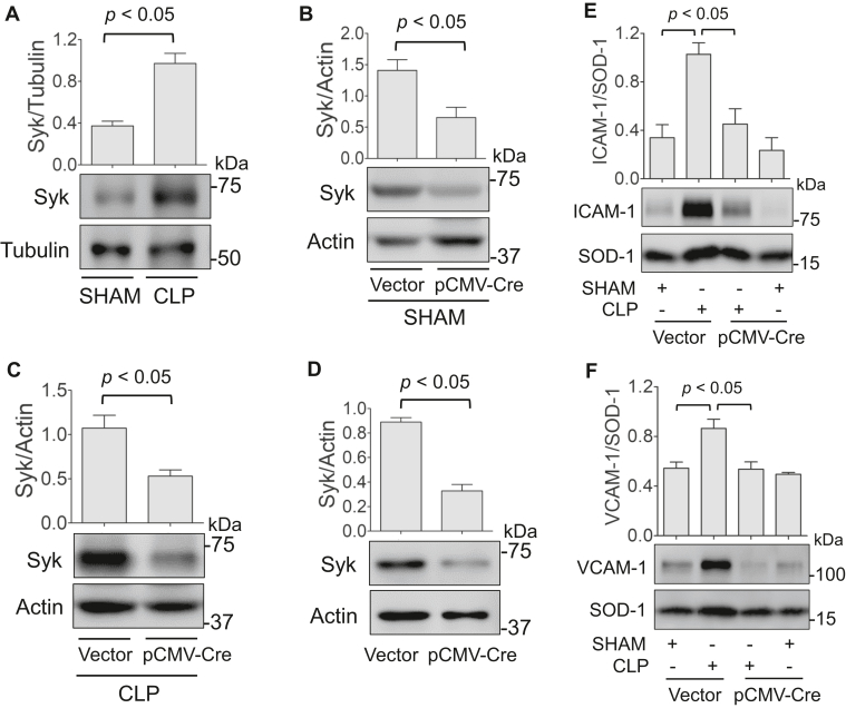
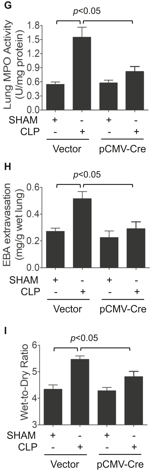
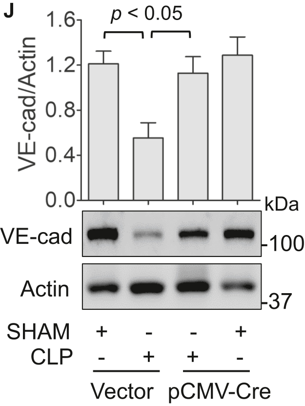
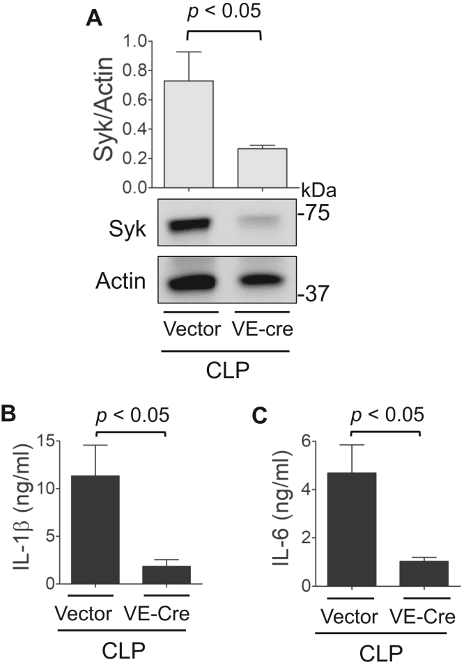
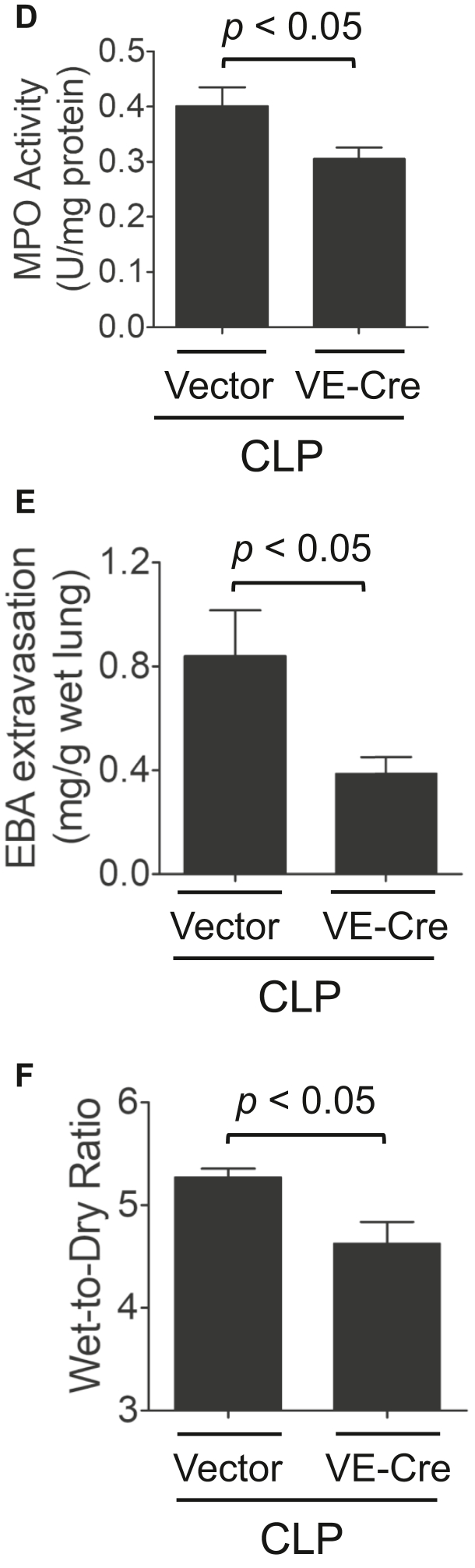
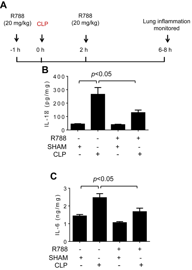
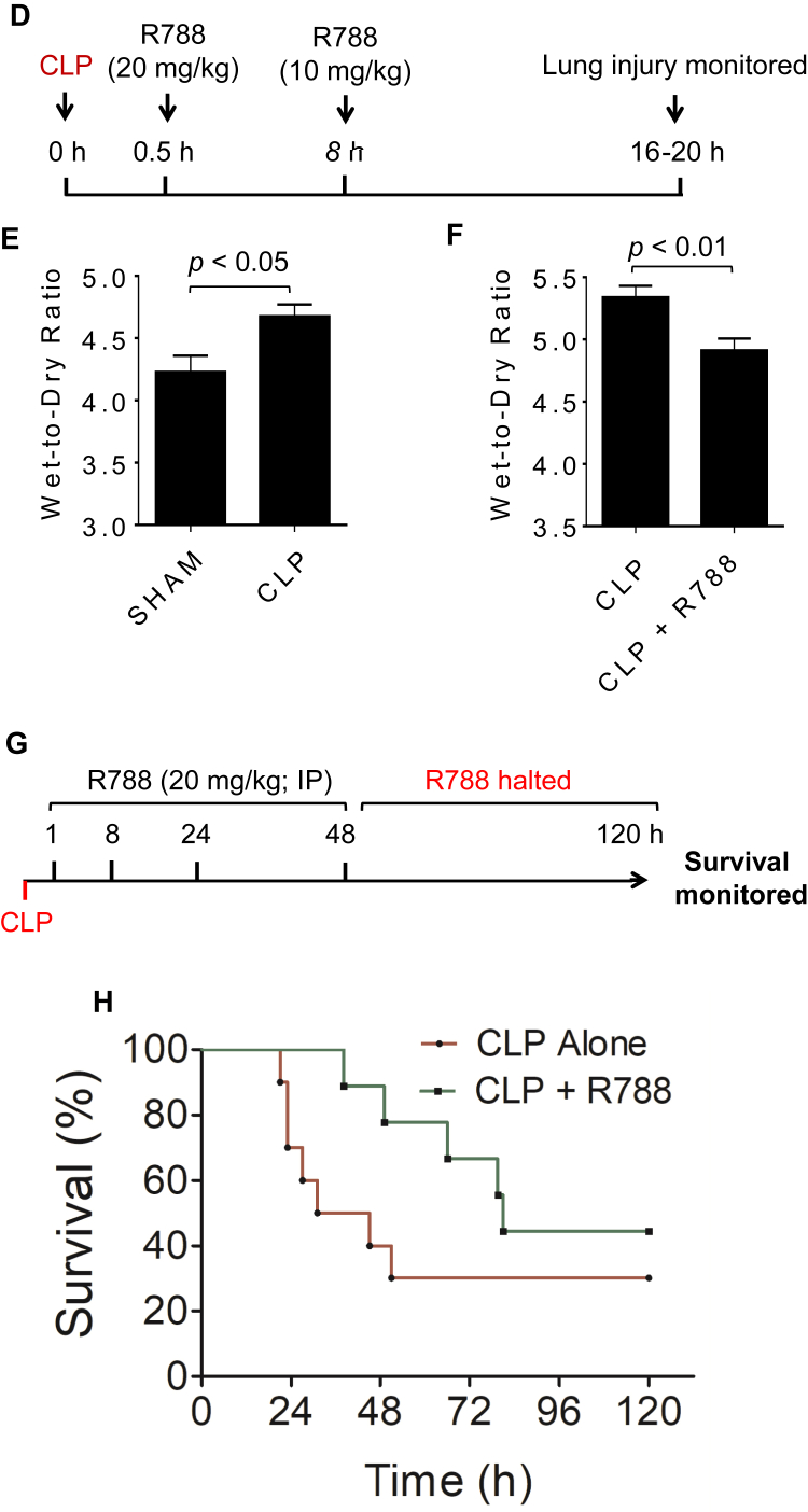
Increased endothelial cell (EC) permeability is a cardinal feature of acute lung injury/acute respiratory distress syndrome (ALI/ARDS). Tyrosine phosphorylation of VE-cadherin is a key determinant of EC barrier disruption. However, the identity and role of tyrosine kinases in this context are incompletely understood. Here we report that Spleen Tyrosine Kinase (Syk) is a key mediator of EC barrier disruption and lung vascular leak in sepsis. Inhibition of Syk by pharmacological or genetic approaches, each reduced thrombin-induced EC permeability. Mechanistically, Syk associates with and phosphorylates VE-cadherin to cause EC permeability. To study the causal role of endothelial Syk in sepsis-induced ALI, we used a remarkably efficient and cost-effective approach based on gene transfer to generate EC-ablated Syk mice. These mice were protected against sepsis-induced loss of VE-cadherin and inflammatory lung injury. Notably, the administration of Syk inhibitor R788 (fostamatinib); currently in phase II clinical trial for the treatment of COVID-19, mitigated lung injury and mortality in mice with sepsis. These data identify Syk as a novel kinase for VE-cadherin and a druggable target against ALI in sepsis.

Keywords: Syk; VE-cadherin; endothelial cells; lung vascular leak; sepsis.


Conflict of interest statement

Conflict of interest The authors declare that they have no conflicts of interest with the contents of this article.

Figures

Figure 1
Figure 1
Depletion of Syk protects against thrombin-induced EC barrier disruption. HPAEC were transfected with siRNA-Control (si-Con) or siRNA-Syk (si-Syk). A, after 48 h, total cell lysates were immunoblotted to monitor Syk and Gapdh levels. Bar graph shows the quantification of Syk abundance in EC transfected with si-Con or si-Syk (n = 3/condition). B–D, after 24 h, cells were reseeded on gold electrode plates and allowed to grow for an additional 48 h. The confluent monolayers were then treated with thrombin, and the real-time changes in transendothelial electrical resistance (TER) were determined as a measure of endothelial barrier function. TER values of each monolayer were normalized to baseline values. ∗p < 0.05. Data were analyzed by a two-tailed unpaired Student t test (B). Quantification of normalized TER in EC transfected with si-Con or si-Syk at (C) 0.5 and (D) 1 h after thrombin treatment (n = 4/condition). E, cells transfected with si-Con or si-Syk were reseeded on transwell inserts placed in a 24-well plate and allowed to grow to confluence. The cells were treated with thrombin for 30 min and FITC-Dextran was added directly to the culture media. FITC-Dextran concentrations were determined in the receiver tray. n = 3 to 4/condition. Data were analyzed by 2-tailed unpaired Student’s t test for comparisons of two groups. For comparisons between multiple groups, one-way ANOVA, followed by Tukey post hoc test was used.
Figure 2
Figure 2
Inhibition or Depletion of Syk protects against septic plasma-induced EC barrier disruption.A, confluent EC monolayers grown on gold electrode plates were pretreated either with vehicle or R406 (10 μM) and then treated with 0.1% plasma from 4 different patients with sepsis. TER was measured over time and normalized to baseline values (n = 4/condition). B, HPAEC were transfected with si-Con or si-Syk. After 24 h, cells were reseeded on gold electrode plates and cultured for an additional 48 h. The confluent monolayers were then treated with 0.1% plasma from 4 different patients with sepsis. TER was measured and normalized to baseline values (n = 4/condition). ∗p < 0.05. Data were analyzed by 2-tailed unpaired Student’s t test.
Figure 3
Figure 3
Depletion or inhibition of Syk inhibitsthrombin-induceddisassembly ofVE-cadherinat AJs and gap formation.A, HPAEC grown on coverslips were transfected with si-Con or si-Syk. After 48 h, cells were treated with thrombin (5 U/ml) for 15 min and non-permeabilized cells were stained with anti-VE-cadherin antibody (green) to mark cell surface VE-cadherin and DAPI (blue) to mark nuclei. Images are representative of three experiments. B, the percent of cells with disrupted adherens junctions (AJs) in (A) was counted as described in the “Experimental procedures” section. Error bars represent mean ± S.E. (n = 3 with 40–50 cells were counted in every fields for each conditions). C, the percent gap area in (A) was calculated as described in the “Experimental procedures”. Error bars represent mean ± S.E. (n = 3 fields analyzed per condition). Scale bars: 25 μm. D–F, HPAEC were pretreated with either vehicle or the Syk inhibitor R406 (10 μM) for 1 h and then challenged with thrombin (5 U/ml) for 15 min and non-permeabilized cells were stained with anti-VE-cadherin (green) to mark cell surface VE-cadherin and DAPI (blue) to mark nuclei. Images are representative of three experiments (D). E, the percent of cells with disrupted adherens junctions (AJs) in (D) was counted as described in the “Experimental procedures” section. Error bars represent mean ± S.E. (n = 3 with 40–50 cells were counted in every fields for each conditions). F, the percent gap area in (D) was calculated as described in the “Experimental procedures”. Error bars represent mean ± S.E. (n = 3 fields analyzed per condition). Scale bars: 25 μm. One-way ANOVA, followed by Tukey post hoc test was used for statistical analysis.
Figure 3
Figure 3
Depletion or inhibition of Syk inhibitsthrombin-induceddisassembly ofVE-cadherinat AJs and gap formation.A, HPAEC grown on coverslips were transfected with si-Con or si-Syk. After 48 h, cells were treated with thrombin (5 U/ml) for 15 min and non-permeabilized cells were stained with anti-VE-cadherin antibody (green) to mark cell surface VE-cadherin and DAPI (blue) to mark nuclei. Images are representative of three experiments. B, the percent of cells with disrupted adherens junctions (AJs) in (A) was counted as described in the “Experimental procedures” section. Error bars represent mean ± S.E. (n = 3 with 40–50 cells were counted in every fields for each conditions). C, the percent gap area in (A) was calculated as described in the “Experimental procedures”. Error bars represent mean ± S.E. (n = 3 fields analyzed per condition). Scale bars: 25 μm. D–F, HPAEC were pretreated with either vehicle or the Syk inhibitor R406 (10 μM) for 1 h and then challenged with thrombin (5 U/ml) for 15 min and non-permeabilized cells were stained with anti-VE-cadherin (green) to mark cell surface VE-cadherin and DAPI (blue) to mark nuclei. Images are representative of three experiments (D). E, the percent of cells with disrupted adherens junctions (AJs) in (D) was counted as described in the “Experimental procedures” section. Error bars represent mean ± S.E. (n = 3 with 40–50 cells were counted in every fields for each conditions). F, the percent gap area in (D) was calculated as described in the “Experimental procedures”. Error bars represent mean ± S.E. (n = 3 fields analyzed per condition). Scale bars: 25 μm. One-way ANOVA, followed by Tukey post hoc test was used for statistical analysis.
Figure 4
Figure 4
Syk is involved inthrombin-inducedphosphorylation and cleavage ofVE-cadherin.A and B, HPAEC were treated with thrombin (5 U/ml) for the indicated time points. Total cell lysates were analyzed by immunoblotting to determine the phosphorylation status of VE-cadherin at (A) Y658 and (B) Y685. Total VE-cadherin was used as a loading control. n = 3/condition. C and D, HPAEC were transfected with si-Con or si-Syk for 48 h and then treated with thrombin (5 U/ml) for 25 min. Total cell lysates were analyzed by Western blot for (C) pY658 and (D) pY685. n = 3/condition. E–G, HPAEC were transfected with si-Con or si-Syk for 48 h and treated with thrombin and total cell lysates were analyzed by Western blot for VE-cadherin cleavage (E). Quantification of the cleaved fragments (∼90 kDa and ∼35 kDa) (F and G). n = 3/condition.
Figure 5
Figure 5
The kinase activity of Syk is necessary forthrombin-inducedphosphorylation and cleavage ofVE-cadherin.A and B, HPAEC were transfected with a kinase-defective mutant of Syk (Syk-KD) or pcDNA3 vector for 48 h and then treated with thrombin (5 U/ml) for 25 min. Total cell lysates were analyzed by Western blot for (A) pY658, total VE-cadherin, Syk and tubulin, and (B) pY685 and total VE-cadherin. n = 3/condition. Low exposures of the Syk blots were taken in order to visualize the overexpressed Syk-KD but not the endogenous Syk protein. C–E, HPAEC were transfected with pcDNA3 or Syk-KD plasmid for 48 h and treated with thrombin and total cell lysates were analyzed by Western blot for VE-cadherin cleavage (C). Quantification of the cleaved fragments (∼90 kDa and ∼35 kDa) (D and E). n = 3/condition. One-way ANOVA, followed by Tukey post hoc test was used for the statistical analysis.
Figure 6
Figure 6
Thrombin induces association of Syk withVE-cadherin.A and B, HPAEC were treated with thrombin (5 U/ml) for 25 min. Total cell lysates were subjected to IP using anti-VE-cadherin antibody and immunoblotted (IB) with antibodies to Syk and VE-cadherin (A). Quantification of VE-cadherin association with Syk (B). n = 3/condition. C and D, HPAEC were treated with thrombin (5 U/ml) for 25 min. Total cell lysates were subjected to immunoprecipitation (IP) using anti-Syk antibody and immunoblotted (IB) with antibodies to VE-cadherin and Syk (C). Quantification of Syk association with VE-cadherin (D). n = 3/condition. Data were analyzed by a 2-tailed unpaired Student’s t test.
Figure 7
Figure 7
Endothelial Syk mediates sepsis-induced ALI.A, Syk level is increased in the lung of mice with sepsis. Mice were subjected to CLP or SHAM surgery as described in Methods. After 12 h, lung homogenates were used to determine the levels of Syk by immunoblotting. n = 5/group. B–D, EC-specific deletion of Syk in mouse lung via cationic liposomes-mediated gene transfer. Cationic liposomes containing pCMV-Cre construct in which CRE expression is driven by CMV promoter were delivered into Sykfl/fl mice via retroorbital injection. B and C, after 8 to 10 days, mice were subjected to SHAM or CLP surgery. After 12 h, lung homogenates were analyzed by immunoblotting to determine the levels of Syk in the lungs of (B) SHAM or (C) CLP mice. n = 4/group. D, after 8 to 10 days, lung microvascular endothelial cells (LMVEC) were isolated from mice using anti-CD31 beads. EC-specific loss of Syk was confirmed by immunoblotting. n = 3/group. E–J, deletion of Syk in the lung endothelium via pCMV-Cre reduces sepsis-induced inflammatory lung injury. Sykfl/fl mice were transduced with pCMV-Cre via cationic liposomes. After 8 to 10 days, mice were subjected to CLP for 12 h and lungs were analyzed for (E) ICAM-1 and (F) VCAM-1 levels by Western blot (n = 3–4/group; (G) MPO activity as a measure of neutrophil infiltration in the lung (n = 5/group); (H) Evans blue albumin (EBA) extravasation to determine lung vascular leakage (n = 5/group; (I) lung wet-to-dry weight ratio to assess pulmonary edema (n=5–7/group); and (J) VE-cadherin level to determine lung vascular integrity (n = 3–4/group). Data were analyzed by a 2-tailed unpaired Student’s t test for comparisons of 2 groups. For comparisons between multiple groups, one-way ANOVA, followed by Tukey post hoc test was used.
Figure 7
Figure 7
Endothelial Syk mediates sepsis-induced ALI.A, Syk level is increased in the lung of mice with sepsis. Mice were subjected to CLP or SHAM surgery as described in Methods. After 12 h, lung homogenates were used to determine the levels of Syk by immunoblotting. n = 5/group. B–D, EC-specific deletion of Syk in mouse lung via cationic liposomes-mediated gene transfer. Cationic liposomes containing pCMV-Cre construct in which CRE expression is driven by CMV promoter were delivered into Sykfl/fl mice via retroorbital injection. B and C, after 8 to 10 days, mice were subjected to SHAM or CLP surgery. After 12 h, lung homogenates were analyzed by immunoblotting to determine the levels of Syk in the lungs of (B) SHAM or (C) CLP mice. n = 4/group. D, after 8 to 10 days, lung microvascular endothelial cells (LMVEC) were isolated from mice using anti-CD31 beads. EC-specific loss of Syk was confirmed by immunoblotting. n = 3/group. E–J, deletion of Syk in the lung endothelium via pCMV-Cre reduces sepsis-induced inflammatory lung injury. Sykfl/fl mice were transduced with pCMV-Cre via cationic liposomes. After 8 to 10 days, mice were subjected to CLP for 12 h and lungs were analyzed for (E) ICAM-1 and (F) VCAM-1 levels by Western blot (n = 3–4/group; (G) MPO activity as a measure of neutrophil infiltration in the lung (n = 5/group); (H) Evans blue albumin (EBA) extravasation to determine lung vascular leakage (n = 5/group; (I) lung wet-to-dry weight ratio to assess pulmonary edema (n=5–7/group); and (J) VE-cadherin level to determine lung vascular integrity (n = 3–4/group). Data were analyzed by a 2-tailed unpaired Student’s t test for comparisons of 2 groups. For comparisons between multiple groups, one-way ANOVA, followed by Tukey post hoc test was used.
Figure 8
Figure 8
Deletion of Syk in the lung endotheliumviapVECad-Crereducessepsis-inducedinflammatory lung injury. Sykfl/fl mice were transduced with pVECad-Cre (pVE-Cre) as described in the Methods. Mice were subjected to CLP for 12 h and lungs were analyzed for (A) Syk, (B) IL-1β, (C) IL-6, (D) MPO activity, (E) EBA extravasation, and (F) lung wet-to-dry weight ratio (n = 5). Data were analyzed by 2-tailed unpaired Student’s t test for comparisons of 2 groups. For comparisons between multiple groups, one-way ANOVA, followed by Tukey post hoc test was used.
Figure 8
Figure 8
Deletion of Syk in the lung endotheliumviapVECad-Crereducessepsis-inducedinflammatory lung injury. Sykfl/fl mice were transduced with pVECad-Cre (pVE-Cre) as described in the Methods. Mice were subjected to CLP for 12 h and lungs were analyzed for (A) Syk, (B) IL-1β, (C) IL-6, (D) MPO activity, (E) EBA extravasation, and (F) lung wet-to-dry weight ratio (n = 5). Data were analyzed by 2-tailed unpaired Student’s t test for comparisons of 2 groups. For comparisons between multiple groups, one-way ANOVA, followed by Tukey post hoc test was used.
Figure 9
Figure 9
Syk is a viable therapeutic target to control sepsis-induced ALI and mortality.A–H, protective effect of Syk inhibition against sepsis-induced lung inflammation. Mice were administered with R788 (20 mg/kg: IP) or saline 1 h prior to and 2 h post CLP as depicted in (A). At 6 to 8 h after CLP, lungs were analyzed for (B) IL-1 β and (C) IL-6 levels by ELISA. (n= 5–6/group). D–F, therapeutic administration of Syk inhibitor R788 reduces sepsis-induced lung injury. Mice were administered with R788 (20 mg/kg; IP) or saline at 0.5 and 8 h post CLP as depicted in (D). At 16 to 20 h after CLP, lungs were analyzed for lung wet-to-dry weight ratio in the absence (E) or presence (F) of R788. (n= 4–6/group in [E] and n = 9/group in [F]). G and H, therapeutic administration of Syk inhibitor R788 improves survival in sepsis. Mice were administered with R788 (20 mg/kg; IP) or saline at the indicated times post CLP as depicted in (G). After 48 h of CLP, R788 administration was halted (G) to determine if the protective effect of R788 was reversed due to its short half-life (∼2 h) in mice. Survival rates (H) were determined over a period of 120 h. (n = 9–10/group). One mouse in CLP + R788 group which died after CLP procedure was excluded from the analysis. Mice receiving R788 show significant improvement in survival (p < 0.05 up to 65 h post-CLP) but this effect is gradually lost after halting the drug (p > 0.05 starting from 66 h post-CLP). p value was determined by log-rank test.
Figure 9
Figure 9
Syk is a viable therapeutic target to control sepsis-induced ALI and mortality.A–H, protective effect of Syk inhibition against sepsis-induced lung inflammation. Mice were administered with R788 (20 mg/kg: IP) or saline 1 h prior to and 2 h post CLP as depicted in (A). At 6 to 8 h after CLP, lungs were analyzed for (B) IL-1 β and (C) IL-6 levels by ELISA. (n= 5–6/group). D–F, therapeutic administration of Syk inhibitor R788 reduces sepsis-induced lung injury. Mice were administered with R788 (20 mg/kg; IP) or saline at 0.5 and 8 h post CLP as depicted in (D). At 16 to 20 h after CLP, lungs were analyzed for lung wet-to-dry weight ratio in the absence (E) or presence (F) of R788. (n= 4–6/group in [E] and n = 9/group in [F]). G and H, therapeutic administration of Syk inhibitor R788 improves survival in sepsis. Mice were administered with R788 (20 mg/kg; IP) or saline at the indicated times post CLP as depicted in (G). After 48 h of CLP, R788 administration was halted (G) to determine if the protective effect of R788 was reversed due to its short half-life (∼2 h) in mice. Survival rates (H) were determined over a period of 120 h. (n = 9–10/group). One mouse in CLP + R788 group which died after CLP procedure was excluded from the analysis. Mice receiving R788 show significant improvement in survival (p < 0.05 up to 65 h post-CLP) but this effect is gradually lost after halting the drug (p > 0.05 starting from 66 h post-CLP). p value was determined by log-rank test.

Similar articles

Cited by

References

    1. Levitt J.E., Matthay M.A. Treatment of acute lung injury: historical perspective and potential future therapies. Semin. Respir. Crit. Care Med. 2006;27:426–437. - PubMed
    1. Bhattacharya J., Matthay M.A. Regulation and repair of the alveolar-capillary barrier in acute lung injury. Annu. Rev. Physiol. 2013;75:593–615. - PubMed
    1. Matthay M.A., Zemans R.L., Zimmerman G.A., Arabi Y.M., Beitler J.R., Mercat A., et al. Acute respiratory distress syndrome. Nat. Rev. Dis. Primers. 2019;5:18. - PMC - PubMed
    1. Erickson S.E., Martin G.S., Davis J.L., Matthay M.A., Eisner M.D. Recent trends in acute lung injury mortality: 1996-2005. Crit. Care Med. 2009;37:1574–1579. - PMC - PubMed
    1. Batah S.S., Fabro A.T. Pulmonary pathology of ARDS in COVID-19: a pathological review for clinicians. Respir. Med. 2021;176 - PMC - PubMed

Publication types