

Reporting of Facet Joint Inflammation in Lumbar Spine MRI Studies in Patients With Low Back Pain
- PMID: 38229507
- PMCID: PMC11042993
- DOI: 10.1097/BRS.0000000000004923
Reporting of Facet Joint Inflammation in Lumbar Spine MRI Studies in Patients With Low Back Pain
Abstract
Study design: Retrospective.
Objective: We aimed to assess the frequency of facet joint inflammatory features noted in routine radiology reports of lumbar spine magnetic resonance imaging (MRI) studies among patients with chronic low back pain.
Summary of background data: Facet joint arthropathy is one of the most common causes of chronic low back pain. It may encompass various inflammatory imaging characteristics, such as facet joint effusion, bone marrow edema, and soft tissue edema. The extent to which radiology reports mention inflammatory features of the lumbar facet joints and the accuracy of these reports have not been investigated.
Materials and methods: The authors performed a chart review on 49 subjects with previous facet-related interventions ( i.e . medial branch blocks or intra-articular facet joint injection) and MRI available in the medical record. One senior musculoskeletal radiologist and a musculoskeletal radiology fellow graded the inflammatory features using a published facet joint inflammation grading system [Gold Standard (GS)]. The authors identified the inflammatory markers mentioned in the radiology reports and calculated the sensitivity and positive predictive value of the radiology reports compared with GS readings.
Results: Compared with the GS, the sensitivity of radiology reports for facet joint effusion, bone marrow, and soft tissue edema ranged from 6% to 22%, and the positive predictive value ranged from 25% to 100%. L4/5 had the highest number of cases with inflammatory features noted on the reports.
Conclusion: Inflammatory findings, such as facet joint effusion, bone marrow edema, and soft tissue edema, are not commonly identified in radiology reports. Further investigations are needed to determine the clinical importance of MRI-detected lumbar facet joint inflammatory features as a potential mechanism of nociception and as a predictor of outcomes following injections or other therapies.
Copyright © 2024 Wolters Kluwer Health, Inc. All rights reserved.
Conflict of interest statement
The authors report no conflicts of interest.
Figures


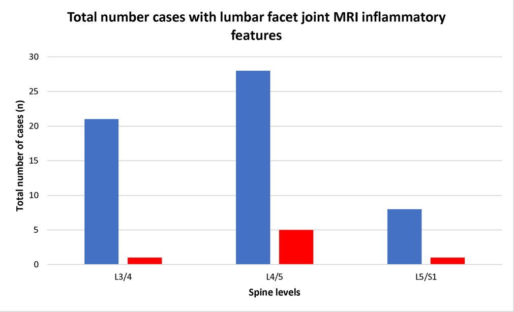
 = Gold standard reader
  = Radiology report
Similar articles
-
Comprehensive Grading System of Inflammatory Features of the Lumbar Facet Joints on Magnetic Resonance Imaging.Spine (Phila Pa 1976). 2024 Mar 1;49(5):332-340. doi: 10.1097/BRS.0000000000004846. Epub 2023 Oct 5. Spine (Phila Pa 1976). 2024. PMID: 37798843 Free PMC article.
-
Prevalence and frequency of subchondral bone marrow edema in the lumbar facet joints of asymptomatic and symptomatic individuals.Skeletal Radiol. 2020 Jul;49(7):1141-1147. doi: 10.1007/s00256-020-03400-4. Epub 2020 Feb 26. Skeletal Radiol. 2020. PMID: 32103296
-
Grading Systems of Lumbar Facet Joint Inflammatory Changes on Magnetic Resonance Imaging: A Scoping Review.Spine (Phila Pa 1976). 2023 May 1;48(9):636-644. doi: 10.1097/BRS.0000000000004609. Epub 2023 Feb 27. Spine (Phila Pa 1976). 2023. PMID: 36856452 Free PMC article.
-
Predictability of the effects of facet joint infiltration in the degenerate lumbar spine when assessing MRI scans.J Orthop Surg Res. 2017 Nov 21;12(1):180. doi: 10.1186/s13018-017-0685-x. J Orthop Surg Res. 2017. PMID: 29162138 Free PMC article.
-
Predictors of Outcomes After Lumbar Intra-Articular Facet Joint Injections and Medial Branch Blocks: A Scoping Review.Spine (Phila Pa 1976). 2023 Oct 15;48(20):1455-1463. doi: 10.1097/BRS.0000000000004776. Epub 2023 Jul 20. Spine (Phila Pa 1976). 2023. PMID: 37470372 Free PMC article.
References
-
- Kjaer P, Leboeuf-Yde C, Korsholm L, et al. Magnetic resonance imaging and low back pain in adults: a diagnostic imaging study of 40-year-old men and women. Spine 2005;30:1173–80. - PubMed
-
- Manchikanti L, Singh V, Pampati V, et al. Evaluation of the relative contributions of various structures in chronic low back pain. Pain Physician 2001;4:308–16. - PubMed
MeSH terms
Grants and funding
LinkOut - more resources
Full Text Sources
Medical
Research Materials
