Case Reports
. 2023 Dec 28;19(3):1100-1105.
doi: 10.1016/j.radcr.2023.12.020. eCollection 2024 Mar.

Breast metastases from extra-mammary cancers: A report of 3 challenging cases and literature review

Affiliations
Case Reports

Breast metastases from extra-mammary cancers: A report of 3 challenging cases and literature review

Caroline Chabot et al. Radiol Case Rep. .

Abstract

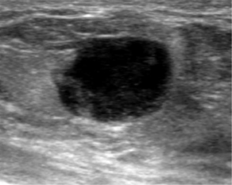
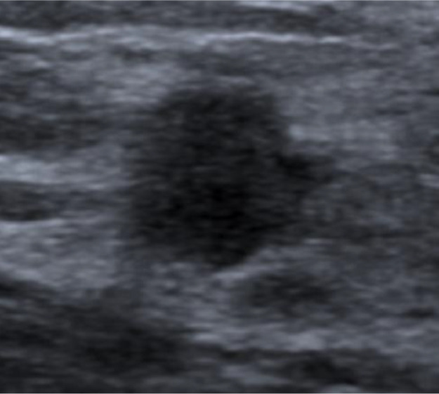
We report 3 cases of patients with a history of extra-mammary cancer who presented with breast nodules, leading to diagnostic challenges and occasional misleading imaging findings. These cases highlight the significance of radiologists considering breast metastases as a potential component of the differential diagnosis when assessing patients with a history of cancer who exhibit palpable breast nodules. Furthermore, these cases underscore the importance of integrating various imaging techniques with histological and immunohistochemical analyses of the lesions to achieve precise diagnoses, ultimately ensuring the highest quality of care for these patients.

Keywords: Breast imaging; Breast metastases; Extra-mammary cancer.


Figures

Fig 1:
Fig. 1
Bilateral mammography in mediolateral oblique incidence displaying multiple rounded opacities in all 4 breast quadrants (indicated by dashed arrows) along with 2 clusters of microcalcifications located in the equatorial outer region and upper outer quadrant of the right breast (indicated by solid arrows).
Fig 2:
Fig. 2
Ultrasound image revealing a hyperechoic nodular lesion located in the upper outer quadrant of the right breast (10:00) and characterized by slightly microlobulated margins within the breast parenchyma.
Fig 3:
Fig. 3
(A) Histological appearance of a breast metastasis (H and E), exhibiting fragments of adipo-connective tissue (indicated by asterisks) infiltrated by neoplastic proliferation composed of marrow-sized cells. These cells have pale eosinophilic cytoplasm and round nuclei, occasionally enlarged and nucleolated (indicated by dashed arrows). (B) Immunohistochemical analysis showing positivity for cytokeratins AE1/AE3, indicating a cancerous lesion. (C) Immunohistochemical expression of the P63 marker, suggesting squamous cell carcinoma. (D) Lack of immunohistochemical expression of the P16 marker, which is typically associated with HPV infection. Scale is 100µm.
Fig 4:
Fig. 4
Right mammography captured in mediolateral oblique (left) and craniocaudal (right) incidence, displaying 2 rounded opacities measuring 10 mm in diameter. These opacities are located in the lower inner quadrant (indicated by the arrows). Notably, there are no microcalcification clusters or signs of adenomegaly.
Fig 5:
Fig. 5
Ultrasound image revealing the presence of a hypoechoic mass in the lower inner quadrant (4:00) of the right breast with relatively indistinct contours and heterogeneous internal content.
Fig. 6:
Fig. 6
PET-CT scan revealing moderate hypermetabolic activity (maximal Standardized Uptake Value - SUV of 2,8) of a nodule located at the junction of the upper quadrants of the left breast (indicated by the arrow).
Fig. 7:
Fig. 7
Ultrasound image displaying an oval-shaped hypoechoic lesion at the junction of the upper quadrants of the left breast, measuring 10 mm x 8 mm, characterized by slightly blurred contours and without any signs of hypervascularization.

References

    1. Jian-Yong Z, Guang-Ping Z, Xue W, Shi-Min Z, Zhen-Guo Z. Breast metastasis of cervical cancer: a case report and systematic literature review. Front Oncol. 2022;12 doi: 10.3389/fonc.2022.974592. - DOI - PMC - PubMed
    1. Koch A., Richter-Marot A., Wissler M.P., Baratte A., Mathelin C. Métastases mammaires de cancers d'origine extra-mammaire : état des lieux et difficultés diagnostiques. Gynécol Obstét Fertil. 2013;41(11):653–659. doi: 10.1016/j.gyobfe.2013.09.013. ISSN 1297-9589. - DOI - PubMed
    1. Khouchani M, Benchakroun N, Tahri A, Tawfiq N, Jouhadi H, Acharki A., et al. Métastase intramammaire d’un cancer vulvaire : à propos d’un cas avec revue de la littérature. Cancer/Radiothérapie. 2008;12(Issue 2):120–125. doi: 10.1016/j.canrad.2007.11.001. ISSN 1278-3218. - DOI - PubMed
    1. Li J, Wahab R, Brown AL, Guarnieri B, Lewis K, Mahoney MC, et al. Extramammary metastases to the breast. Radiographics. 2023;43:10. - PubMed
    1. Moreno-Astudillo L, Villaseñor-Navarro Y, Sánchez-Goytia V, Porras-Reyes F, Lara-Mercado A, Sollozo-Dupont I. A case series of breast metastases from different extramammary malignancies and their literature review. Case Rep Radiol. 2019;2019 doi: 10.1155/2019/9454201. - DOI - PMC - PubMed

Publication types

LinkOut - more resources