. 2023 Nov 30:10:041310.
doi: 10.1063/5.0162640.

Graphene nanocomposites for real-time electrochemical sensing of nitric oxide in biological systems

Affiliations

Graphene nanocomposites for real-time electrochemical sensing of nitric oxide in biological systems

Tanveer A Tabish et al. Appl Phys Rev. .

Abstract

Nitric oxide (NO) signaling plays many pivotal roles impacting almost every organ function in mammalian physiology, most notably in cardiovascular homeostasis, inflammation, and neurological regulation. Consequently, the ability to make real-time and continuous measurements of NO is a prerequisite research tool to understand fundamental biology in health and disease. Despite considerable success in the electrochemical sensing of NO, challenges remain to optimize rapid and highly sensitive detection, without interference from other species, in both cultured cells and in vivo. Achieving these goals depends on the choice of electrode material and the electrode surface modification, with graphene nanostructures recently reported to enhance the electrocatalytic detection of NO. Due to its single-atom thickness, high specific surface area, and highest electron mobility, graphene holds promise for electrochemical sensing of NO with unprecedented sensitivity and specificity even at sub-nanomolar concentrations. The non-covalent functionalization of graphene through supermolecular interactions, including π-π stacking and electrostatic interaction, facilitates the successful immobilization of other high electrolytic materials and heme biomolecules on graphene while maintaining the structural integrity and morphology of graphene sheets. Such nanocomposites have been optimized for the highly sensitive and specific detection of NO under physiologically relevant conditions. In this review, we examine the building blocks of these graphene-based electrochemical sensors, including the conjugation of different electrolytic materials and biomolecules on graphene, and sensing mechanisms, by reflecting on the recent developments in materials and engineering for real-time detection of NO in biological systems.


Conflict of interest statement

Conflict of Interest The authors have no conflicts to disclose.

Figures

Fig. 1
Fig. 1. Representative schematics of Shibuki- and solid-type electrochemical NO sensors.
A Shibuki-style sensor uses silver and platinum as a reference and working electrode, respectively, whereas in the solid sensor, carbon fiber acts as the working electrode with an internal or external reference electrode of silver or silver chloride. A film of oxidized TMHPP-Ni [tetrakis(3-methoxy-4-hydroxyphenyl)-nickel porphyrin] on the surface of the carbon fiber improves the sensitivity toward NO. The use of Nafion® on the electrode avoids electrode fouling and is selectively permeable to NO, excluding interference from other molecules such as ascorbic acid and nitrite. Reproduced with permission from Brown and Schoenfisch, Chem. Rev. 119, 11551–11575 (2019). Copyright 2019 American Chemical Society.
Fig. 2
Fig. 2. Summary of structural models of various derivatives of graphene.
(a) Graphene, which is a single layer sp2 hybridized carbon sheet packed in a hexagonal lattice structure. Graphene has excellent properties including but not limited to high charge mobility, high specific surface area, excellent optical, electrical and mechanical characteristics and superior conductivity. (b) Graphene oxide (GO) is an oxidized form of graphene synthesized via chemical exfoliation of graphite which possesses a large number of oxygen-centered functional groups including oxygen-containing groups such as hydroxyl, epoxide, carbonyl, and carboxylic groups on its surface. The existence of functional groups offers advantages such as high chemical reactivity and hydrophilicity. (c) Reduced graphene oxide (rGO) is a reduced form of GO which has defects on its edges and basal planes. The reduction of functional groups onto the surface of GO introduces defects and pores within the graphene network. rGO shows high adsorption capacity toward different molecules due to its low oxygen content, hydrophobicity, high surface area and defects. And (d) three-dimensional (3D) graphene foam is highly porous network of graphene arranged in a micro size sheets. 3D graphene networks are found in different forms such as foam, hydrogel, aerogel etc. they offer superior porosity, specific surface area, and electrical conductivity in comparison to GO and rGO. Reproduced with permission from Tabish et al., Redox Biol. 15, 34–40 (2018). Copyright 2018 Author(s), licensed under a Creative Commons Attribution (CC BY) license.
Fig. 3
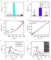
Fig. 3. Electrochemical sensing performance of the graphene nanocomposite rGO–CeO2 composite/GCE for the detection of NO.
(a) and (b) show specificity for NO even in the presence of potentially interfering biomolecules, ascorbic acid (AA), uric acid (UA), and dopamine (DA). (c) Amperometric response of the modified electrode with different NO concentrations at an applied potential of 0.8 V. (d) Calibration curve of modified rGO–CeO2/GCE electrode for NO, exhibiting a wide linear range of 18.0 nM to 5.6 μM, a sensitivity of 1676.06 mA cm−2 M−1 and a low detection limit of 9.6 nM. (e) Real-time detection of NO secreted from cultured A549 cells stimulated by different amounts of acetylcholine (ACh) in cell culture medium with a cell density of 5 x 10⁴ ml−1 on rGO–CeO2 sensor. Ach is an endogenous neurotransmitter that stimulates NO release from endothelial cells, whereas hemoglobin (Hb) is a strong scavenger of NO and is used as a control. (f) Real-time detection of NO from A549 cells in cell culture medium with different cell densities. The insets are microscope photographs of A549 cells (n = 5). Reproduced with permission from Hu et al., Biosens. Bioelectron. 70, 310–317 (2015). Copyright 2015 Elsevier.
Fig. 4
Fig. 4. DNA-templated AuNPs on nitrogen-doped graphene sheets for electrochemical detection of NO.
(a) Current–time responses of laminin/AuNPs-ctDNA-nitrogen doped graphene sheets (NGS)/screen-printed carbon electrode (SPCE) to 2,10, 20, 50, 80,100, 200, 300, 400, 500 nM of NO. (b) Calibration curve of the amperometric response vs the concentration of NO from 2 to 500 nM (n = 3). (c) Scanning electron microscopy image of MCF-7 cancer cells cultured on laminin/AuNPs-ctDNA-NGS/SPCE. (d) Current–time response curves of laminin/AuNPs-ctDNA-NGS/SPCE to (a) the addition of 4 mM L-Arg in the absence of cells, (b) the addition of 4 mM L-Arg and 4 mM L-NAME in the presence of cultured MCF-7 cells. A significant current response was only observed when 4 mM L-Arg was added to cultured MCF-7 cells (c). The red arrows indicated the injection of the drugs. L-Arginine (L-Arg) was used as a donor to generate NO, and L-NAME was used to inhibit the generation of NO. Reproduced with permission from Dou et al., Anal. Chem. 91, 2273–2278 (2019). Copyright 2019 American Chemical Society.
Fig. 5
Fig. 5. RGD-peptide functionalized graphene film for real-time detection of NO in HUVECs.
(a) Illustration of RGD-peptide functionalized graphene film with HUVECs. (b) Real-time detection of NO released from HUVECs when treated with 1 mM acetylcholine to stimulate NO release (red), 0.5 mM acetylcholine (blue), and 1 mM acetylcholine with 1 mM L-NAME (green) demonstrating concentration changes and NO specificity when treated with a NO inhibitor (L-NAME). Reproduced with permission from Guo et al., ACS Nano 6, 6944–6951 (2012). Copyright 2012 American Chemical Society.
Fig. 6
Fig. 6
Schematic diagram of (a) a sensitive acupuncture needle microsensor for real-time monitoring of NO in acupoints of rats. (b) FGPC/AuNPs/acupuncture needle and (c) real-time NO measurement in acupoint ST 36 stimulated by L-arginine in the presence of NO synthase (NOS) under physiological conditions to produce NO. The functionalized needle is used for real-time detection of NO for different acupoints of rats, identifying in vivo NO detection. Reproduced with permission from Tang et al., Sci. Rep. 7, 6446 (2017). Copyright 2017 Nature Springer.

Similar articles

Cited by

References

    1. Ignarro LJ, Buga GM, Wood KS, Byrns RE, Chaudhuri G. Endothelium-derived relaxing factor produced and released from artery and vein is nitric oxide. Proc Natl Acad Sci U S A. 1987;84:9265–9269. - PMC - PubMed
    1. Lundberg JO, Weitzberg E. Nitric oxide signaling in health and disease. Cell. 2022;185:2853–2878. - PubMed
    1. Knott AB, Bossy-Wetzel E. Nitric oxide in health and disease of the nervous system. Antioxid Redox Signaling. 2009;11:541–554. - PMC - PubMed
    1. Farah C, Michel LYM, Balligand J-L. Nitric oxide signalling in cardiovascular health and disease. Nat Rev Cardiol. 2018;15:292–316. - PubMed
    1. Forstermann U, Sessa WC. Nitric oxide synthases: Regulation and function. Eur Heart J. 2012;33:829–837. - PMC - PubMed

LinkOut - more resources