Review
. 2023 Dec 26;12(12):1760-1773.
doi: 10.21037/gs-23-288. Epub 2023 Dec 22.

Current status of autologous breast reconstruction in Europe: how to reduce donor site morbidity

Affiliations
Review

Current status of autologous breast reconstruction in Europe: how to reduce donor site morbidity

Moustapha Hamdi et al. Gland Surg. .

Abstract

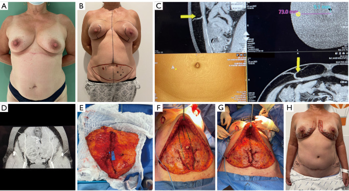
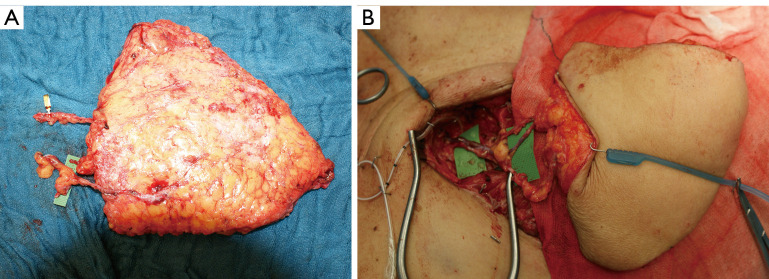
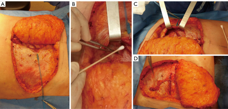
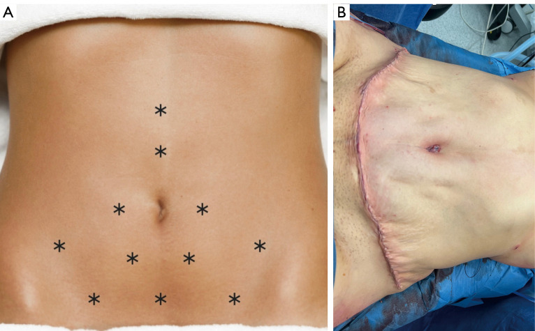
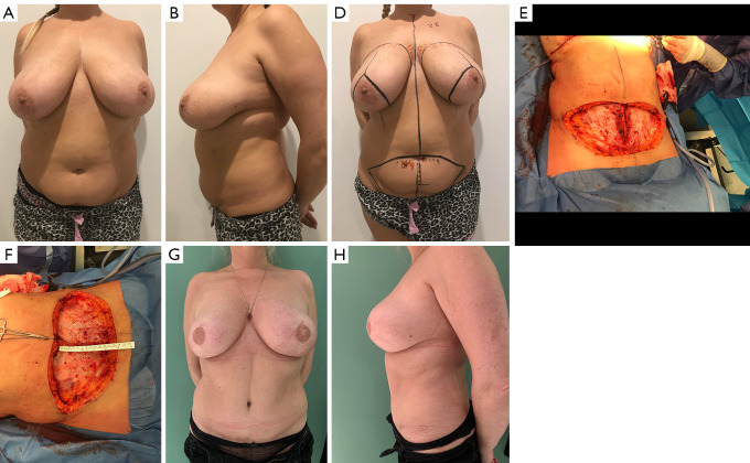
Autologous reconstruction techniques for breast reconstruction have significantly evolved in the last few decades in Europe. In the search of reducing the donor site morbidity, surgeons explored the possibilities to preserve the rectus muscle and its function, and a transition to deep inferior epigastric perforator (DIEP) flaps was started in the nineties. Throughout the years, and especially in the last decade, we have increasingly implemented aesthetic refinements for donor site handling in DIEP flap breast reconstruction. In our practice, autologous breast reconstruction provides an opportunity to effectively remodel the donor site, minimising functional morbidity, and maximising aesthetic satisfaction. To achieve this, careful patient selection, pre-operative preparation, meticulous intra-operative dissection, and a clear post-operative protocol are essential. The main goal in autologous breast reconstruction, and its biggest advantage, is to offer the patient a natural look and feel of the reconstructed breast. A second goal is to minimize the number of procedures needed to reach the desired breast shape, size, and volume. In most patients, the number of operations ranges between one and three. The third main goal is to minimize the donor site morbidity, both functionally and aesthetically. Functionally, this implies preserving as much of the rectus abdominis muscle as possible, limiting the fascia incision, preserving the motor branches to the muscle, ensuring an adequate fascial closure, and repairing the rectus diastasis is present. Aesthetically, we aim to have a low position of the scar, an aesthetically pleasing location of the umbilicus, and limited or no lateral skin excess or so called "dogears". In this clinical practice review article, we provide an overview of current autologous reconstruction methods, with a focus on minimising donor site morbidity and enhancing the aesthetic result of the donor site. We discuss key concepts in autologous reconstruction and provide surgical pearls for performing the procedure effectively with optimal reconstructive and aesthetic result.

Keywords: Autologous reconstruction; abdominoplasty deep inferior epigastric perforator flap (abdominoplasty DIEP flap); aesthetic donor site closure.


Conflict of interest statement

Conflicts of Interest: All authors have completed the ICMJE uniform disclosure form (available at https://gs.amegroups.com/article/view/10.21037/gs-23-288/coif). The series “Hot Topics in Breast Reconstruction World Wide” was commissioned by the editorial office without any funding or sponsorship. M.H. reports that he serves as the National Expert for the Superior Council in Belgian Minister of Health (MoH) and is a consultant to Polytech for scientific activities. The authors have no other conflicts of interest to declare.

Figures

Figure 1
Figure 1
A 56-year-old patient with difficult screening for breast cancer requiring multiple biopsy procedures. A decision for prophylactic mastectomy was made with implant-based reconstruction. However, following the initial reconstruction, the patient decided for autologous breast reconstruction using free DIEP flaps. (A) Before bilateral mastectomies and implant insertion. (B) The patient marking for implants removal and bilateral DIEP flap breast reconstruction. (C) CT-angio scan image of the right dominant perforator. The arrows show the chosen perforator, on axial, sagittal and coronal views, on which the flap will be harvested. (D) The left side has two average size perforators. (E) Left harvested DIEP flap with two perforators. (F) The deep fascia was closed then the muscle plicature was marked. (G) The muscle facia was done in two layers: first with separate non-absorbable suture, then with a running barbed absorbable suture. (H) The outcome at 3-month postoperatively. Fat grafting was planned with dog-ear scar correction. DIEP, deep inferior epigastric perforator; CT, computed tomography.
Figure 2
Figure 2
The dissection of the SIEV with enough length in order to reach a recipient vein. (A) The SIEV was dissected and preserved. (B) The SIV was hooked to the one of the collateral deep inferior epigastric vein after performing the main anastomosis to the internal mammary vessels. SIEV, superficial inferior epigastric vein; SIV, superficial inferior vein.
Figure 3
Figure 3
The surgical perforator dissection. (A) The perforator is freed for muscle and deep fascia and muscle is retracted using Lone Star Elastic Hook. (B) A cotton Q-tip is very useful to free perforator and intercostal nerves from the rectus muscle. (C) The main pedicle is dissected under the rectus abdominis muscle using surgical retractors without extending the deep fascia incision. (D) The perforator/pedicle dissection with 6 cm fascia-incision which is usually closed towards the midline in order to be included in the rectus abdominis plicature suture.
Figure 4
Figure 4
The umbilicoplasty with routine position of the progressive tension suture in closure of the abdominoplasty DIEP flap. (A) The location of the progressive tension sutures. This is illustrated by the asterisk marks, which show the exact placement of the interrupted progressive tension sutures. (B) The aspect of the donor site at the end of the surgery. DIEP, deep inferior epigastric perforator.
Figure 5
Figure 5
A 47-year-old patient with BRCA1 genetic mutation. A bilateral skin-sparing mastectomy was done with immediate DIEP flap breast reconstruction. (A) Preoperative anterior view shows large ptotic breasts. (B) Lateral preoperative view. (C) Planning of the bilateral skin sparing mastectomies with inverted T incision. The perforators are marked on the abdominal and flap was designed with low incision line. (D) Donor site after harvesting both flaps based on one perforator each side. The fascia incision kept short and parallel to the midline. (E) Plicature of the rectus muscles done after fascia closure. (F) The results of the abdominoplasty flap breast reconstruction at 3 years. (G) The lateral view shows significant improvement in the abdominal contouring. DIEP, deep inferior epigastric perforator.

Similar articles

Cited by

References

    1. Holmström H. The free abdominoplasty flap and its use in breast reconstruction. An experimental study and clinical case report. Scand J Plast Reconstr Surg 1979;13:423-7. 10.3109/02844317909013092 - DOI - PubMed
    1. Robbins TH. Rectus abdominis myocutaneous flap for breast reconstruction. Aust N Z J Surg 1979;49:527-30. 10.1111/j.1445-2197.1979.tb05869.x - DOI - PubMed
    1. Hartrampf CR, Scheflan M, Black PW. Breast reconstruction with a transverse abdominal island flap. Plast Reconstr Surg 1982;69:216-25. 10.1097/00006534-198202000-00006 - DOI - PubMed
    1. Mühlbauer W, Olbrisch R. The latissimus dorsi myo-cutaneous flap for breast reconstruction. Chir Plastica 1977;4:27-34.
    1. Lejour M, Dome M. Abdominal wall function after rectus abdominis transfer. Plast Reconstr Surg 1991;87:1054-68. 10.1097/00006534-199106000-00007 - DOI - PubMed