

Image-based multiplex immune profiling of cancer tissues: translational implications. A report of the International Immuno-oncology Biomarker Working Group on Breast Cancer
- PMID: 38230434
- PMCID: PMC11288342
- DOI: 10.1002/path.6238
Image-based multiplex immune profiling of cancer tissues: translational implications. A report of the International Immuno-oncology Biomarker Working Group on Breast Cancer
Abstract
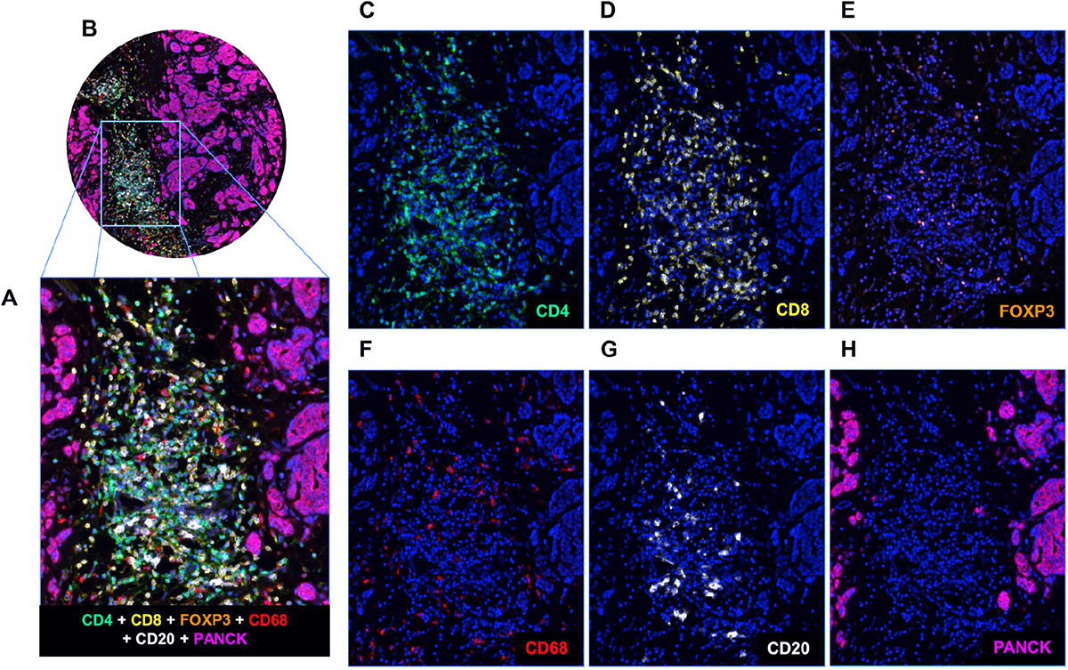
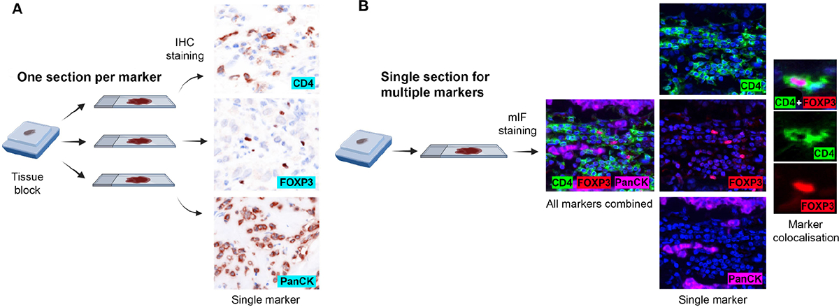
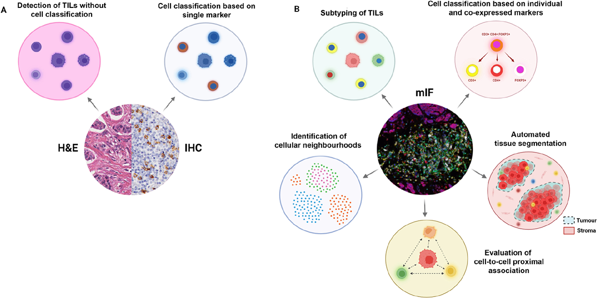
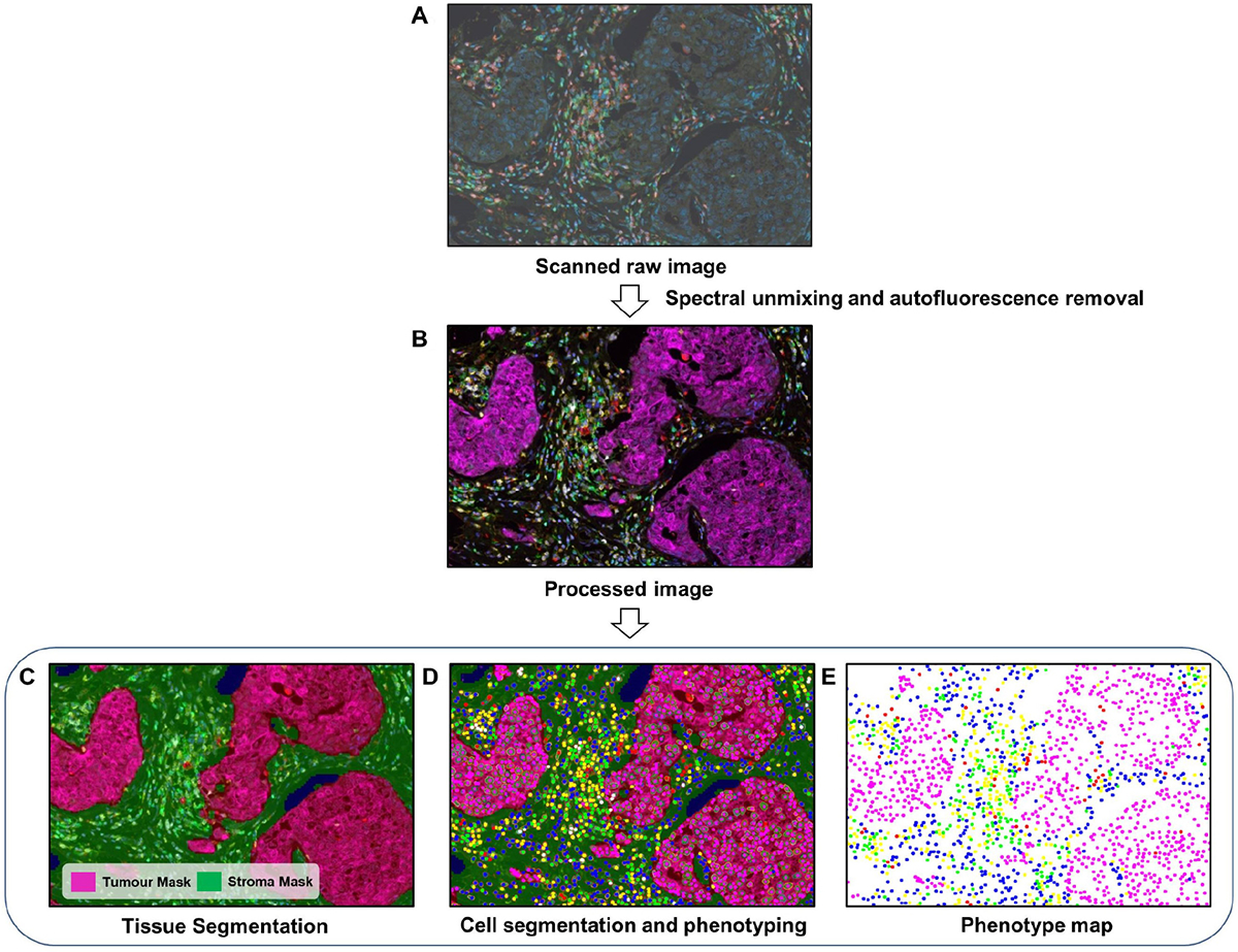
Recent advances in the field of immuno-oncology have brought transformative changes in the management of cancer patients. The immune profile of tumours has been found to have key value in predicting disease prognosis and treatment response in various cancers. Multiplex immunohistochemistry and immunofluorescence have emerged as potent tools for the simultaneous detection of multiple protein biomarkers in a single tissue section, thereby expanding opportunities for molecular and immune profiling while preserving tissue samples. By establishing the phenotype of individual tumour cells when distributed within a mixed cell population, the identification of clinically relevant biomarkers with high-throughput multiplex immunophenotyping of tumour samples has great potential to guide appropriate treatment choices. Moreover, the emergence of novel multi-marker imaging approaches can now provide unprecedented insights into the tumour microenvironment, including the potential interplay between various cell types. However, there are significant challenges to widespread integration of these technologies in daily research and clinical practice. This review addresses the challenges and potential solutions within a structured framework of action from a regulatory and clinical trial perspective. New developments within the field of immunophenotyping using multiplexed tissue imaging platforms and associated digital pathology are also described, with a specific focus on translational implications across different subtypes of cancer. © 2024 The Authors. The Journal of Pathology published by John Wiley & Sons Ltd on behalf of The Pathological Society of Great Britain and Ireland.
Keywords: cancer prognosis; clinical integration; digital image analysis; multiplex imaging; multiplex immunofluorescence; multiplex immunohistochemistry; therapy response; tumour immune profiling; tumour infiltrating lymphocytes.
© 2024 The Authors. The Journal of Pathology published by John Wiley & Sons Ltd on behalf of The Pathological Society of Great Britain and Ireland.
Conflict of interest statement
DBP: Speaker’s Bureau: Genentech, Novartis, Clinical Care Options, Oncocyte; research support: WindMIL, Brooklyn Immunotherapeutics, Merck, Bristol Myers Squibb, IMV; consulting: Merck, Biotheranostics, Puma, Gilead, Lilly, Sanofi, NGM Bio, Sanford Burnham Prebys, AstraZeneca. GB: Speaker’s fee from MSD and Novartis, advisory boards for Roche and MSD, consultant for MSD, Novartis, and Roche, travel and conference support from Roche, MSD, and Gilead. JSR-F is an Associate Editor of The Journal of Pathology and has received personal/consultancy fees from Goldman Sachs, Bain Capital, REPARE Therapeutics, Saga Diagnostics, and PaigeAl, membership of the scientific advisory boards of VolitionRx, REPARE Therapeutics, and Paige. Al, membership of the Board of Directors of Grupo Oncoclinicas, and ad hoc membership of the scientific advisory boards of AstraZeneca, Merck Daiichi Sankyo, Roche Tissue Diagnostics, and Personalis, outside the scope of this study. AIH: Research fund from Visiopharm A/S. ZK: Paid advisory role for Eli Lilly and AstraZeneca Canada. JT: Employee of Visiopharm A/S. KRMB: Scientific Advisory Board for CDI Labs, research funding form Carevive. FC: Chair of the Scientific and Medical Advisory Board of TRIBVN Healthcare, France, and advisory board fees from TRIBVN Healthcare, France in the last 5 years. Shareholder of Aiosyn BV, the Netherlands. LADC: Participation in the Tempus Algorithm Advisors program. AC: Contracted researcher for Oncoinvent AS and Novocure and a consultant for Sotio a.s. and Epics Therapeutics SA. ME: Egyptian missions sector. SBF: Expert advisory panel for AXDEV Group. JMG: Employee and stockholder of Roche/Genentech. SG: Research funding from Regeneron Pharmaceuticals, Boehringer Ingelheim, Bristol Myers Squibb, Celgene, Genentech, EMD Serono, Pfizer, and Takeda, unrelated to the current work; named co-inventor on an issued patent for multiplex immunohistochemistry to characterise tumours and treatment responses. The technology is filed through Icahn School of Medicine at Mount Sinai (ISMMS) and is currently unlicensed. NH: Patent on a technology to measure immune infiltration in cancer to predict treatment outcome (W02012038068A2). MGH: Consultant for PaigeAl, VolastraTx, and advisor for PathPresenter. JH: Speaker’s honoraria or advisory board remunerations from Roche, Novartis, AstraZeneca, Eli Lilly, and MSD. Co-founder and shareholder of Stratipath AB. KK Employee and stockholder of Roche. GA: Employee of Merck & Co Inc. AIK: Honorarium from Roche, MSD, and Pfizer, member of the Advisory Board of Pfizer. A-VL Institutional grants from AstraZeneca and personal grants from AstraZeneca (travel and honorarium from advisory board), MSD (honorarium from advisory board), and Daiichi Sankyo (travel). XL: Eli Lilly Company, Advisor, Cancer Expert Now, Advisor, Champions Oncology, Research fund. AM: Equity holder in Picture Health, Elucid Bioimaging and Inspirata Inc, advisory board of Picture Health, Aiforia Inc and SimBioSys, consultant for SimBioSys, sponsored research agreements with AstraZeneca, Boehringer-lngelheim, Eli-Lilly, and Bristol Myers Squibb, technology licensed to Picture Health and Elucid Bioimaging involvement in three different R01 grants with Inspirata Inc DKM: Consulting: Astrazeneca, Lilly USA LLC Hologic. Sponsored Research: Merck Agendia. SM: Scientific Committee Study member. Roche, data and safety monitoring member of clinical trials: Sensorion, Biophytis, Servier, IQVIA, Yuhan, Kedrion. FuAAM: Research studentship funding from GSK DAM: Speaker fees from AstraZeneca, Eli Lilly, and Takeda, consultancy fees from AstraZeneca, Thermo Fisher, Takeda, Amgen, Janssen, M/M Software, Bristol Myers Squibb, and Eli Lilly, educational support from Takeda and Amgen. FP-L: Personal financial interests: AbbVie, Agendia, Amgen, Astellas, AstraZeneca, Bayer, BMS, Daiichi-Sankyo, Eisai, Exact Science, GSK lllumina, Incyte, Janssen, Lilly, MERCK lifa, Merck-MSD, Myriad, Novartis, Pfizer, Pierre-Fabre, Roche, Sanofi, Seagen, Takeda, Veracyte, Servier. Institutional financial interests: AstraZeneca, Bayer, BMS, MSD, Myriad, Roche, Veracyte. Congress invitations: AbbVie, Amgen, AstraZeneca, Bayer, BMS, Gilead, MSD, Novartis, Roche, Lilly, Pfizer. NMR: Co-Founder, Director and CSO of Histofy Ltd, UK AS: Advisory Board/Speaker’s Bureau: Aignostics, AstraZeneca, Bayer, BMS, Eli Lilly, lllumina, Incyte, Janssen, MSD, Novartis, Pfizer, Roche, Seagen, Takeda, and Thermo Fisher; grants from Bayer, Bristol Meyers Squibb, Chugai, and Incyte. TT: Employee of Tempus Labs. JT: Shareholder of EllogonAI BV. TT: Speaker’s fee from Pfizer. JvdL: Member of the advisory boards of Philips, the Netherlands and ContextVision, Sweden, research funding from Philips, the Netherlands, ContextVision, Sweden, and Sectra, Sweden in the last 5 years. Chief scientific officer and shareholder of Aiosyn BV, the Netherlands. TW: Collaboration with TRIBUN Health on automatic grading of biopsies for head and neck cancer, patent on the prediction of homologous recombination deficiency in breast cancer. YW: Employee of CellCarta. HYW: Advisory faculty of AstraZeneca. YY: Speaker/consultant for Roche and Merck PS: Consultant (uncompensated) to Roche-Genentech. SL Research funding to institution from Novartis, Bristol Meyers Squibb, Merck Puma Biotechnology, Eli Lilly, Nektar Therapeutics, AstraZeneca, Roche-Genentech, and Seattle Genetics. Consultant (not compensated) to Seattle Genetics, Novartis, Bristol Meyers Squibb, Merck, AstraZeneca, Eli Lilly, Pfizer, and Roche-Genentech. Consultant (paid to her institution) to Aduro Biotech, Novartis, GlaxoSmithKline, Roche-Genentech, AstraZeneca, Silverback Therapeutics, GI Therapeutics, PUMA Biotechnologies, Pfizer, Gilead Therapeutics, Seattle Genetics, Daiichi-Sankyo, Amunix, Tallac therapeutics, Eli Lilly, and Bristol Meyers Squibb. RS: Non-financial support from Merck and Bristol Myers Squibb, research support from Merck Puma Biotechnology and Roche; personal fees from Roche, Bristol Myers Squibb and Exact Sciences for advisory boards. WMG: Co-founder, shareholder and part-time Chief Scientific Officer of OncoAssure Limited, shareholder in Deciphex and member of the Scientific Advisory Board of Carrick Therapeutics.
Figures
References
Publication types
MeSH terms
Substances
Grants and funding
- 1U01CA239055-01/CA/NCI NIH HHS/United States
- UH3 CA225021/CA/NCI NIH HHS/United States
- C45982/A21808/CRUK_/Cancer Research UK/United Kingdom
- 1U01CA248226-01/CA/NCI NIH HHS/United States
- R01CA208236-01A1/CA/NCI NIH HHS/United States
- U01CA269181/CA/NCI NIH HHS/United States
- 1U54CA254566-01/CA/NCI NIH HHS/United States
- R01CA26820701A1/CA/NCI NIH HHS/United States
- U01 CA239055/CA/NCI NIH HHS/United States
- R01CA202752-01A1/CA/NCI NIH HHS/United States
- R01 CA220581/CA/NCI NIH HHS/United States
- U24 CA215109/CA/NCI NIH HHS/United States
- R37 CA225655/NH/NIH HHS/United States
- 1R43EB028736-01/EB/NIBIB NIH HHS/United States
- U24 CA224319/CA/NCI NIH HHS/United States
- R01CA216579-01A1/CA/NCI NIH HHS/United States
- R01CA220581-01A1/CA/NCI NIH HHS/United States
- R01 CA216579/CA/NCI NIH HHS/United States
- R33 CA263705/CA/NCI NIH HHS/United States
- R01 CA202752/CA/NCI NIH HHS/United States
- R01 CA208236/CA/NCI NIH HHS/United States
- R01CA268287A1/CA/NCI NIH HHS/United States
- U01 DK124165/DK/NIDDK NIH HHS/United States
- R37 CA225655/CA/NCI NIH HHS/United States
- KCL-BCN-Q3/CRUK_/Cancer Research UK/United Kingdom
- U01 CA248226/CA/NCI NIH HHS/United States
- R01CA249992-01A1/CA/NCI NIH HHS/United States
- P50 CA116201/CA/NCI NIH HHS/United States
- P50 CA247749/CA/NCI NIH HHS/United States
- I01 BX004121/BX/BLRD VA/United States
- CA263705/NH/NIH HHS/United States
- R43 EB028736/EB/NIBIB NIH HHS/United States
- U01 CA269181/CA/NCI NIH HHS/United States
- CRUK/07/012/CRUK_/Cancer Research UK/United Kingdom
- R01 CA257612/CA/NCI NIH HHS/United States
- DK124165/NH/NIH HHS/United States
- U54 CA254566/CA/NCI NIH HHS/United States
- CA224319/NH/NIH HHS/United States
- CA196521/NH/NIH HHS/United States
- R01 CA268287/CA/NCI NIH HHS/United States
- R01CA257612-01A1/CA/NCI NIH HHS/United States
- P30 CA196521/CA/NCI NIH HHS/United States
- R01 CA249992/CA/NCI NIH HHS/United States
LinkOut - more resources
Full Text Sources
Medical
