Review
. 2024 Feb;56(1):80-96.
doi: 10.1080/03602532.2024.2303507. Epub 2024 Jan 17.

The aminosalicylate - folate connection

Affiliations
Review

The aminosalicylate - folate connection

Robert E London. Drug Metab Rev. 2024 Feb.

Abstract

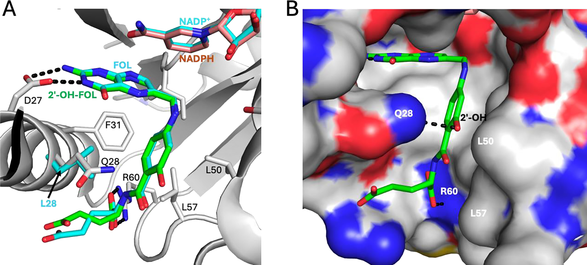
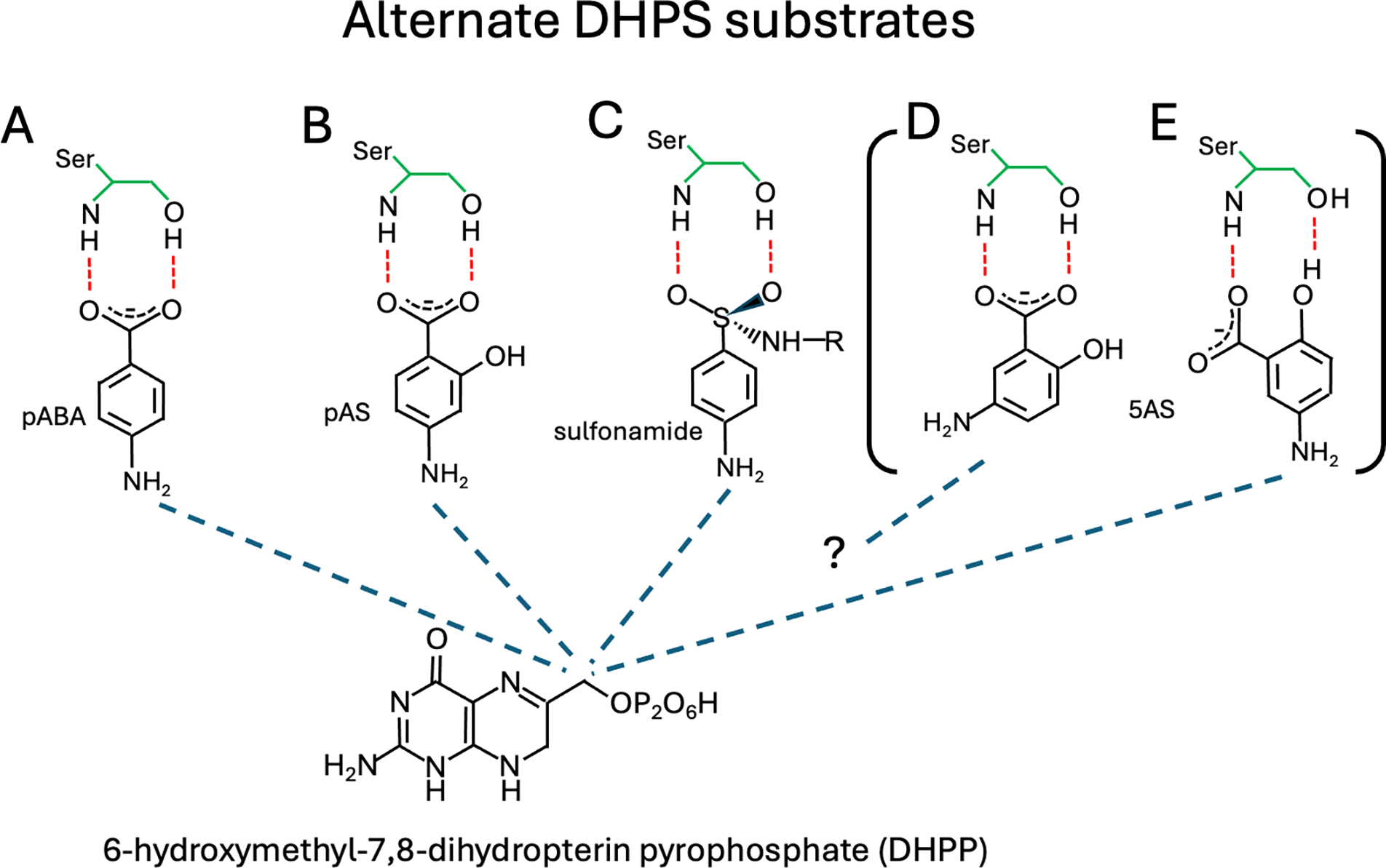
Two aminosalicylate isomers have been found to possess useful pharmacological behavior: p-aminosalicylate (PAS, 4AS) is an anti-tubercular agent that targets M. tuberculosis, and 5-aminosalicylate (5AS, mesalamine, mesalazine) is used in the treatment of ulcerative colitis (UC) and other inflammatory bowel diseases (IBD). PAS, a structural analog of pABA, is biosynthetically incorporated by bacterial dihydropteroate synthase (DHPS), ultimately yielding a dihydrofolate (DHF) analog containing an additional hydroxyl group in the pABA ring: 2'-hydroxy-7,8-dihydrofolate. It has been reported to perturb folate metabolism in M. tuberculosis, and to selectively target M. tuberculosis dihydrofolate reductase (mtDHFR). Studies of PAS metabolism are reviewed, and possible mechanisms for its mtDHFR inhibition are considered. Although 5AS is a more distant structural relative of pABA, multiple lines of evidence suggest a related role as a pABA antagonist that inhibits bacterial folate biosynthesis. Structural data support the likelihood that 5AS is recognized by the DHPS pABA binding site, and its effects probably range from blocking pABA binding to formation of a dead-end dihydropterin-5AS adduct. These studies suggest that mesalamine acts as a gut bacteria-directed antifolate, that selectively targets faster growing, more folate-dependent species.

Keywords: 5-aminosalicylate; Ulcerative colitis; inflammatory bowel disease; mesalamine; p-aminosalicylate; tuberculosis.


Conflict of interest statement

Competing Interests

The Author declares that there are no competing interests associated with the manuscript.

Figures

Figure 1.
Figure 1.. Structures of dihydrofolate, PAS-derived metabolites and dihydropterin adducts formed by DHPS.
A) Dihydrofolate, with positions numbered according to https://iupac.qmul.ac.uk/misc/folic.html. B) 2’-hydroxy-7,8-dihydrofolate metabolite formed in the presence of PAS; C) 5-formyl-2’-hydroxyTHF metabolite identified by Wacker et al. (Wacker, Kolm, and Ebert 1958) in PAS-treated Enterococcus stei. D) 7,8-dihydropterin adduct with sulfathiazole; E) 7,8-dihydropterin adduct with 5AS postulated to form if DHPS is able to utilize 5AS as a substrate.
Figure 2.
Figure 2.. Structures of the mtDHFR complex with 5’-hydroxyfolate.
A) Structure of mtDHFR (gray cartoon with selected sidechains shown using stick representation) with 5’-hydroxyfolate (green stick representation), and NADPH (salmon stick representation) (PDB: 6DDW, (Hajian et al. 2019)). The positions of folate and NADP+ (cyan) derived from an overlay with the ecDHFR-FOL-NADP+ ternary complex (PDB: 1RX2, (Sawaya and Kraut 1997)) are included. The Leu28 sidechain (cyan) of the ecDHFR structure also is included for comparison. B) mtDHFR shown as a surface representation with bound 2’-hydroxyfolate (green stick model) illustrates the accommodation of the 2’-hydroxyl group. The positions of the Arg60 and Gln28 sidechains are indicated. Figures were generated from pdb files using the PyMol Molecular Graphics System, Schrodinger, Inc., NY,NY).
Figure 3.
Figure 3.. Comparison of DHFR substrate and product complexes.
A) Aligned ecDHFR complexes containing folate (green stick representation) (PDB: 1RX2, (Sawaya and Kraut 1997)), DHF (cyan stick representation) (PDB: 6CXK, (Cao et al. 2018)), and THF (gold stick representation) (PDB: 6CW7, (Cao et al. 2018)). The ecDHFR structure of 1RX2 is shown as a gray cartoon with selected folate-interacting sidechains included. NADP+ (salmon stick representation) present in structure 1RX2 is also included. The DHF and THF hydrogen atoms are included in panel A to indicate the reduction state. B) The overlay shown in panel A after rotation of the THF in order to reduce the steric conflict with nicotinamide. In addition to rotating the entire molecule, a small additional rotation about the C6-C9 bond indicated by the red arrow in panel A was introduced to reduce steric conflict. Dashed black lines indicate H-bonds and the dashed red line indicates the distance between THF C-7 and nicotinamide C-5 before (panel A) and after (panel B) ligand rotation. As noted in the text, the THF rotation significantly reduces enzyme interactions with the pABG group. C) Modeled complex of mtDHFR (PDB: 6DDW (Hajian et al. 2019)) with rotated 2’-hydroxyTHF (gold stick representation) and NADP+ (salmon stick representation). Rotation of the 2’-hydroxyTHF product in the mtDHFR binding site reduces the unfavorable steric interaction between the reduced pterin and the bound nicotinamide, and can allow a favorable H-bond between the 2’-hydroxyTHF 2’-hydroxyl group and Arg60. Color coding is as in panel A. Figures were generated from pdb files using the PyMol Molecular Graphics System, Schrodinger, Inc., NY, NY).
Figure 4.
Figure 4.. Schematic of ligand interactions with the ecDHPS pABA binding site.
The examples illustrate interactions with a DHPS that utilizes Ser for PABA orientation, and identifies the reactive amine and dihydropterin C-9 atoms with dashed blue lines. A) pABA; B) p-aminosalicylate; C) p-aminobenzenesulfonamide; D) 5AS with orientation determined by carboxylate-Ser interaction (inhibitory mode); E) 5AS with an alternate H-bond interaction that supports a ring orientation rotated by ~ 60° from that in panel D, placing the amino group in the catalytically active position. Actual ligand orientations also depend on additional active site contacts.
Figure 5.
Figure 5.. Predicted inhibition of DHPS by 5AS.
A) Structure of the ternary pvDHPS domain complex (gray stick model showing selected sidechains) with 6-hydroxymethylpterinpyrophosphate (PtPP, beige) and pABA (green) (PDB: 5Z79, (Yogavel et al. 2018)), where PtPP is a catalytically inert DHPP analog. H-bonds are indicated by dashed black lines, and the reactive pABA amino nitrogen and the PtPP C-9 atom are connected by a dashed red line. B) Structure shown in panel A after in silico modification of pABA to 5AS. The additional hydroxyl group is positioned to H-bond with the Gly551 carbonyl and the 5-amino group to H-bond with the PtPP bridging oxygen atom. C) Structure of ypDHPS-DHP+−pyrophosphate-pABA complex (PDB: 3TYZ, (Yun et al. 2012)), color-coded as in panel A. In this example, Ser222 acts as the positioning residue, however the DHPP substrate has been cleaved to yield the reactive DHP+ species plus pyrophosphate. Figures were generated from pdb files using the PyMol Molecular Graphics System, Schrodinger, Inc., NY, NY).
Figure 6.
Figure 6.. 5AS docked with bacterial DHPS.
A) CB-docked 5AS (gold) in pvDHPS ternary complex (PDB: 5Z79, (Yogavel et al. 2018)) after removal of the ligands. The PtPP (pink) and pABA (cyan) ligands in the reported structure were added back for the figure. B) CB-docked 5AS (gold) in ecDHPS-pteroate product complex (PDB: 5U10, (Dennis et al. 2018) after removal of pteroate (Pter, cyan), which has been added back in the figure for comparison. In this structure, pyrophosphate is not present and loop 1 is disordered, removing some close contacts with 5AS. H-bonds are shown as dashed black lines, and potential reactive amino and C-9 atoms are connected with a dashed red line. Figures were generated from pdb files using the PyMol Molecular Graphics System, Schrodinger, Inc., NY, NY).
Figure 7.
Figure 7.. Interaction of 5AS with DHPS.
After binding of substrate DHPP and 5AS to DHPS, the DHPP pyrophosphate group is cleaved, creating a DHP+ carbocation (Yun et al. 2012). To some extent, reaction of DHP+ with 5AS may produce a 7,8-dihydropterin-5-aminosalicylate adduct, particularly if 5AS adopts an alternate orientation (e.g. as shown in Figure 4E) in which the 5-amino group is oriented optimally for the chemical reaction. Alternatively, in the carboxylate-determined orientation the 5AS amino position is less optimal for reaction, so that reaction of the DHP+ with water may occur, yielding 6-hydroxymethyl-7,8-dihydropterin (HMDHP). The HMDHP can then be pyrophosphorylated by HPPK (hydroxymethydihydropterin pyrophosphate kinase), reforming the DHPP substrate. Dashed black lines indicate H-bonds and dashed red lines indicate potential reactive nuclei.

References

    1. Allgayer H 2003. ‘Review article: mechanisms of action of mesalazine in preventing colorectal carcinoma in inflammatory bowel disease’, Aliment Pharmacol Ther, 18 Suppl 2: 10–4. - PubMed
    1. Allgayer H, Sonnenbichler J, Kruis W, and Paumgartner G. 1985. ‘Determination of the pK values of 5-aminosalicylic acid and N-acetylaminosalicylic acid and comparison of the pH dependent lipid-water partition coefficients of sulphasalazine and its metabolites’, Arzneimittelforschung, 35: 1457–9. - PubMed
    1. Andersen T, Hvid M, Johansen C, Stengaard-Pedersen K, Hetland ML, Horslev-Petersen K, Junker P, Ostergaard M, and Deleuran B. 2015. ‘Interleukin-23 in early disease development in rheumatoid arthritis’, Scand J Rheumatol, 44: 438–42. - PubMed
    1. Ariens EJ, and Simonis AM. 1955. ‘The biosynthesis of folic-acid homologues from amino-carboxy-pyridine and other para-aminobenzoic-acid substitutes by E. coli’, Schweiz Z Pathol Bakteriol, 18: 71–80. - PubMed
    1. Auletta AE, Gery AM, Parmar A, Davis J, Mishra L, and Mead JA. 1974. ‘The effect of folate and folate analogs upon dihydrofolate reductase and DNA synthesis in kidneys of normal mice’, Life Sci, 14: 1541–53. - PubMed

Publication types

LinkOut - more resources