. 2024 Dec;15(1):2301244.
doi: 10.1080/21505594.2023.2301244. Epub 2024 Jan 17.

Nervous necrosis virus induced vacuolization is a Rab5- and actin-dependent process

Affiliations

Nervous necrosis virus induced vacuolization is a Rab5- and actin-dependent process

Jiaxin Liu et al. Virulence. 2024 Dec.

Abstract

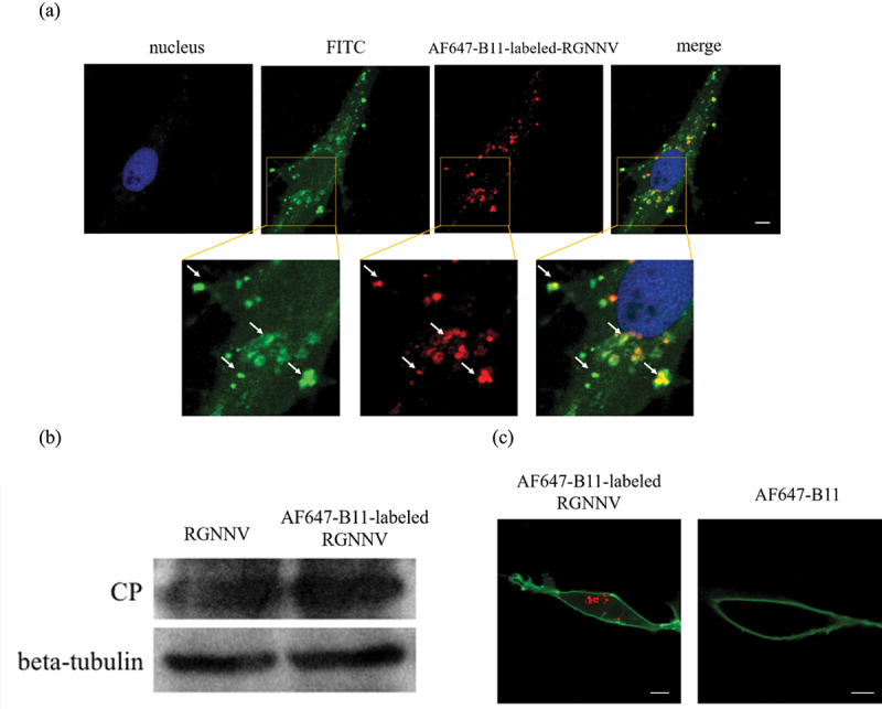
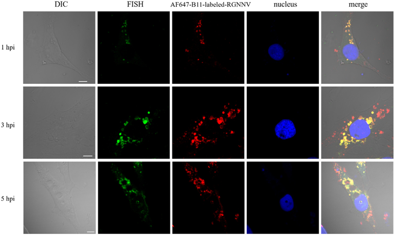
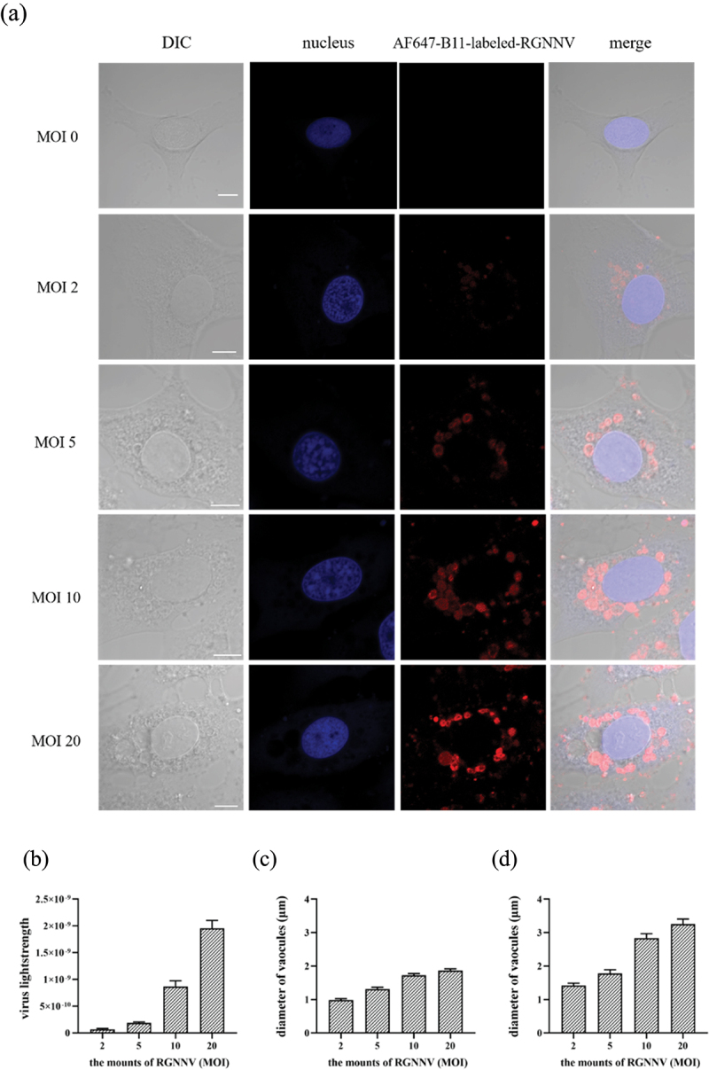
Cytoplasmic vacuolization is commonly induced by bacteria and viruses, reflecting the complex interactions between pathogens and the host. However, their characteristics and formation remain unclear. Nervous necrosis virus (NNV) infects more than 100 global fish species, causing enormous economic losses. Vacuolization is a hallmark of NNV infection in host cells, but remains a mystery. In this study, we developed a simple aptamer labelling technique to identify red-spotted grouper NNV (RGNNV) particles in fixed and live cells to explore RGNNV-induced vacuolization. We observed that RGNNV-induced vacuolization was positively associated with the infection time and virus uptake. During infection, most RGNNV particles, as well as viral genes, colocalized with vacuoles, but not giant vacuoles > 3 μm in diameter. Although the capsid protein (CP) is the only structural protein of RGNNV, its overexpression did not induce vacuolization, suggesting that vacuole formation probably requires virus entry and replication. Given that small Rab proteins and the cytoskeleton are key factors in regulating cellular vesicles, we further investigated their roles in RGNNV-induced vacuolization. Using live cell imaging, Rab5, a marker of early endosomes, was continuously located in vacuoles bearing RGNNV during giant vacuole formation. Rab5 is required for vacuole formation and interacts with CP according to siRNA interference and Co-IP analysis. Furthermore, actin formed distinct rings around small vacuoles, while vacuoles were located near microtubules. Actin, but not microtubules, plays an important role in vacuole formation using chemical inhibitors. These results provide valuable insights into the pathogenesis and control of RGNNV infections.

Keywords: RGNNV; Rab5; aptamer; imaging; vacuolization.


Conflict of interest statement

No potential conflict of interest was reported by the author(s).

Figures

Figure 1.
Figure 1.
Imaging aptamer-labelled RGNNV particles (a) AF647-B11-labelled RGNNV (red) colocalizes with the anti-CP antibody labelling (green). GS cells were infected with AF647-B11-labelled RGNNV and fixed at 1 hpi for further IFA. The nucleus was stained with Hoechst 33,342. (b) AF647-B11 (200 nM) exerted no significant effects on RGNNV replication. GS cells were separately infected with RGNNV or AF647-B11-labelled RGNNV and collected at 36 hpi for western blotting. (c) AF647-B11 enters GS cells only when incubated with RGNNV particles. GS cells were separately incubated with AF647-B11-labelled RGNNV or AF647-B11 and fixed at 1 hpi. Cells were stained with DiO to highlight the cell membrane (green). Scale bar = 5 μm.
Figure 2.
Figure 2.
The time course of RGNNV-induced cytoplasmic vacuoles (a) imaging of RGNNV and vacuoles at different hpi. GS cells were incubated with AF647-B11-labelled RGNNV followed by fixation at 0, 1, 3, and 5 hpi for imaging. The nucleus was stained with Hoechst 33,342. (b) Three-dimensional (3D) images of vacuoles in RGNNV infected cells. GS cells were incubated with AF647-B11-labelled RGNNV and fixed at 5 hpi for imaging. Successive Z-stacks spaced by 200 nm were recorded to construct 3D images. The xz image plane was used for recording and was approximately 1 μm thick. small vacuoles (<3 μm) were indicated by the black arrows, and large vacuoles (>3 μm) were indicated by white arrowheads. Red fluorescence represented RGNNV. (c) Quantification analysis of internalized RGNNV levels; > 80 cells were randomly selected and analysed by MATLAB. (d-e) quantitative analysis of the diameters of all vacuoles/cell (d) and the biggest vacuoles/cell (e) during RGNNV infection. Vacuole diameters were analysed by image J. Scale bar = 5 μm.
Figure 3.
Figure 3.
CP gene colocalized with vacuoles during RGNNV infection. GS cells were infected with AF647-B11-labelled RGNNV (red) synchronously, and fixed at 1, 3, and 5 hpi. A specific FISH probe (green) was used to target viral CP. Scale bar = 5 μm.
Figure 4.
Figure 4.
Analysis of internalized virus and vacuolization after RGNNV infection at different MOIs (a) GS cell imaging with different RGNNV MOIs at 5 hpi. GS cells were incubated with AF647-B11-labelled RGNNV (MOI = 0, 2, 5, 10, 20) as described. Cells were fixed 5 hpi. The nucleus was stained with Hoechst 33,342. (b) Quantification analysis of internalized RGNNV at different MOIs; > 80 cells were randomly selected and analysed by MATLAB. (c-d) quantitative analysis of the diameters of all vacuoles/cell (c) and the biggest vacuoles/cell (d) infected with different RGNNV MOIs. Vacuole diameters were analysed by image J software. Scale bar = 5 μm.
Figure 5.
Figure 5.
CP overexpression did not induce vacuoles. GS cells were transfected with pEGFP-C1 or pEGFP-CP. At 24 hpi, cells were fixed and nuclei stained with Hoechst 33,342. Scale bar = 5 μm.
Figure 6.
Figure 6.
Rab5 is highly correlated with NNV induced vacuolar fusion (a) the obvious colocalization between Rab5 and vacuoles bearing RGNNV. GS cells transfected with pEGFP-Rab5 were incubated with AF647-B11-labelled RGNNV at 4 °C for 20 min and then immediately transferred to 28 °C to initiate infection. The cells were fixed with 4% paraformaldehyde at 0.5, 1.5 hpi. GS cells transfected with pEGFP-Rab5 were used as the control. (b) The real-time tracking of dynamic fusion of early endosomes bearing RGNNV. GS cells transfected with pEGFP-Rab5 were incubated with AF647-B11-labelled RGNNV and images captured by CLSM. Red fluorescence represented RGNNV, and green fluorescence represented Rab5. Scale bar = 5 μm.
Figure 7.
Figure 7.
Vacuoles induced by RGNNV are related to Rab5. (a) Imaging of RGNNV and vacuoles in cells transfected with si-control (NC) or si-Rab5 (siRab5). GS cells were transfected with NC or siRab5 respectively. At 24 h post transfection, the cells were incubated with AF647-B11-labelled RGNNV, and then were fixed with 4% paraformaldehyde at 3 hpi. (b) Rab5 knockdown did not affect RGNNV entry. >80 cells were randomly selected and analysed by MATLAB program. (c-d) Rab5 knockdown significantly reduced the diameter of all vacuoles/cell (c) and diameters of the biggest vacuoles/cell (d). (e) Interactions between CP and Rab5 by IP. GS cells were transfected with pEGFP-C1 or pEGFP-Rab5. At 24 h post-transfection, cells were incubated with RGNNV and collected at 4 hpi and 24 hpi for IP. (f) Rab5 knockdown inhibited virus replication. GS cells were transfected with pEGFP-C1 or pEGFP-Rab5. At 24 h post-transfection, after cells were incubated with RGNNV, they were collected at 24 hpi for qRT-PCR and WB (n = 3, mean ± standard error of the mean (SEM)). *p < 0.05. Scale bar = 5 μm.
Figure 8.
Figure 8.
Colocalization between actin and vacuoles bearing RGNNV. GS cells were infected with AF647-B11-labelled RGNNV (red) at 28 °C for 0.5, 1, 3, 5 h, fixed, and incubated with Alexa Fluor 488 phalloidin (green) for 30 min to indicate actin. Scale bar = 5 μm.
Figure 9.
Figure 9.
The vacuoles bearing RGNNV were colocalized with microtubules. GS cells were infected with AF647-B11-labelled RGNNV (red) at 28°C for 0.5, 1, 3, 5 h, and then fixed with 4% paraformaldehyde. Anti-β-tubulin was used to indicate microtubules (green) by IFA. The images were captured by CLSM. Scale bar = 5 μm.
Figure 10.
Figure 10.
RGNNV-induced vacuoles were analysed when actin or microtubules were disrupted. (a) Imaging of RGNNV and vacuoles in cells untreated or treated with cyto D or nocodazole. GS cells were pretreated with 2 μM cyto D for 1 h or 4 μM nocodazole for 2 h before infection with AF647-B11-labelled-RGNNV. (b) Cyto D or nocodazole significantly affected RGNNV entry. More than 80 cells were randomly selected and analysed by MATLAB program. (c-d) the quantitative analysis of the diameter of (c) all vacuoles and (d) the biggest vacuole in cyto D- or nocodazole- treated cells during RGNNV infection. Vacuole diameter was analysed using image J software. Scale bar = 5 μm.

Similar articles

Cited by

References

    1. Dan J, Nie L, Jia X, et al. Visualization of the oncolytic alphavirus M1 life cycle in cancer cells. Virol Sin. 2021;36(4):655–17. doi: 10.1007/s12250-020-00339-7 - DOI - PMC - PubMed
    1. Kujala P, Ikaheimonen A, Ehsani N, et al. Biogenesis of the Semliki Forest virus RNA replication complex. J Virol. 2001;75(8):3873–3884. doi: 10.1128/JVI.75.8.3873-3884.2001 - DOI - PMC - PubMed
    1. Luo Y, Motamedi N, Magaldi TG, et al. Interaction between simian virus 40 Major capsid protein VP1 and cell surface ganglioside GM1 triggers vacuole formation, mBio. MBio. 2016;7(2):e00297. doi: 10.1128/mBio.00297-16 - DOI - PMC - PubMed
    1. Bandin I, Souto S.. Betanodavirus and VER disease: a 30-year research review. Pathogens. 2020;9(2):106. doi: 10.3390/pathogens9020106 - DOI - PMC - PubMed
    1. Cheng CA, Luo JM, Chiang MH, et al. Nervous necrosis virus coat protein mediates Host translation shutoff through nuclear translocalization and degradation of polyadenylate binding protein. J Virol. 2021;95(17):e0236420. doi: 10.1128/JVI.02364-20 - DOI - PMC - PubMed

LinkOut - more resources