Review
. 2024 Jan 17;166(1):17.
doi: 10.1007/s00701-024-05902-9.

Acute evacuation of 54 intracerebral hematomas (aICH) during the microsurgical clipping of a ruptured middle cerebral artery bifurcation aneurysm-illustration of the individual clinical courses and outcomes with a serial brain CT/MRI panel until 12 months

Affiliations
Review

Acute evacuation of 54 intracerebral hematomas (aICH) during the microsurgical clipping of a ruptured middle cerebral artery bifurcation aneurysm-illustration of the individual clinical courses and outcomes with a serial brain CT/MRI panel until 12 months

Anniina H Autio et al. Acta Neurochir (Wien). .

Abstract

Purpose: In aneurysmal intracerebral hemorrhage (aICH), our review showed the lack of the patient's individual (i) timeline panels and (ii) serial brain CT/MRI slice panels through the aICH evacuation and neurointensive care until the final brain tissue outcome.

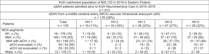
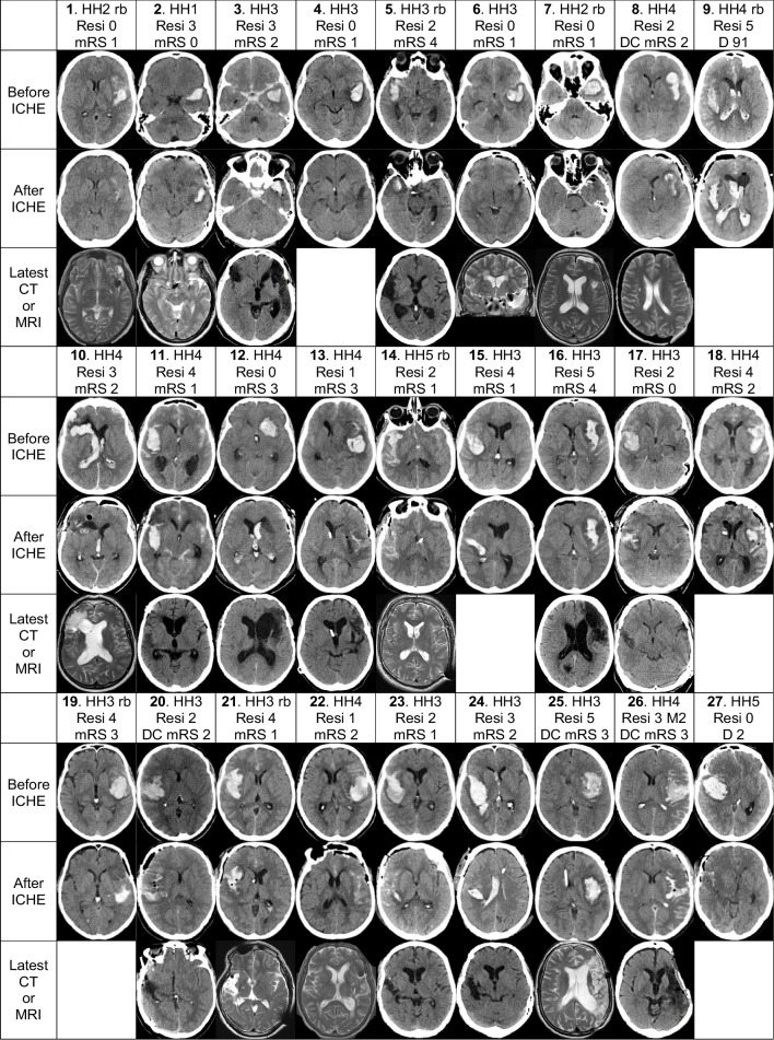
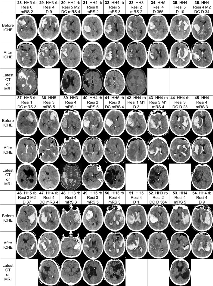
Methods: Our retrospective cohort consists of 54 consecutive aICH patients from a defined population who acutely underwent the clipping of a middle cerebral artery bifurcation saccular aneurysm (Mbif sIA) with the aICH evacuation at Kuopio University Hospital (KUH) from 2010 to 2019. We constructed the patient's individual timeline panels since the emergency call and serial brain CT/MRI slice panels through the aICH evacuation and neurointensive care until the final brain tissue outcome. The patients were indicated by numbers (1.-54.) in the pseudonymized panels, tables, results, and discussion.

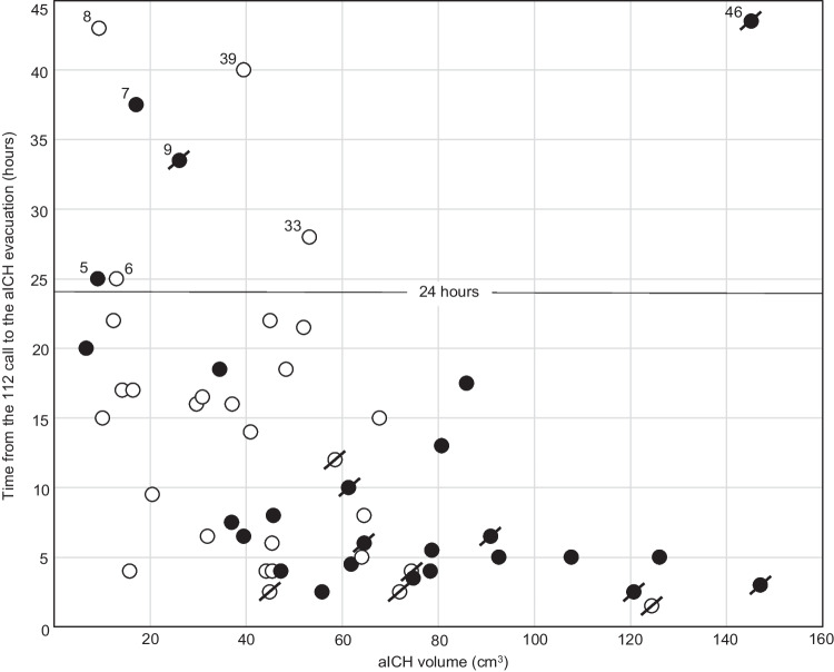
Results: The aICH volumes on KUH admission (median 46 cm3) plotted against the time from the emergency call to the evacuation (median 8 hours) associated significantly with the rebleeds (n=25) and the deaths (n=12). The serial CT/MRI slice panels illustrated the aICHs, intraventricular hemorrhages (aIVHs), residuals after the aICH evacuations, perihematomal edema (PHE), delayed cerebral injury (DCI), and in the 42 survivors, the clinical outcome (mRS) and the brain tissue outcome.

Conclusions: Regarding aICH evacuations, serial brain CT/MRI panels present more information than words, figures, and graphs. Re-bleeds associated with larger aICH volumes and worse outcomes. Swift logistics until the sIA occlusion with aICH evacuation is required, also in duty hours and weekends. Intraoperative CT is needed to illustrate the degree of aICH evacuation. PHE may evoke uncontrollable intracranial pressure (ICP) in spite of the acute aICH volume reduction.

Keywords: Aneurysmal intracerebral hematoma; Brain tissue outcome; EMS Emergency medical services; Individual serial brain imaging panel; Microsurgical evacuation and clipping; Perihematomal edema.


Conflict of interest statement

The authors declare no competing interests.

Figures

Fig. 1
Fig. 1
Flowchart. A total of 613 consecutive patients were acutely admitted—within 24 h from the CT diagnosis of the first subarachnoid hemorrhage (SAH) – to the neurointensive care at the Kuopio University Hospital (KUH) between 2010 and 2019 from a defined population. A total of 146 patients had a saccular intracranial aneurysm (sIA) on the middle cerebral artery bifurcation (Mbif), with 83 / 146 (57%) causing an intracerebral hematoma (aICH). The 176 clinical date point timelines were re-constructed until the death (n= 56 / 176) or the last follow-up. The final study population consists of the 54 patients with the evacuation of the aICH during the microsurgical clipping of the Mbif sIA. Abbreviations: aICH, aneurysmal intracerebral hematoma; aSAH, aneurysmal subarachnoid hemorrhage; CT, computed tomography; HH, Hunt and Hess scale; KUH, Kuopio University Hospital; M1, middle cerebral artery M1 trunk segment; Mbif, middle cerebral artery bifurcation; Mdist, middle cerebral artery peripheral segments; SAH, subarachnoid hemorrhage; sIA, saccular intracranial aneurysm
Fig. 2
Fig. 2
Individual CT/MRI panels of the 54 patients. The serial CT/MRI scan panels of the 54 patients with an acute intracerebral hematoma (aICH) from a saccular intracranial aneurysm (sIA) on the middle cerebral artery (MCA) bifurcation (Mbif) (Flowchart in Fig 1). After the 112 call, the 54 aSAH patients were transferred by the emergency medical services (EMS) to the neurointensive care in the Kuopio University Hospital (KUH). A total of 25 (46%) patients had a clinical or CT-verified re-bleed (rb) before the sIA occlusion. All 54 patients (Patients 1.–54.) underwent the microsurgical clipping of the ruptured Mbif sIA with the intention to remove the aICH. Of the 54 patients, 17 (32%) had also an intraventricular hematoma (aIVH). For each patient, two axial CT slices were selected: CT 1. before the clipping and CT 2. after the clipping. The 54 CT 1. slices are arranged according to the increasing largest area of the aICH on admission. The latest available slice (CT or MRI) was selected for the survivors at about 12 months (not available in the Patients 4. 15. 18. 19.). The white data box contains: the patient number (1.–54.); the Hunt and Hess scale (HH) on admission; rb, re-bleed before sIA occlusion; decompressive craniectomy (DC; n=11); mRS 0 to 5 at about 12 months for the 42 survivors; D and days to death for the 12 deceased patients. The white box also contains the visual estimate of the aICH volume reduction (Resi) as sizes of the residuals (0–5) graded as follows: 0—no residual (n=9); 1—patchy small residual (n=4); 2—patchy residual (n=9); 3—small solid residual (n=10); 4—partial solid residual (n=16); 5—all solid left (n=6). M1 denotes the M1 occlusion in two patients (42. 43.) and M2 denotes the M2 branch occlusion in four patients (26. 30. 36. 46.) in the postoperative angiography. Abbreviations: aICH, aneurysmal intracerebral hematoma; aIVH, aneurysmal intraventricular hematoma; CT, computed tomography; D, deceased; DC, decompressive craniectomy; EMS, emergency medical services; HH, Hunt and Hess scale; ICHE, aICH evacuation; KUH, Kuopio University Hospital; M1, middle cerebral artery M1 trunk segment; M2, middle cerebral artery M2 branch segments; Mbif, middle cerebral artery bifurcation; MCA, middle cerebral artery; MRI, magnetic resonance imaging; mRS, modified Rankin Scale; rb, re-bleed before the sIA occlusion; Resi, visual estimate of the aICH volume reduction in the first CT after the evacuation; sIA, saccular intracranial aneurysm
Fig. 2
Fig. 2
Individual CT/MRI panels of the 54 patients. The serial CT/MRI scan panels of the 54 patients with an acute intracerebral hematoma (aICH) from a saccular intracranial aneurysm (sIA) on the middle cerebral artery (MCA) bifurcation (Mbif) (Flowchart in Fig 1). After the 112 call, the 54 aSAH patients were transferred by the emergency medical services (EMS) to the neurointensive care in the Kuopio University Hospital (KUH). A total of 25 (46%) patients had a clinical or CT-verified re-bleed (rb) before the sIA occlusion. All 54 patients (Patients 1.–54.) underwent the microsurgical clipping of the ruptured Mbif sIA with the intention to remove the aICH. Of the 54 patients, 17 (32%) had also an intraventricular hematoma (aIVH). For each patient, two axial CT slices were selected: CT 1. before the clipping and CT 2. after the clipping. The 54 CT 1. slices are arranged according to the increasing largest area of the aICH on admission. The latest available slice (CT or MRI) was selected for the survivors at about 12 months (not available in the Patients 4. 15. 18. 19.). The white data box contains: the patient number (1.–54.); the Hunt and Hess scale (HH) on admission; rb, re-bleed before sIA occlusion; decompressive craniectomy (DC; n=11); mRS 0 to 5 at about 12 months for the 42 survivors; D and days to death for the 12 deceased patients. The white box also contains the visual estimate of the aICH volume reduction (Resi) as sizes of the residuals (0–5) graded as follows: 0—no residual (n=9); 1—patchy small residual (n=4); 2—patchy residual (n=9); 3—small solid residual (n=10); 4—partial solid residual (n=16); 5—all solid left (n=6). M1 denotes the M1 occlusion in two patients (42. 43.) and M2 denotes the M2 branch occlusion in four patients (26. 30. 36. 46.) in the postoperative angiography. Abbreviations: aICH, aneurysmal intracerebral hematoma; aIVH, aneurysmal intraventricular hematoma; CT, computed tomography; D, deceased; DC, decompressive craniectomy; EMS, emergency medical services; HH, Hunt and Hess scale; ICHE, aICH evacuation; KUH, Kuopio University Hospital; M1, middle cerebral artery M1 trunk segment; M2, middle cerebral artery M2 branch segments; Mbif, middle cerebral artery bifurcation; MCA, middle cerebral artery; MRI, magnetic resonance imaging; mRS, modified Rankin Scale; rb, re-bleed before the sIA occlusion; Resi, visual estimate of the aICH volume reduction in the first CT after the evacuation; sIA, saccular intracranial aneurysm
Fig. 3
Fig. 3
The 54 patients’ aICH volumes in relation to the time elapsed to the aICH evacuation. The 54 patients with their aICH volumes on admission and their individual times from the 112 call to the aICH evacuation during the microsurgical clipping of the ruptured Mbif sIA. The x-axis presents the 54 aICH volumes (cm3) and the y-axis presents the corresponding times (hours). The 54 aICH volumes distributed as follows 7–31–46–75–147 cm3 and the 54 times 1–4–8–18–43 hours (shortest – 25% quartile – median – 75% quartile – longest). In a total of 8 patients, the times exceeded 24 hours (horizontal line), in association with a small aICH (Patients 5. 6. 7. 8. 9. 33.), good condition (Patient 39.), or poor initial condition (Patient 46.). The time to the sIA clipping significantly shortened with the increasing aICH volume (p < 0,001). Re-bleeds (black circle) occurred in the 25 (46%) patients, and 7 (28%) of them deceased ( / ). No re-bleeds (white circle) occurred in the 29 (54%) patients, and 5 (17%) of them deceased ( / ). The re-bleeds associated significantly with the increasing aICH volume (p = 0,006). Abbreviations: aICH, aneurysmal intracerebral hematoma; cm3, cubic centimeter; Mbif, middle cerebral artery bifurcation; sIA, saccular intracranial aneurysm
Fig. 4
Fig. 4
The 54 patients arranged according to their largest aICH area and aICH residual grade. The 54 patients with an acute aneurysmal intracerebral hematoma (aICH) evacuated during the clipping of the ruptured middle cerebral artery (MCA) bifurcation (Mbif) saccular intracranial aneurysm (sIA). The patients (1.–54.) are arranged according to the increasing largest area of the aICH in the preoperative CT scan (Fig 2). The residual aICH volume (Resi) in the first postoperative CT scan was visually graded (0 to 5) as follows: 0 – no residual (n=9; 17%); 1 – patchy small residual (n=4; 7%); 2 – patchy residual (n=9; 17%); 3 – small solid residual (n=10; 19%); 4 – partial solid residual (n=16; 30%); 5 – all solid left (n=6; 11%). The outcome until 12 months is indicated by the dot colour: death (black, n=12, patient numbers in bold) and alive (white, n=42). Abbreviations: aICH, aneurysmal intracerebral hematoma; CT, computed tomography; Mbif, middle cerebral artery bifurcation; MCA, middle cerebral artery; Resi, visual estimate of the aICH volume reduction in the first CT after the evacuation; sIA, saccular intracranial aneurysm

Similar articles

Cited by

References

    1. Arnold TC, Freeman CW, Litt B, Stein JM (2023) Low-field MRI: Clinical promise and challenges. J Magn Reson Imaging. 10.1002/jmri.28408 - PMC - PubMed
    1. Autio AH, Paavola J, Tervonen J, Lång M, Huuskonen TJ, Huttunen J, Kärkkäinen V, von Und Z, Fraunberg M, Lindgren AE, Koivisto T, Kurola J, Jääskeläinen JE, Kämäräinen OP (2023) Should individual timeline and serial CT/MRI panels of all patients be presented in acute brain insult cohorts? A pilot study of 45 patients with decompressive craniectomy after aneurysmal subarachnoid hemorrhage. Acta Neurochir. 10.1007/s00701-022-05473-7 - PMC - PubMed
    1. Autio AH, Paavola J, Tervonen J, Lång M, Huuskonen TJ, Huttunen J, Kärkkäinen V, von Und Z, Fraunberg M, Lindgren AE, Koivisto T, Jääskeläinen JE, Kämäräinen OP (2021) Clinical condition of 120 patients alive at 3 years after poor-grade aneurysmal subarachnoid hemorrhage. Acta Neurochir. 10.1007/s00701-021-04725-2 - PMC - PubMed
    1. Baldvinsdóttir B, Kronvall E, Ronne-Engström E, Enblad P, Lindvall P, Aineskog H, Friðriksson S, Klurfan P, Svensson M, Alpkvist P, Hillman J, Eneling J, Nilsson OG (2023) Adverse events associated with microsurgial treatment for ruptured intracerebral aneurysms: a prospective nationwide study on subarachnoid haemorrhage in Sweden. J Neurol Neurosurg Psychiatry. 10.1136/jnnp-2022-330982 - PubMed
    1. Bohnstedt BN, Nguyen HS, Kulwin CG, Shoja MM, Helbig GM, Leipzig TJ, Payner TD, Cohen-Gadol AA (2013) Outcomes for clip ligation and hematoma evacuation associated with 102 patients with ruptured middle cerebral artery aneurysms. World Neurosurg. 10.1016/j.wneu.2012.03.008 - PubMed

Publication types