Clinical Trial
. 2024 Apr 1;35(4):483-494.
doi: 10.1681/ASN.0000000000000311. Epub 2024 Jan 17.

Urinary CD4 + T Cells Predict Renal Relapse in ANCA-Associated Vasculitis

Affiliations
Clinical Trial

Urinary CD4 + T Cells Predict Renal Relapse in ANCA-Associated Vasculitis

Luka Prskalo et al. J Am Soc Nephrol. .

Abstract

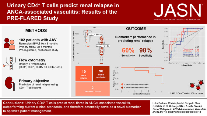
Significance statement: Early identification of patients at risk of renal flares in ANCA vasculitis is crucial. However, current clinical parameters have limitations in predicting renal relapse accurately. This study investigated the use of urinary CD4 + T lymphocytes as a predictive biomarker for renal flares in ANCA vasculitis. This study, including urine samples from 102 patients, found that the presence of urinary CD4 + T cells was a robust predictor of renal relapse within a 6-month time frame, with a sensitivity of 60% and a specificity of 97.8%. The diagnostic accuracy of urinary CD4 + T cells exceeded that of ANCA titers, proteinuria, and hematuria. Monitoring urinary CD4 + T lymphocytes could help assess the risk of future renal relapse, enabling early preventive measures and tailored treatment strategies.

Background: In ANCA-associated vasculitis, there is a lack of biomarkers for predicting renal relapse. Urinary T cells have been shown to differentiate active GN from remission in ANCA-associated vasculitis, but their predictive value for renal flares remains unknown.

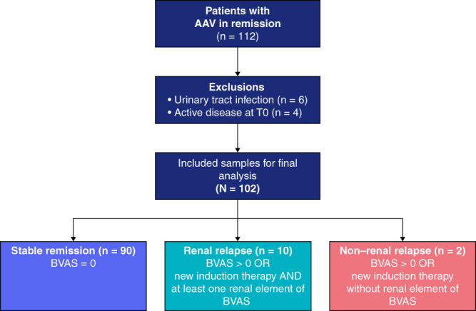
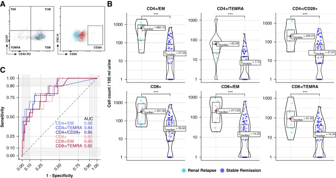
Methods: The PRE-FLARED study was a prospective multicenter biomarker study including 102 individuals with ANCA-associated vasculitis in remission aimed to predict renal relapse by quantifying urinary CD4 + T-cell subsets using flow cytometry at baseline and monitoring clinical outcomes over a 6-month follow-up.

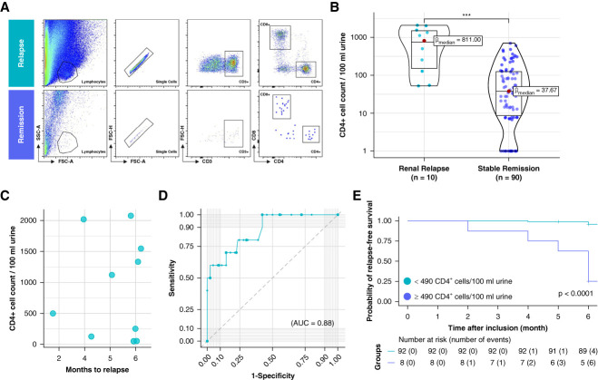
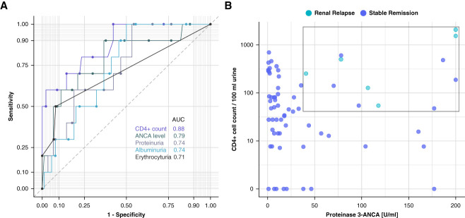
Results: Among the participants, ten experienced renal relapses, two had non-renal flares, and 90 remained in stable remission. The median baseline urinary CD4 + T-cell count was significantly higher in patients who relapsed compared with those in remission. Receiver operating characteristic curve analysis of urinary CD4 + T-cell counts showed an area under the curve value of 0.88 for predicting renal flares, outperforming ANCA titers, hematuria, and proteinuria. Using a cutoff of 490 CD4 + T cells per 100 ml urine, the sensitivity and specificity in identifying patients with future renal flares were 60% and 97.8%, respectively. In a post hoc analysis, combining urinary CD4 + T-cell counts with proteinase-3 ANCA levels suggested improved predictive performance in the PR3 + subgroup. In addition, the number of urinary CD4 + T cells showed a limited correlation with a decline in GFR and an increase in proteinuria over the follow-up period.

Conclusions: This study concluded that urinary CD4 + T-cell counts could identify patients with ANCA-associated vasculitis at a substantial risk of renal relapse within 6 months. Combining these counts with ANCA levels further improved the prediction of relapse.

Clinical trial registry name and registration number: Urinary T Lymphocytes Predict Renal Flares in Patients With Inactive ANCA-associated Glomerulonephritis (PRE-FLARED), NCT04428398 .


Conflict of interest statement

M. Bieringer reports employment with Helios Klinikum Berlin-Buch and speakers bureau for Vifor Pharma Deutschland GmbH. K.-U. Eckardt reports employment with Charité - Universitätsmedizin Berlin; consultancy for Akebia, AstraZeneca, Boehringer Ingelheim, GSK, Novartis, and Otsuka; research funding from Bayer, Evotec, and Travere; honoraria from Akebia, AstraZeneca, Bayer, Boehringer Ingelheim, GSK, Novartis, Sanofi, and Otsuka; and role on the Editorial Boards of BMJ and Kidney International. P. Enghard reports employment with Charité; consultancy for GlaxoSmithKline Advisory Board; ownership interest in Gilead (stocks); research funding from Hansa Biopharm; and honoraria from Akademie der Nieren, AstraZeneca, BDI, GlaxoSmithKline, NAWBerlin, and NephroUpdate. P. Enghard has submitted a patent for conserving urinary cells for subsequent analysis using flow cytometry. P. Freund reports employment with Charité and Alexianer St. Hedwig Kliniken Berlin; consultancy for Aware Health GmbH; and patents or royalties for US2022/0003748A1 and EP2019/086433. P. Freund has submitted a patent for conserving urinary cells for subsequent analysis using flow cytometry. E. Grothgar, J. Klocke, and L. Wagner report employment with Charité Universitätsmedizin Berlin. F. Hiepe reports consultancy for AstraZeneca; ownership interest in Methuselah Bio AG; research funding from Sanofi; patents or royalties from Methuselah Bio AG; advisory or leadership roles for AstraZeneca and Methuselah Bio AG; and speakers bureau for AstraZeneca. D. Metzke reports employment with Charité and patents or royalties from Charité. D. Metzke has submitted a patent for conserving urinary cells for subsequent analysis using flow cytometry. L. Prskalo reports report employment with Charité Universitätsmedizin Berlin and has received funding from the Berlin Institute of Health and the German Academic Exchange Service (BIH-MD-TRENAL Stipend). A.D. Salama reports consultancy for Hansa Medical and Vifor; research funding from Chiesi; honoraria from AnaptysBio, AstraZeneca, Hansa Medical, and Vifor Pharmaceuticals; patent application number 2301306.3 February 2023: Treatment of Granulomatous diseases; and advisory or leadership role for UK Kidney Association. U. Schneider reports employment with Charité Universitaetsmedizin Berlin; consultancy for Boehringer-Ingelheim, Germany, and Vifor, Germany; honoraria from AstraZeneca, Germany, Boehringer-Ingelheim, Germany, GSK, Germany, and Vifor Pharma, Germany; and speakers bureau for Boehringer-Ingelheim, Germany, GSK, Germany, and Sobi, Germany. A. Schreiber reports employment with Charite Berlin; consultancy for Alexion, Hansa Biopharm, Otsuka, Sanofi, Stadapharm, Travere, and Vifor Pharma; research funding from Biontech, eleva GmbH, and Lempo; advisory or leadership roles for Alexion, Hansa Biopharm, Otsuka, Sanofi, and Travere; and other interests or relationships with Dt. Gesellschaft für Nephrologie. C.M. Skopnik reports employment with Charité Universitaetsmedizin Berlin and has submitted a patent for conserving urinary cells for subsequent analysis using flow cytometry. All remaining authors have nothing to disclose.

Figures

None
Graphical abstract
Figure 1
Figure 1
PRE-FLARED study population. A total of 102 patients with AAV in remission were included in this study. After 6-month follow-up, patients were grouped into stable remission, renal relapse, or non–renal relapse. AAV, ANCA-associated vasculitis; BVAS, Birmingham Vasculitis Activity Score.
Figure 2
Figure 2
Urinary CD4+ T lymphocytes predict renal relapse in patients with ANCA-associated vasculitis in remission during a 6-month follow-up. (A) Flow cytometric gating strategy of T-lymphocyte subsets shown in one patient with subsequent renal relapse and one patient in continued stable remission. (B) Comparison of absolute CD4+ T-cell counts per 100 ml of urine between renal relapse and stable remission. (C) Selection of patients with renal relapse. Shown are the corresponding urinary CD4+ T-cell counts and the timing of renal relapse over the follow-up period. (D) ROC curve analysis showing the diagnostic performance of urinary CD4+ T lymphocytes in predicting renal relapse in patients with ANCA-associated vasculitis. (E) Kaplan–Meier analysis on renal relapse-free survival. AUC value, 0.88. Significance levels: ns, no significance, ***P < 0.001. AUC, area under the curve; FSC, forward scatter; ROC, receiver operating characteristic; SSC, side scatter.
Figure 3
Figure 3
Predictive performance of different T-lymphocyte phenotypes. (A) Gating strategy of flow cytometric analysis for detection of additional T-lymphocyte populations. FMO control is shown in blue, and full stain is displayed in red. (B) Selection of T-cell populations with significant difference in absolute cell counts per 100 ml of urine between renal relapse and stable remission. (C) ROC curve analysis of selected T-cell populations to distinguish renal relapse from stable remission. Corresponding ROC curves and AUC values shown in distinct color shades, in pink CD4+ subpopulations, in green CD8+ subpopulations. Significance levels: ns, ***P < 0.001. FMO, fluorescence-minus-one; ns, no significance; TCM, T central memory cells; TEM, T effector memory cells; TEMRA, T effector memory cells reexpressing CD45RA; TNV, naive T cells.
Figure 4
Figure 4
Relationship between change in kidney function and urinary CD4+ T lymphocytes. (A) Spearman correlation scatter plot between CD4+ cell counts per 100 ml urine and loss of GFR in ml/min from baseline to follow-up time point after 6 months. Black line represents linear regression with 95% CI. In addition to renal relapse and stable remission, patients with non–renal relapse were also considered for analysis (N=102). (B) Spearman correlation between CD4+ cell counts per 100 ml urine and change in UPCR in mg/g from baseline to follow-up time point at 6 months. Black line represents linear regression with 95% CI. Tow outliers are also displayed with the respective value for change in protein/creatinine ratio indicated. CI, confidence interval; UPCR, urine protein-to-creatinine ratio.
Figure 5
Figure 5
Enhanced diagnostic performance of urinary CD4+ T lymphocytes over current clinical screening parameters, with improved diagnostic yield in PR3+ subgroup analysis, at the 6-month follow-up. (A) ROC curve analysis to distinguish renal relapse from stable remission of CD4+ T-cell biomarker compared with serum ANCA levels, proteinuria, albuminuria, and erythrocyturia. ROC curves and AUC values are shown in different shades. (B) Selection of PR3+ subgroup (n=72). All relapses cluster in the depicted upper left quadrant. PR3, proteinase-3.

Similar articles

Cited by

References

    1. Kitching AR Anders H-J Basu N, et al. . ANCA-associated vasculitis. Nat Rev Dis Primers. 2020;6(1):71. doi:10.1038/s41572-020-0204-y - DOI - PubMed
    1. Hoffman GS Kerr GS Leavitt RY, et al. . Wegener granulomatosis: an analysis of 158 patients. Ann Intern Med. 1992;116(6):488–498. doi:10.7326/0003-4819-116-6-488 - DOI - PubMed
    1. Jennette JC, Falk RJ. Small-vessel vasculitis. N Engl J Med. 1997;337(21):1512–1523. doi:10.1056/NEJM199711203372106 - DOI - PubMed
    1. Falk RJ, Hogan S, Carey TS, Jennette JC. Clinical course of anti-neutrophil cytoplasmic autoantibody-associated glomerulonephritis and systemic vasculitis. The Glomerular Disease Collaborative Network. Ann Intern Med. 1990;113(9):656–663. doi:10.7326/0003-4819-113-9-656 - DOI - PubMed
    1. Fauci AS, Haynes BF, Katz P, Wolff SM. Wegener’s granulomatosis: prospective clinical and therapeutic experience with 85 patients for 21 years. Ann Intern Med. 1983;98(1):76–85. doi:10.7326/0003-4819-98-1-76 - DOI - PubMed

Substances

Associated data

Grants and funding

LinkOut - more resources