Review
. 2023 Nov 22;15(23):4483.
doi: 10.3390/polym15234483.

On the Mechanism of the Ionizing Radiation-Induced Degradation and Recycling of Cellulose

Affiliations
Review

On the Mechanism of the Ionizing Radiation-Induced Degradation and Recycling of Cellulose

Richard List et al. Polymers (Basel). .

Abstract

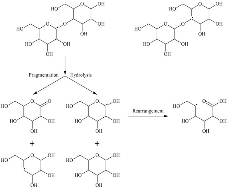
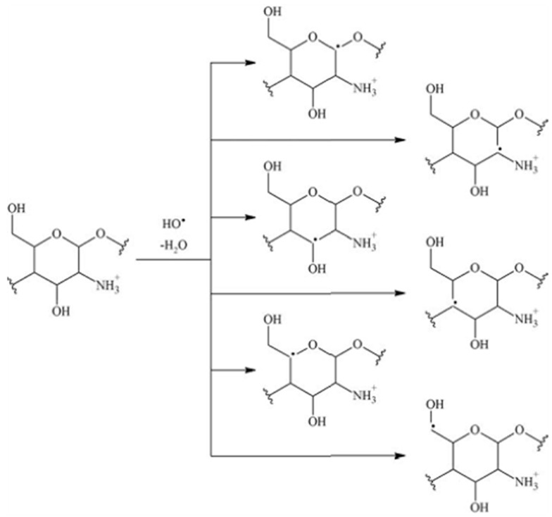
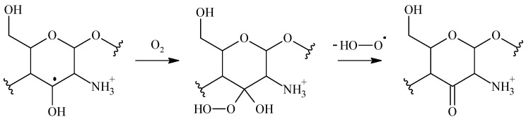
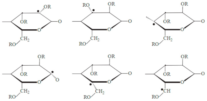
The use of ionizing radiation offers a boundless range of applications for polymer scientists, from inducing crosslinking and/or degradation to grafting a wide variety of monomers onto polymeric chains. This review in particular aims to introduce the field of ionizing radiation as it relates to the degradation and recycling of cellulose and its derivatives. The review discusses the main mechanisms of the radiolytic sessions of the cellulose molecules in the presence and absence of water. During the radiolysis of cellulose, in the absence of water, the primary and secondary electrons from the electron beam, and the photoelectric, Compton effect electrons from gamma radiolysis attack the glycosidic bonds (C-O-C) on the backbone of the cellulose chains. This radiation-induced session results in the formation of alkoxyl radicals and C-centered radicals. In the presence of water, the radiolytically produced hydroxyl radicals (OH) will abstract hydrogen atoms, leading to the formation of C-centered radicals, which undergo various reactions leading to the backbone session of the cellulose. Based on the structures of the radiolytically produced free radicals in presence and absence of water, covalent grafting of vinyl monomers on the cellulose backbone is inconceivable.

Keywords: cellulose ionizing-radiation; degradation; recycling; structure.


Conflict of interest statement

The authors declare no conflict of interest.

Figures

Figure 1
Figure 1
Schematic of the scissioning of an organic molecule after electron beam irradiation. The schematic shows the formation of two radicals, represented by dots, and the reactions that lead to radical decay. Figure taken from Chaudhary et al. [91].
Figure 2
Figure 2
Schematic of the radiation-induced curing of a monomer (M) through polymerization and crosslinking reactions. Figure taken from Chaudhary et al. [91].
Figure 3
Figure 3
Schematic of radiation-induced grafting of monomer (M) onto polymer (A). Figure taken from Chaudhary et al. [91].
Figure 4
Figure 4
Schematic of the cleavage of the glycosidic bond and the chain scission of cellobiose during solid-state irradiation. Figure taken from Al-Assaf et al. [104].
Figure 5
Figure 5
Chemical structures of radicals formed upon HO attack on a polysaccharide molecule [104].
Figure 6
Figure 6
Rearrangement, hydrolysis, and fragmentation reactions during radiolysis of chitosan in oxygen-free aqueous solution [104].
Figure 7
Figure 7
Schematic of the peroxyl radical reaction in chitosan [104].
Figure 8
Figure 8
Schematic of the identified primary cellulose radicals formed after irradiation of cellulose at 10 kGy. Sample was irradiated at 77 K and heated up to 293 K. The structures were determined based on the EPR measurements, which tracked the transformation of the radicals as the temperature was increased. Figure taken from Wach et al. [150].
Figure 9
Figure 9
EPR spectra of irradiated microcrystalline cellulose (MCC) at different total doses (a) 1200 kGy, (b) 1000 kGy, (c) 800 kGy, (d) 600 kGy, (e) 400 kGy, (f) 200 kGy, (g) 0 kGy. The spectrum for the unirradiated (0 kGy, sample (g)) shows no radicals on the sample. As expected, as the dose is increased, a more intense signal is observed, with the sample irradiated at 1200 kGy (sample (a)) having the largest signal. Figure taken from Liu et al. [139].
Figure 10
Figure 10
Monomer unit of cellulose with the C atoms of the pyranose ring identified according to convention. Figure is used to illustrate the origin of the signals obtained in the EPR spectrum.
Figure 11
Figure 11
EPR signal intensity as a function of dose for flax fibers irradiated from 0 to 100 kGy. The concentration of radicals can be determined by integrating the EPR spectrum. Thus, it is directly proportional to the signal intensity shown in this figure. Figure taken from Taibi et al. [162].
Figure 12
Figure 12
Molecular weight distribution of unbleached kraft pulp (UBKP) after electron beam irradiation at varying doses. At low does (from 2.5 to 5.0 kGy), a bimodal distribution is observed. At 25 kGy, the plot shows a transition which looks like a bump in the bell curve. After 50 kGy, there is increased degradation, which appears in the figure as a single distribution plot. Figure taken from Sarosi et al. [118].
Figure 13
Figure 13
XRD spectra of irradiated cellulose at different doses: (a) 0 kGy, (b) 10 kGy, (c) 50 kGy, (d) 100 kGy, (e) 300 kGy, and (f) 500 kGy. The characteristic peaks of the cellulose at 16.6°, and 22.7° can be seen at all doses. Figure taken from Sun et al. [179].
Figure 14
Figure 14
(a) 1D and (b) 2D, 13C NMR spectra of bacterial cellulose. The 1D spectra has been labeled with the respective C atom positions according to the cellulose structure. Figure taken from Masuda et al. [190].
Figure 15
Figure 15
Raman spectra, normalized by the 1096 cm−1 peak, of the samples used for calibration with their calculated CrIs shown on the right. The samples were mixtures derived from cotton microcrystalline cellulose. Figure taken from Agarwal et al. [204].

References

    1. Peng B.L., Dhar N., Liu H.L., Tam K.C. Chemistry and Applications of Nanocrystalline Cellulose and Its Derivatives: A Nanotechnology Perspective. Can. J. Chem. Eng. 2011;89:1191–1206. doi: 10.1002/cjce.20554. - DOI
    1. Keshk S., Sameshima K. Influence of Lignosulfonate on Crystal Structure and Productivity of Bacterial Cellulose in a Static Culture. Enzym. Microb. Technol. 2006;40:4–8. doi: 10.1016/j.enzmictec.2006.07.037. - DOI
    1. Klemm D., Heublein B., Fink H.-P., Bohn A. Cellulose: Fascinating Biopolymer and Sustainable Raw Material. Angew. Chem. Int. Ed. 2005;44:3358–3393. doi: 10.1002/anie.200460587. - DOI - PubMed
    1. Brown R.M. The Biosynthesis of Cellulose. J. Macromol. Sci. Part A. 1996;33:1345–1373. doi: 10.1080/10601329608014912. - DOI
    1. Ross P., Mayer R., Benziman M. Cellulose Biosynthesis and Function in Bacteria. Microbiol. Rev. 1991;55:35–58. doi: 10.1128/mr.55.1.35-58.1991. - DOI - PMC - PubMed

LinkOut - more resources