

Chitosan-Polyvinyl Alcohol Nanocomposites for Regenerative Therapy
- PMID: 38232016
- PMCID: PMC10708655
- DOI: 10.3390/polym15234595
Chitosan-Polyvinyl Alcohol Nanocomposites for Regenerative Therapy
Abstract
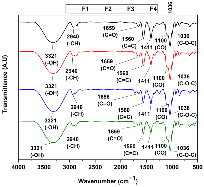
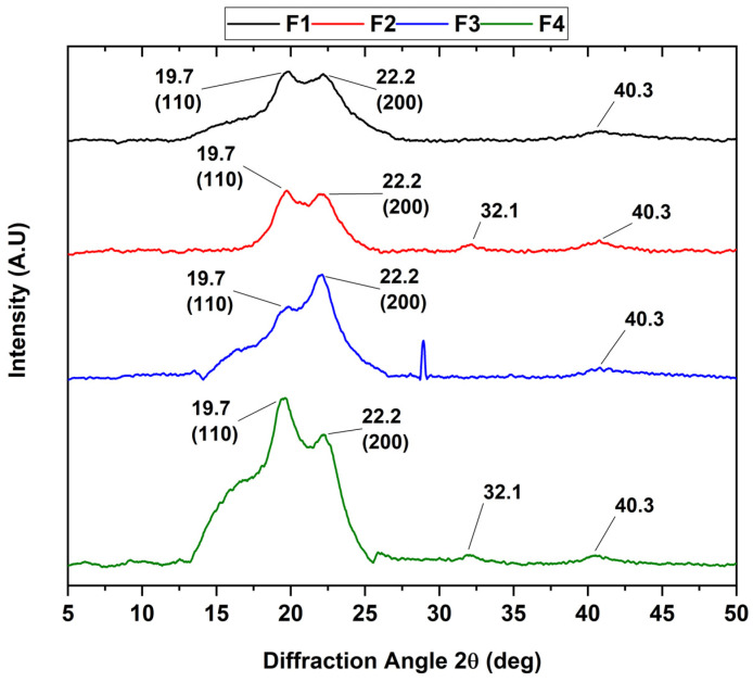
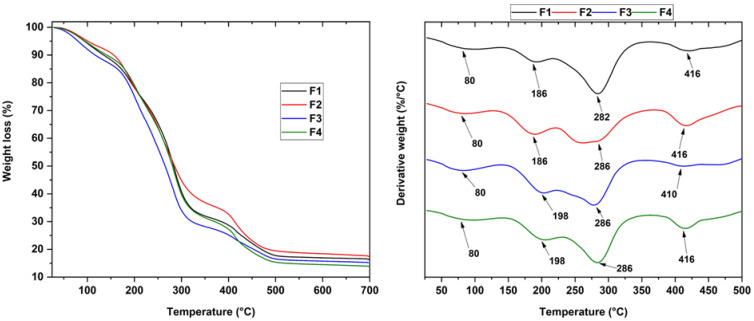
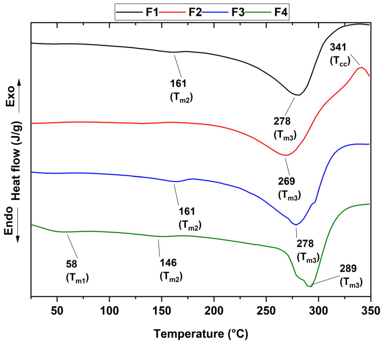
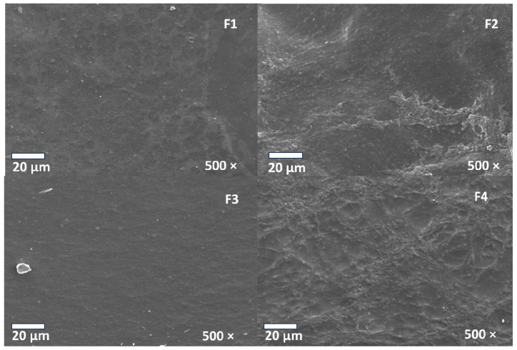
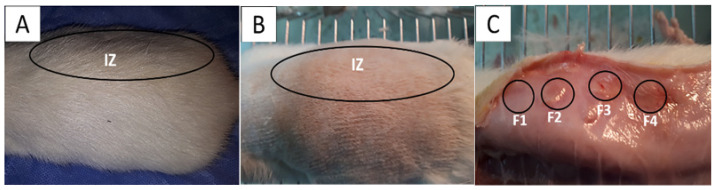
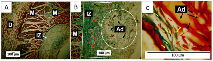
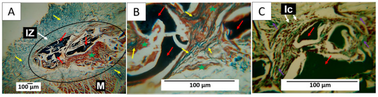
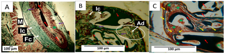
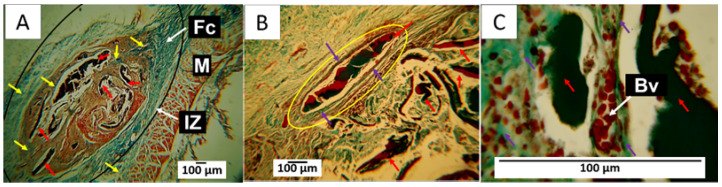
Tissue accidents provide numerous pathways for pathogens to invade and flourish, causing additional harm to the host tissue while impeding its natural healing and regeneration. Essential oils (EOs) exhibit rapid and effective antimicrobial properties without promoting bacterial resistance. Clove oils (CEO) demonstrate robust antimicrobial activity against different pathogens. Chitosan (CS) is a natural, partially deacetylated polyamine widely recognized for its vast antimicrobial capacity. In this study, we present the synthesis of four membrane formulations utilizing CS, polyvinyl alcohol (PVA), and glycerol (Gly) incorporated with CEO and nanobioglass (n-BGs) for applications in subdermal tissue regeneration. Our analysis of the membranes' thermal stability and chemical composition provided strong evidence for successfully blending polymers with the entrapment of the essential oil. The incorporation of the CEO in the composite was evidenced by the increase in the intensity of the band of C-O-C in the FTIR; furthermore, the increase in diffraction peaks, as well as the broadening, provide evidence that the introduction of CEO perturbed the crystal structure. The morphological examination conducted using scanning electron microscopy (SEM) revealed that the incorporation of CEO resulted in smooth surfaces, in contrast to the porous morphologies observed with the n-BGs. A histological examination of the implanted membranes demonstrated their biocompatibility and biodegradability, particularly after a 60-day implantation period. The degradation process of more extensive membranes involved connective tissue composed of type III collagen fibers, blood vessels, and inflammatory cells, which supported the reabsorption of the composite membranes, evidencing the material's biocompatibility.
Keywords: chitosan composites; clove essential oil; nanobioglass; scaffolds; subdermal tissue regeneration.
Conflict of interest statement
The authors declare no conflict of interest.
Figures
Similar articles
-
Histology Assessment of Chitosan-Polyvinyl Alcohol Scaffolds Incorporated with CaO Nanoparticles.Molecules. 2025 Jan 12;30(2):276. doi: 10.3390/molecules30020276. Molecules. 2025. PMID: 39860146 Free PMC article.
-
Polycaprolactone (PCL)-Polylactic Acid (PLA)-Glycerol (Gly) Composites Incorporated with Zinc Oxide Nanoparticles (ZnO-NPs) and Tea Tree Essential Oil (TTEO) for Tissue Engineering Applications.Pharmaceutics. 2022 Dec 22;15(1):43. doi: 10.3390/pharmaceutics15010043. Pharmaceutics. 2022. PMID: 36678672 Free PMC article.
-
Preparation, characterization and antimicrobial activity of polyvinyl alcohol/gum arabic/chitosan composite films incorporated with black pepper essential oil and ginger essential oil.Int J Biol Macromol. 2020 May 15;151:366-375. doi: 10.1016/j.ijbiomac.2020.02.176. Epub 2020 Feb 18. Int J Biol Macromol. 2020. PMID: 32084477
-
Overview of bacterial cellulose composites: a multipurpose advanced material.Carbohydr Polym. 2013 Nov 6;98(2):1585-98. doi: 10.1016/j.carbpol.2013.08.018. Epub 2013 Aug 15. Carbohydr Polym. 2013. PMID: 24053844 Review.
-
Polyvinyl Alcohol-Chitosan Scaffold for Tissue Engineering and Regenerative Medicine Application: A Review.Mar Drugs. 2023 May 17;21(5):304. doi: 10.3390/md21050304. Mar Drugs. 2023. PMID: 37233498 Free PMC article. Review.
Cited by
-
Bryophyte-Bioinspired Nanoporous AAO/C/MgO Composite for Enhanced CO2 Capture: The Role of MgO.Nanomaterials (Basel). 2024 Apr 10;14(8):658. doi: 10.3390/nano14080658. Nanomaterials (Basel). 2024. PMID: 38668152 Free PMC article.
-
Impact of Carbon-Based Nanoparticles on Polyvinyl Alcohol Polarizer Features: Photonics Applications.Nanomaterials (Basel). 2024 Apr 23;14(9):737. doi: 10.3390/nano14090737. Nanomaterials (Basel). 2024. PMID: 38727331 Free PMC article.
-
Lyophilized Polyvinyl Alcohol and Chitosan Scaffolds Pre-Loaded with Silicon Dioxide Nanoparticles for Tissue Regeneration.Molecules. 2024 Aug 14;29(16):3850. doi: 10.3390/molecules29163850. Molecules. 2024. PMID: 39202929 Free PMC article.
References
-
- O’Brien F.J. Biomaterials & scaffolds for tissue engineering. Mater. Today. 2011;14:88–95. doi: 10.1016/S1369-7021(11)70058-X. - DOI
-
- Shelke N.B., James R., Laurencin C.T. Polysaccharide biomaterials for drug delivery and regenerative engineering. Polym. Adv. Technol. 2014;25:448–460. doi: 10.1002/pat.3266. - DOI
Grants and funding
LinkOut - more resources
Full Text Sources
Miscellaneous
